SlideShare a Scribd company logo
1 of 228
BY
DR. Aamir Mehmood Butt
fcps
Incharge department of
prosthodontics,Lumhs
WHAT IS A DENTAL IMPLANT?
Dental implant is an artificial titanium fixture
(similar to those used in orthopedics)
which is placed surgically into the jaw bone to
substitute for a missing tooth and its root(s).
OR
 A permucosal device which is biocompatible
and biofunctional and is placed within mucosa
or, on or within the bone associated with the
oral cavity to provide support for fixed or
removable prosthetics.
Types of dental implant
1. Mucosal Insert
2. Endodontic Implant (Stabilizer)
3. Sub-periosteal implant
4. Endosteal or Endosseous implant
 Plate-form implant
 Ramus-frame implant
 Root-form implant
5. Transosseous implant
Mucosal Insert
Endodontic Implant (Stabilizer)
 Endodontic stabilizer implants are
endosteal implants.
 threaded post that passes at least 5
mm beyond the apex of the tooth
root into available bone.
 providing additional abutment
support for restorative dentistry.
 Five millimeters of available apical
bone is the minimum that can
increase the crown-root ratio to an
extent sufficient to affect positively
the prognosis of the tooth.
Sub-periosteal implant
 introduced in the 1940.
 longest period of clinical
application.
 shaped to ride on the
residual bony ridge of
either the upper or lower.
 have been used in
completely edentulous as
well as partially
edentulous upper and
lower jaws. However, the
best results have been
achieved in treatment of
the edentulous lower jaw.
Sub-periosteal implant…
Indications
 Usually a severely
resorbed, completely
edentulous, lower jaw
bone which does not
offer enough bone
height to
accommodate Root
form Implants as
anchoring devices.
Endosteal or Endosseous implant
 Plate-form implant
 Ramus-frame implant
 Root-form implant
Plate-form implant
 Their name is derived from their flat,
blade-like (or plate-like) portion,
which is the part that gets embedded
into the bone.
 Used where the residual bone ridge
of the jaw is either too thin (due to
resorption) & Difficult to place
conventional Root form Implants or
certain vital anatomical structures
prevent conventional implants from
being placed.
 Bone grafting procedure, which re-
establishes the lost bone have
reduced the use of this form
Ramus-frame implant
 Ramus-frame Implants belong in the category of
endosseous implants, although their appearance
might not suggest that at first.
 These implants are designed for the edentulous lower
jaw only and are surgically inserted into the jaw bone
in three different areas: the left and right back area of
the jaw (the approximate area of the wisdom teeth),
and the chin area in the front of the mouth.
 The part of the implant that is visible in the mouth
after the implant is placed looks similar to that of the
Subperiosteal Implant.
Dr,salah hegazy
Dr,salah hegazy
 Indications:
Usually a severely resorbed, edentulous lower
jaw bone, which does not offer enough bone
height to accommodate Root form Implants as
anchoring devices. These implants are usually
indicated when the jaws are even resorbed to the
point where Subperiosteal Implants will not
suffice anymore.
Ramus-frame implant
Dr Aamir Butt
An additional advantage that comes with this
type of implant is a tripodial stabilization of the
lower jaw. A jaw as thin as the one shown above
can easily fracture at its thinnest part. The
Ramus-frame Implant, once integrated (after a
three month waiting period) will also stabilize
and protect the jaw somewhat from fracturing.
Dr Aamir Butt
The Ramus-frame Implant usually comes in a standard pre-shaped form and
needs to be custom-fitted to the patient's individual jaw dimension, as shown
below:
Dr,salah hegazy
Ramus-frame implant
C.Root form implant
Since the introduction of the Osseointegration
concept and the Titanium Screw by Dr.
Branemark, these implants have become the
most popular implants in the world today.
Root form Implants come in a variety of
shapes, sizes, and materials and are being
offered by many different companies
worldwide. Some clinicians regard them to be
the Standard of Care in Oral Implantology.
These implants can be placed wherever a
tooth or several teeth are missing, when
enough bone is available to accommodate
them. However, even if the bone volume is not
sufficient to place Root form Implants, Bone
grafting procedures within reasonable limits
should be initiated, in order to benefit from
these implants.
Root form implant shape:
Other variations dwell on the shape of the Root
form implant. Some are screw-shaped, others are
cylindrical, or even cone-shaped or any
combination thereof.
Today, the most accepted material for dental implants
is high grade Titanium—either CP Titanium or an
alloy thereof. The titanium alloy implants tend to be
stronger than the CP titanium implants. The bone
integration shows no difference to the two different
types of titanium.
Some implants have an outer coating of
Hydroxyapatite (HA). Other implants have their
surface altered through plasma spraying, or beading
process. This was developed to increase the surface
area of the titanium implant and, thus, in theory, give
them more stability. These surface treatments were
also offered as an alternative to the HA coatings,
which on some implants have shown to break loose or
even dissolve after a few years.
6. Transosseous implant
These implants are not in use that much
any more, because they necessitate an
extraoral surgical approach to their
placement, which again translates into
general anesthesia, hospitalization and higher
cost, but not necessarily higher benefits to the
patient.
In any case, these implants are used in
mandibles only and are secured at the lower
border of the chin via bone plates. These were
originally designed to have a secure implant
system, even for very resorbed lower jaws.
A typical Transosseous Implant. The plate on the
bottom is firmly pressed against the bottom part of
the chin bone, whereas the long screw posts go
through the chin bone, all the way to the top of the jaw
ridge inside the mouth. The two attachments that will
eventually protrude through the gums can be used to
attach an overdenture-type prosthesis.
The plate
long screw posts
The two attachments
History and Present status of Implant Dentistry
Ancient Implants
 16 th Dark stone
( Egyptian-South American)
 17 th Carved ivory teeth
Early Implants
 1809Gold implant
 e.20th Lead, iridium, tantalum, stainless steel,
and cobalt alloy
 1913 hollow basket
iridium + gold wires
(Greenfield)
Early Implants
 1937Adams’s submergible threaded cylindrical
implant with round bottom
 1938 Strock’s (long term) threaded vitallium
implant
(cobalt+chrome+molybdenum)
 The modern implants appear to be variants or
composites of some of the designs of early
implants
Subperiosteal Implants
 Placing implants on and around bone rather than in it
 1943 Dahl of Sweden placed with 4 projecting posts
 Direct bone impression
 Cobalt-chrome-molybdenum casting
 CT-generated CAD-CAM model
One-stage pins and screws
 Early 1960s pin, screw, and cylinder shaped implants
 One piece and not submerged
 Did not osseo-integration
 Fibrous peri-implant membrane
 Shock-absorbing claim
Blade Implants
 1967 Linkow blade implant-in narrow ridge
 Required shared support with natural teeth
 1970 Roberts and Roberts – Ramus blade
implant (titanium)
Transosteal Implants
 1975Small introduced transosteal mandibular staple
bone plate
 Limited to mandible only
Transosteal Implants
 1970Cranin - single transosteal implant
 1989 Bosker – transmandibular implant
 The First Dental Implant Consensus Conference,
sponsored by the National Institutes of Health (NIH)
and Harvard University in 1978, was a landmark event.
“ Dental Implants: Benefits and Risks”
 The Toronto Conference opened the door to prompt
widespread recognition of the Branemark implant.
 The discovery of osseointegration has been one of the
most significant scientific break throughs in dentistry.
Endosteal root-form implants
 1978Two-stage threaded titanium root-form implant
was first presented in North America by Branemark
(Toronto conference)
 Terms “fixture”
 First fixture was placed in 1965
 Well-documented, long term prospective study
Present Status
Three Basic:
“ In Bone”
“Through Bone”
“On Bone”
“In Bone”
 1. Ramus concepts (Harold and Ralph Roberts)
 2. Pin concepts (J. Scialom Michelle Chercheve)
 3. Disk concepts (Gerard Scorteci)
 4. Plateform concepts (Harold + Roberts/Linkow)
 5. Cylindrical or root form concepts
Present Status
 Many other root-forms have been introduced.
 Body shaped competition
 Surface competition – roughness
 Varieties competition
 Connection competition
Dental Implants
Implant material should have suitable:
 Mechanical strength,
 Biocompatibility,
 Structural Biostability in physiologic environments.
I. Modulus of elasticity
II. Tensile strength
III. Compressive strength
IV. Elongation
V. Metallurgy
BIOCOMPATIBILITY
 “The ability of an implanted material to undergo
only a minimal amount of deterioration during service,
to produce only a minimal change in the body
environment, and to function satisfactorily in every
other respect.”
KEY FACTORS THAT INFLUENCE THE BENEFITS AND
MAINTENANCE OF BIOCOMPATIBILITY
 Corrosion resistance
 Cytotoxicity of corrosion products
 Metal contamination
Biostability
Based on tissue response and systemic toxicity effects of
the implant:
 Biotolerant
 Bioinert
 Bioactive
Long term effects
 Biotolerant materials, such as polymethylmethacrylate
(PMMA), are usually characterized by thin fibrous
tissue interface.
 Chemical product irritate surrounding tissues.
Long term effects
 Bioinert materials, such as titanium and aluminum
oxide, are characterized by direct bone contact, or
osseointegration, at the interface under favorable
mechanical conditions.
 Non-reactive
Long term effects
 Bioactive materials, such as glass and calcium
phosphate ceramics, have a bone-implant interface
characterized by direct chemical bonding of the
implant with surrounding bone.
 Free calcium and phosphate compounds at the surface.
Tissue response to implant materials
 Most commonly used biomaterials:
 Commercially pure (CP) titanium
 Titanium-aluminum-vanadium alloy (Ti-6Al-4V)
 Cobalt-chromium-molydenum (Co-Cr-Mo) alloy is
most used for subperiosteal implants.
Tissue response to implant materials
 Calcium phosphate ceramics, Hydroxyapatite (HA),
used for augmentation material or coating on surface.
SUCCESS RATES
%
Subperiosteal 39 - 90
Staple 95
Vitreous carbon 50
Blade 65 - 90
Osseointegrated 80 - 100
Advantages & disadvantages of implant
over conventional treatment
 Implants do not involve preparation of the
adjacent teeth, they preserve the residual bone,
and excellent aesthetics can be achieved.
 However, it is expensive, the patient requires
surgery, time consuming, and technically
complex.
INDICATIONS FOR TREATMENT
Factors precluding wear of a removable prosthesis
 Poor anatomy for denture support
 Poor oral muscular coordination
 Poor mucosal tissue tolerance
 Parafunctional habits
 Unrealistic expectations
 Hyperactive gag reflex
 Psychological inability to wear
 Unfavourable number and location of
abutments
INDICATIONS
 Fully edentulous
 Partially edentulous
 Single tooth
INDICATIONS -
FULLY EDENTULOUS
 Poor retention
 Functional disturbances
 Psychological disturbances
Dr,salah hegazy
Diagnosis and
Treatment Planning
 The evaluation of a patient as a suitable
candidate for implants should follow the same
basic format as the standard patient
evaluation, although some areas require
additional emphasis and attention:
I. Medical History.
II. Psychological Status.
III. Dental History.
Dr,salah hegazy
I. Medical History
The patient’s medical history may reveal a number
of conditions that could complicate or even contra-
indicate implant therapy. These include:
1. Bleeding disorders; Paget’s disease; A history of
radiation therapy in the maxilla or mandible region;
Uncontrolled diabetes; Epilepsy that presents with
more than one grand mal seizure per month;
2. In addition, there are a host of systemic medical
conditions, including steroid therapy,
hyperthyroidism, and adrenal gland dysfunction
3. Substance abuse including tobacco and alcohol
Diabetes
 7% population is affected
 Type I (insulin depandent) & Type II which effects
older age group & more common.
 Blood glucose less than 150mg/dl with HbA1c value is
7. can be manage with normal protocols i:e
Early morning appointment
Stress reduction protocols
Infection control measures
Intravenous glucose for lengthy procedures
Do not prescribe steroids
For insulin controlled diabetes implant may be
contraindicated . This may not be the case for diet
Adrenal gland disorder
 Epinephrine, nor epinephrine, corticosteroids &
mineralocorticoids are affected.
 Complicate the implant placement by:
Inhibiting the response to inflammation
,pain & swelling
Steroids reduced the protein synthesis &
leukocytic activity that effects the
healing process & incresed tendency to
infection
Thyroid disorders
Large endocrine gland responsible for T3 & T4
hormones level in blood
Sensitivity to Epinephrine in LA & retraction cords
Stress related to implant surgery increase the
catecholomine level that leads to thyrotoxicosis or
thyroid storm symptoms includes:
Fever
Hypertension
arrhythmias
Hematological disorders
 ANEMIA & POLYCYTHEMIA
 Anemia characterized by reduced Hb level
 Almost associated with every other blood disoder
 Most common form is Iron deficiency anemia
For implants special considerations are required
including:
 Suppressed bone marrow maturation
 Increased trabecular pattern & reduced density of bone
therefore more time for osseointegration is required
LIVER DISEASES
 Cirrhosis is the third leading cause of death
 Alcohol, viruses are the common causes of liver
damage.
 Reduced formation of fibrinogen & clotting proteins
 Vit: K
 Qualitative & quantitative defect of platelet
 1.5 times Increased PT contra indicate the implant
placement.

OSTEOPOROSIS
 Disease of bone metabolism.
 Bone mineral density less than 2.5 standard deviation
of the young healthy women.
 Common in post menopausal women because of low
estrogen level
 Implant treatment need special considerations:
Implant body with greater width & threads plus some
surface coating to improve bone formation is selected
More healing time
Progressive loading of implant
Hormonal therapy does not effect the prognosis
OSTEOMALACIA
 Vit: D deficiency
 Oral findings are;
Dec: trabecular bone
Indistinct lamina dura
Inc: chronic periodontitis
 Treatment includes: supplement oral vit: D (50,000
IU)
 Don’t Give implant during active phase of the disease
Hyperparathyroidism
 Hormonal problem
 Sever skeletal depletion
 Alveolar bone involvement is earlier than others bones
 Ground glossy appearance b/c of altered trabecular
pattern
 Loose teeth
 Loss of lamina dura
 Implant is contraindicated in active disease
Fibrous Dysplasia
 Bone is replaced with the mass of fibrous connective
tissue.
 Twice as common in women as men.
 May effect single or multiple bones
 Ground glass appearance
 Movement of teeth
 Inc: in trabeculation
 Implant is used following the excision & stabilization
of bone in an affected area
Osteitis Deformans (Paget's Disease)
 Metabolic disease
 Slow apposition & resorption of bone
 Characterized by:
 Lion face
 Bone pain
 Diastemas of teeth
 High level of serum alkaline phosphatase level
 Normal serum calcium level
 Implant is contraindicated
Multiple Myeloma
 Plasma cell neoplasm originates in bone marrow.
 Causes sever hypercalcemia, immune suppression,
anemia, thrombocytopenia & widesprad bone
destruction.
 Found b/w 40 – 70 years of age.
 Orally ( paresthesia, swelling, tooth mobility, gingival
enlargement)
 Plasma cell malignancy
 Case report has described a successful placement of
implants in this disease ( Sager RD 1990)
Osteomyelitis
 Acute or chronic inflammatory bone disease.
 Bacterial in nature
 Radiographically…… poorly defined radiolucent area
with isolated segments of bone.
 Caused by… odontogenic, periodontal infections,
trauma, implants, immuno-compromised state &
hypovascularized bone,
 Common in mandible .
 Treated by surgical drainage & I/V antibiotics
 Relative contraindication to dental implants
Osteogenesis imperfecta
 Inherent bone disease,
 characterized by poor bone quality & fragility.
 Bone fractures with poor healing .
 Thin cortical & trabecular pattern of bone .
 Dental implants need prolong healing time.
Cement-Osseous Dysplasia
 Fibro-osseous lesion
 Mandibular anterior region is affected.
 Common in middle age women
 Implants are only restricted in sclerotic phase of
disease where bone is hypovascularized
Prosthetic joints
 450,000 joint arthroplasties are performed every year
in USA.
 Dental implants may be used with other prosthetic
implants
 The most critical period is up to 2 years after joints
placement where hematogenous infection can spread
b/c of dental implant placement.
 Prophylactic antibiotic can prevent hematogenous
infection .
Ectodermal Dysplasia
 Genetically inherent disorder affects 1 per 100,000 live
births.
 May be X= linked or Autosomal
 Characterized by hypodontia, hypohydrosis &
hypotrichosis.
 Intra orally anodontia is common feature.
 Conventional prosthodontics does not fullfill the
functional, esthetic & psychological requirements b/c
of anatomical variations.
 success Rate of implants in Preadolescencents:
Age 7 – 11 yrs, 87% …….. Age 12 – 17 yrs, 90%......
Age above 17 yrs 97%
Ectodermal Dysplasia
 Vertical growth results in submersion of implants
need prosthetic revision or possible use of longer
abutments
Sjogren's Syndrome
 Autoimmune disease
 Xerostomia & xeropthalmia
 Healing response & integration of implants is not
affected.
 non tissue borne prostheses reduced the prosthetic
complications in these pt:
Systemic Lupus Erythematosus
 Autoimmune disorder
 Dematological manifestation ( malar rash )
 Oral vesicu lo-bulous lesions.
 Treated with steroids & immunosuppresive drugs
 No direct contraindications
scleroderma
 Chronic disease chracterized by deposits of collagen
that cause musculoskeletal, pulmonary & GI
involvement.
 Detal implants with fixed prostheses is recommended
since pt: is not be able to retrieve a removable
prostheses.
Rheumatoid Arthritis
 Chronic inflammatory autoimmune disease
 Affects muscles & joints
 Loss of mobility & dexterity
 Implants with fix restoration is indicated however
special attention should be given to treatment
medications as steroids may contraindicate the
implant placement.
Human Immunodeficiency Virus
 HIV is a retrovirus resposible for AIDS.
 Immune system get depressed
 Pt: suffer life threatening opportunistic infections
 Implant therapy is successful in HIV pts: however
current status of immune system & medications toxic
effects must be evaluated.
Tobacco/smoking
 Detrimental effects of tobacco
1. Vasoconstriction
2. Tissue hypoxia
3. Collagen deposition
4. Prostacyclin formation
5. Inc: platelate aggregation
6. Polymorphonuclear neutrophil dysfunction.
7. Inc: fibrinogen .
8. Blood viscosity
9. Systemic Inc: in epinephrine. Norepinephrine
10. Dec: calcium absorption & wound healing
Tobacco/smoking
 Pt should be informed abt detrimental effects of
implants
 Recommend ceasation of smoking 2 weeks before
surgery & continue 8 weeks after the surgery.
 Smoking is not the absolute contraindication
Alcohol use
 Ethyl alcohol most widely mood altering drug in
world.
 Associated with :
dec: osteoblasts functions
Dec bone formation
Dec wound healing
Inc parathyroid hormone secretion which lead to dec
bone density
pregnancy
 Implants are contraindicated
 Need medications & radiographs
IRRADIATION
 Radiotherapy results in:
Progressive fibrosis of blood vessels & soft tissues
Xerostomia
Dec: bone healing quality.
Tissues get hypovascularized, hypoxic, hypocellular
These detrimental effects the wound repair & healing
significantly .
Implant Placement after Radiotherapy
 The ability of implant to osseointegrate with the
irradiated bone depends on:
 Area of irradiation
 Radiation dosage
 Time elapsed since radiation exposure
Radiation dosage…..
 Dose > 50Gy …………..71% survival rate.
 Dose <50Gy……………. 84% survival rate.
Dose >120Gy……………very low success rate B/C ORN
The time B/w radiotherapy &
implant placement
 Controversial…..?
 More the time elapsed better will be the prognosis.
 Different periods to initiate the implant treatment are
recommended:
 3 – 6 months ( King MA 1979)
 12 months (Albrekttson T 1988)
 & 24 months (Taylor TD 1993)
II. Psychological Status
 Perception of outcome
 hypercritical
 demanding
 unrealistic expectation
 Time and expense
 Aesthetics
 Maintenance
III. Dental History
It is also vital to evaluate the patient’s chief
complaint, as it may have an equal bearing on
treatment outcome.
For example, the treatment plan
recommended to the patient desiring a more
secure lower denture will be quite different from
the one proposed to the patient seeking a fixed
and rigid appliance.
Implant Guidelines
Diagnostic phase
 Problem list & treatment
considerations
-radiographic analysis
 surgical analysis
 esthetic analysis
Implant Guidelines
Diagnostic phase
 radiographic analysis
 periapical pathology
 radiopaque/radiolucent regions
 adequate vertical bone height
 adequate space above inferior
alveolar nerve or below maxillary
sinus
RADIOLOGY
 Periapical
 OPG
 Lateral Cephalogram
 CT Scan - axial
- coronal
- 3D reconstruction
- Dentascan
 MR
Periapical radiographs
OPG
offers following advantages
OPG………….
Magnification errors
CT SCANE
 Give best cross sectional assessment of an object
 Irradiated portion of the jaw can be determine
 Height of the available bone can be assessed by a
millimeter ruler.
 Metallic restoration interferes the results.
 Both quantity & density of bone can be assessed
 Ideal for pre-maxillary region.
CT Of PRE-MAXILLA
MRI
 Has limited use in implantology
 Expensive
 No radiations
 Qulity of bone is difficult to assess.
Implant Guidelines
surgical analysis -
 implant length/diameter
 determined by quantity of bone apical to
extraction site
 use longest implant safely possible
 diameter dictated by corresponding root
anatomy at crest of bone
Implant Guidelines
surgical analysis
 treatment options
 immediate - place implant at time of tooth
extraction
 delayed immediate - 8-10 week delay
 delayed - 9-10 months or longer
 NOTE : immediate will not allow bone resorption, but delayed
allows bone fill for stabilization
Implant Guidelines
 surgical analysis
 proper surgical technique during implant placement is
critical
 minimal heat generation important
esthetic analysis
 implant emergence profile
 restored implant should appear to
“grow” or emerge from the gingiva
 very natural & desirable in appearance
A direct structural and functional connection
between ordered living bone and the surface of a
load carrying implant
P-I Branemark
IV. Osseointegration
 Definition:
A time-dependant healing process where by
clinically symptomatic rigid fixation of alloplastic
materials is achieved, and maintained, in bone
during functional loading.
(Zarb & Albrektson,1991)
Factors affecting osseointegration
1. Implant biocompatibility
2. Implant design
3. Implant surface
4. Implant bed
5. Surgical technique
6. Loading condition
1. Implant biocompatibility
 Materials used are:
 Cp titanium (commercially pure titanium)
 Titanium alloy (titanium-6aluminum-
4vanadium)
 Zirconium
 Hydroxyapatite (HA), one type of calcium
phosphate ceramic material
5. Plasma sprayed coating
Osseointegration
(A) Hematoma occurs near screw threads
(B) After 3 weeks – Osteoblasts begin forming spongy
bone
(C) After 4 months – spongy bone replaced by
compact bone Lamellar bone – strongest type of
bone, most desired next to implant
(D) Osseointegration failure
2. Implant design (root-form)
 Cylindrical Implant
Some investigators explain the lack of
bone steady state by overload due to
micromovement of the cylindrical design,
whereas others incriminates an
inflammation/infection caused particularly
by the very rough surfaces typical for these
types of implant.
 Threaded Implant
In contrast, Threaded implants have
demonstrated maintenance of a clear steady
state bone response.
To enhance initial stability and increase
surface contact, most implant forms have
3. Implant surface
The Pitch is the number of threads per unit
length, is an important factor in implant
osseointegration. Increased pitch and increased
depth between individual threads allows for
improved contact area between bone and
implant.
Moderately rough surfaces with 1.5µm also,
improved contact area between bone and
implant surface.
Reactive implant surface by anodizing
(Oxide layer) ,acid etching or HA coating
enhanced osseointegration
Bone Quality
 Quality I
Was composed of homogenous compact bone, usually found in
the anterior lower jaw.
 Quality II
Had a thick layer of cortical bone surrounding dense
trabecular bone, usually found in the posterior lower jaw.
 Quality III
Had a thin layer of cortical bone surrounding dense trabecular
bone, normally found in the anterior upper jaw but can also be
seen in the posterior lower jaw and the posterior upper jaw.
 Quality IV
Had a very thin layer of cortical bone surrounding a core of
low-density trabecular bone, It is very soft bone and normally
found in the posterior upper jaw. It can also be seen in the
anterior upper jaw.
According to Lekholm and Zarb.,1985
Bone density
BONE QUALITY
5. Surgical technique
Minimal tissue violence at surgery is essential for
proper osseointegration.
 Careful cooling while surgical drilling is
performed at low rotatory rates
 Use of sharp drills
 Use of graded series of drills
 Proper drill geometry is important, as
intermittent drilling.
 The insertion torque should be of a moderate
level because strong insertion torques may result
in stress concentrations around the implant,
with subsequent bone resorption.
6. Loading condition
A. Delayed / conventional loading :
B. Immediate loading:
C. Early loading
(prosthetic function within two months)
D. Progressive loading
Delayed / conventional loading :
Restoration is placed from 3 – 6 months
This is applicable for two stage
protocol
Immediate loading:
 The biomechanical definition of immediate loading is
also debated: For some researchers,
The concept of immediate loading is
satisfied as soon as the coronal portion of the prosthesis
is inserted, even if it is kept out of occlusion.
 • For others,
The term immediate loading can be applied
only if the prosthesis is subjected to occlusal forces as
soon as it is inserted.
 To qualify as an immediately loaded implant, the
definitive prosthesis must be placed on the same day.
Still others accept a delay in loading of 48 hours to
72 hours.
Following are immediate-loading protocols •
A. Immediate occlusal loading vs immediate
functional loading
B. Immediate functional loading vs immediate
nonfunctional loading
Progressive loading
 Misch first proposed the concept of progressive
or gradual bone loading during prosthetic
reconstruction to decrease crestal bone loss
and early implant failure of endosteal implants
in 1980 based on empirical information.
 98.9% survival at Stage II uncovery followed
with a progressive loading format and found no
early loading failures during the first year of
function.
Progressive loading protocol
Progressive loading protocol (TIME)
 • The macroscopic coarse trabecular bone heals about
50% faster than dense cortical bone.
 • The healing time between the initial and second-stage
surgeries is kept similar for Dl and D2 bone and is 3 to
4 months.
 A longer time is suggested for the initial healing phase
of D3 and D4 bone (5 and 6 months, respectively)
because of the lesser bone contact and decreased
amount of cortical bone to allow for the maturation
of the interface and the development of some lamellar
bone.
Progressive loading protocol (TIME)
Progressive loading protocol (DIET)
 • The patient is limited to a soft diet such as pasta and
fish, from the initial transitional prosthesis delivery
until the initial delivery of the final prosthesis.
 • After the initial delivery of the final prosthesis, the
patient may include meat in the diet, which requires
about 21 psi in bite force.
 The final restoration can bear the greater force without
risk of fracture or uncementation. After the final
evaluation appointment, the patient may include raw
vegetables, which require an average 27 psi of force. A
normal diet is permitted only after evaluation of
the final prosthesis function, occlusion, and
proper cementation.
Progressive loading protocol (OCClUSAl.
MATERIAL)
 Using acrylic as the occlusal material, with the benefit
of a lower impact force than metal or porcelain.
 Either metal or porcelain can be used as the final
occlusal material.
 If parafunction or cantilever length cause concern
relative to the amount of force on the early implant-
bone interface, the dentist may extend the softer diet
and acrylic restoration phase several months. In this
way, the bone has a longer time to mineralize and
organize to accommodate the higher forces.
Progressive loading protocol
(OCCLUSION)
(step 1):
No occlusal contacts are permitted during initial
healing.
(step 2).
The first transitional prosthesis is left out of
occlusion in partially edentulous patients The occlusal
contacts then are similar to those of the final restoration
for areas supported by implants.
(step 3).
However, no occlusal contacts are made on
cantilevers The occlusal contacts of the final restoration
follow the implant protective occlusion concepts.
Progressive loading phases
Progressive loading protocol
(PROSTHESIS DESIGN)
 Its purpose is to splint the implants together, to reduce
stress by the mechanical advantage, and to have
implants sustain masticatory forces solely from
chewing.
 In the second acrylic transitional restoration,
occlusal contacts are placed on the implants with
occlusal tables similar to the final restoration but with
no cantilevers in nonesthetic regions.
 In the final restoration, narrow occlusal tables and
cantilevers are designed with occlusal contacts
following implant-protective occlusion guidelines.
implant-protective occlusion
 Concept was developed by MISCH
 Concept refers to an occlusal plane that is often
unique & specifically designed for the restoration
of endosteal implants, providing an environment
for improved clinical longevity of both implant &
prosthesis
implant-protective occlusion
The salient features are:
 Using wider width of dental implant whenever
possible.
 Anterior teeth should disclude the posterior
teeth.
 Absence of lateral contacts in excursion.
 All occlusal contacts more medial than the natural
teeth.
 A reduced width of occlusal table
V. Biomechanics of osseointegrated
implant.
In all incidences of clinical loading, occlusal forces are
first introduced to the prosthesis and then reach the
bone implant interface via the implant. So far, many
researchers have, therefore, focused on each of these
steps of force transfer to gain insight into the
biomechanical effect of several factors such as
 Force directions and magnitudes,
 Prosthesis type,
 Prosthesis material,
 Implant design,
 Number and distribution of supporting implants,
 Bone density, and
 The mechanical properties of the bone-implant
interface.
Parts
of
Implant
Parts of Implant
1. Implant body or fixture
The implant body is the
component that is placed
within the bone during first
stage of surgery. It could be
threaded or non threaded.
The implant bodies may be
coated with hydroxyapatite
or titanium particles to
incorporate microscopic
component into them.
Prosthetic Component Cont….
2. First stage cover (Healing screw)
During the healing phase ,this screw is
normally placed in the superior
surface of the body.
The functions of this component are:
• Facilitates the suturing of soft
tissue.
• Prevents the growth of tissue
over the edge of the implant.
Prosthetic Component Cont…
3. Second stage permucosal extension or
healing abutment (healing cap)
After a prescribed healing period, a second stage
procedure may be performed to expose the implant
and/or attach atransepithelial portion.
This transepithelial portion is termed a permucosal
extension because it extends the implant above the soft
tissues and results in the development of a permucosal
seal around the implant. This implant component is also
called a healing abutment because stage II uncovery
surgery often uses this device for initial soft tissue healing.
Prosthetic Component Cont….
4. Abutment
It is the portion of the implant
that supports or retains a
prosthesis or implant
superstructure.
Three main categories of implant
abutment are described according
to the method by which the
prosthesis or superstructure is
retained to the abutment.
A. An abutment for screw retention
B. An abutment for cement
retention
C. An abutment for attachments
Prosthetic Component Cont….
5. Impression coping or impression posts
It is a part of the implant that facilitates the transfer
of the intraoral location (of the implant or
abutment) to a similar position on the cast.
Therefore it can also be called as implant body
transfer coping or abutment transfer coping.
It has two types:
 Transfer impression
 Pick type impression
Transfer impression
Pick up type impression
Prosthetic Component Cont . . .
6. Laboratory analogues
An analogue is something that is similar or analogous to
something else. An implant analogue is used in the
fabrication of the master cast to replicate the
retentive portion of the implant body or abutment
(implant body analogue, implant abutment
analogue). After the master impression is obtained
the corresponding analogue( e.g., implant body,
abutment for screw) is attached to the transfer coping
and the assembly is poured in stone to fabricate the
master cast.
Prosthetic Component Cont . . .
7. Prosthesis retaining
screw
A screw retained prosthesis
or superstructure is
secured to the implant
body or abutment with a
prosthetic screw.
IMPLANT SUPER STRUCTURES
A super structure is the prosthetic component fabricated
over the implant after its placement.
Commonly used super structures include
1. Overdentures,
2. Fixed partial dentures/bridges
3. Single Crown
IMPLANT SUPER STRUCTURES
1 - Implant Supported
Over Dentures
They can be either a
complete or a partial
over denture. The
implants are placed on
suitable sites in the
edentulous ridge. The
implant abutments may
either be present
individually or be
connected to one
another with a bar.
IMPLANT SUPER STRUCTURES
2 – Implant Supported
Fixed Partial Dentures
These may be either pure
implant supported or a
combination of implant
and tooth supported
fixed bridges.
IMPLANT SUPER STRUCTURES
3 – Implant Supported
Single Tooth
Replacements
PROSTHETIC OPTIONS
IN
IMPLANT DENTISTRY
C.Misch in 1989 reported five prosthetic options
in implant dentistry.
Of the five, the first three are fixed prosthesis (FP)
that may be partial or complete replacements,
which in turn may be cemented or screw
retained.
The remaining two are removable prosthesis (RP)
that are classified on the support derived.
PROSTHETIC OPTIONS IN
IMPLANT DENTISTRY Cont. . .
• FP-1: Fixed prosthesis; replaces only the crown; looks
like a natural tooth.
PROSTHETIC OPTIONS IN
IMPLANT DENTISTRY Cont. . .
• FP-2: Fixed prosthesis; replaces the crown and a portion
of the root; crown contour appears normal in the occlusal
half but it is elongated or hypercontoured in the gingival
half.
PROSTHETIC OPTIONS IN
IMPLANT DENTISTRY Cont. . .
• FP-3: Fixed prosthesis; replaces missing crowns and
gingival color and the portion of the edentulous side;
prosthesis most often uses denture teeth and acrylic
gingiva, but may be made of porcelain.
PROSTHETIC OPTIONS IN
IMPLANT DENTISTRY Cont. .
• RP-4: Removable prosthesis; overdenture supported
completely by implants.
PROSTHETIC OPTIONS IN
IMPLANT DENTISTRY Cont. .
• RP-5: Removable prosthesis; overdenture supported by
both soft tissue and implants.
 Ideal design of implant
and tissue supported
overdentures;
 Internal of overdenture;
Implant-Supported Fixed
Prostheses
1. Number of implants abutment
2. Location of implants
3. Quality of bone
4. Amount of bone
5. Amount of circumoral activity
 Quasi- general formula i:e
• five implants b/w two mental foramina to support 10 – 12
units fixed mandibular prosthesis.
•& six implants for maxilla
 formula did not address the following considerations:
Arch form ( Flat versus curvature)
Length of implants
Length of cantilever
Occlusal forces
Location of implants
 Implant distribution is more favorable in curved
arches since it provides :
More occlusal units
& optimal cantilever design
 Flat arches are favorable for overdentures
Quality of bone
 D3 & D4 BONE
Quantity of bone
 10 mm of vertical height of the bone is minimally
required
 Bone grafting, sinus lift, frozen bone use to improves
the quantity of bone
Amount of circumoral activity
 This would effect the choice of maxillary prosthesis
 Where high lip line & advance residual ridge
resorption will require the use of visible labial flange
to compensate bone resorption .
 This design demands high value for hygiene
maintenance & preclude fixed option
The most common line of
treatment
Treatment Planning Determinants
1. Changes in Oral Structures in Edentulism
2. Posterior Ridge Anatomy
3. Occlusal Forces
4. Quality, Location and Quantity of Bone
5. Implant Size
6. Implant Location
7. Arch configuration
8. "Mapping" the Mandible
9. Cantilevering
1. Changes in Oral Structures in
Edentulism
With successive denture treatments, it is
common for the vertical dimension of occlusion
to decrease as bone resorbs. This promotes an
increased tendency toward a skeletal Class III
relationship.
Posteriorly, poor ridge height, inadequate
attached gingiva and compromised ridge
shape cause increased horizontal movement of
the prosthesis. This increases the lateral
forces that are brought to bear on the anterior
implants, and will affect bar and prosthesis
design.
2. Posterior Ridge Anatomy
Dr,salah hegazy
Posterior Ridge Anatomy
3. Occlusal Forces
The maximum bite force of subjects with a
mandibular denture supported by implants is
60 to 200% higher than that of subjects with a
conventional denture
Edentulous patients that are predisposed to
clenching and bruxing may be given the
necessary "tools" to begin parafunctional
habits once the implant bar is secured in
place.
The minimum buccal-lingual thickness of
osseous tissue required to successfully place an
implant is 5 mm.
In order to achieve a 5.0 mm "flat" base, either
the anterior ridge crest peak must be removed or
a bone graft must be considered.
4. Quality, Location and Quantity of
Bone
5. Implant Size.
The greater the surface area of the implant-
bone system, the less concentrated the force
transmitted to the crest of bone at the implant
interface. Similarly, the greater the surface area
of the implant-bone system, the better the
prognosis for the implant.
 For each 0.25 mm increase in diameter, the
surface area of a cylinder increases by more
than 10 per cent;
 For each 3.0 mm increase in length , the surface
area of a cylinder increases by more than 10 per
cent.
Dr,salah hegazy
Implant Size
0.25 mm diameter = 3.0 mm length
6. Implant Location
 Ideally, occlusal forces should be directed
along the long axis of the implants. Therefore
,The angle of the osseous ridge crest is a key
determinant of implant angulation.
 the distance between an implant and any
adjacent "landmark" (natural tooth or another
implant), which should be not less than 2.0
mm.
The angle of the osseous ridge crest is a key
determinant of implant angulation.
7. Arch configuration
Mandibular arch forms may be classified as
tapered or square.
 With tapered arch forms, the most posterior
right and left implants in a four-implant
treatment are often placed well around the
"turn" of the arch, creating a "U" shaped design
that is well suited to cantilevering,
 With a square arch, the four implants are
usually placed in a relatively straight line. This
"straight line" bar design is not well suited to
cantilevering.
8. "Mapping" the Mandible
The anterior symphysis can be divided into five
geographic sites:
A point, 6.0 mm anterior to each mental
foramen, determines the most posterior
boundaries, right and left.
Another possible implant location occurs at the
midline.
Two additional sites are chosen on each side of
the midline, spaced equidistantly between the
midline and the respective distal sites.
" Mapping" the Mandible
Factors which helps in determining the appropriate
cantilever than a suggested formula.
The number of implants,
Their respective lengths and locations,
 The quality of bone support,
The posterior ridge anatomy,
Occlusal forces,
And the opposing dentition
9. Cantilevering
suggested formula.
 One method is to draw a line through the
most anterior implant, and another
through the two most posterior implants.
The distance between the two lines can
then be measured. A suggested
maximum cantilever would be 1.5 times
this distance.
Dr,salah hegazy
The distance between the
two lines can then be
measured. A suggested
maximum cantilever would
be 1.5 times this distance.
Dr,salah hegazy
Cantilevering
Treatment Planning
When all the diagnostic information has been
assembled, a variety of available treatment
options must be assessed:
1. One-Implant Overdenture
2. Two-Implant Overdenture
3. Three-Implant Overdenture
4. Four-Implant Overdenture
5. Five-Implant Overdenture
One-Implant Overdentures
Indications:
 The maladaptive or dissatisfied denture
patient who demands greater stability and
oral comfort,
 Elderly patients desiring a more stable
mandibular denture,
 Or, as a minimal implant treatment objective
for the partially edentulous patient with
severely compromised teeth in which
removal would convert a patient to a fully
edentulous state
One-Implant Overdentures
In the two-implant over-denture, an
attachment is used to greatly enhance the
retentive potential of what is essentially a
tissue-supported prosthesis.
If only two implants are placed, which are
13mm long or longer, and they are in
dense bone, they can be left as individual
supporting units with little risk.
. Two-Implant Solitary Overdenture
Dr,salah hegazy
Two-Implant Solitary Overdenture
Dr,salah hegazy
2. Two-Implant Bar Overdenture
If the two implants are 10 mm long or
shorter, or the bone quality is
compromised, then ideally:
 They should be splinted.
 They should be at least 10 mm apart (in
order to allow room for a clip or fastening
mechanism)
 They should be no further than 18 mm
apart in order to limit bar flexure.
Dr,salah hegazy
Two-Implant Bar Overdenture
Dr,salah hegazy
Two-Implant Bar Overdenture
3. Three-Implant Overdenture
The three-implant overdenture is
still essentially a tissue-
supported prosthesis with
enhanced retention supplied by
the attachment/bar complex.
Dr,salah hegazy
Three-Implant Over-denture
4. Four-Implant Overdenture
At this level, the prosthesis begins to
derive a larger part of its support and
retention from the implant/bar complex,
and the importance of tissue support
decreases.
Also, the attachments selected for a four-
implant bar over-denture can be more rigid,
as the torquing forces generated by the
prosthesis will be better tolerated.
This number allows for some "insurance"
in case one implant fails to integrate.
Dr,salah hegazy
Unsplinted Implant Overdenture
Dr,salah hegazy
Implant-Bar Overdenture
5. Five-Implant Overdenture
At this level, a prosthesis can be fabricated
that is completely implant supported and
retained, if the AP spread of the implants is
adequate.
The decision to fabricate a bar over-denture
over five implants, rather than a fixed
detachable restoration, usually relates to the
patients’ ability to maintain proper oral
hygiene.
Dr,salah hegazy
Five-Implant Overdenture
Dr,salah hegazy
Five-Implant Overdenture
PROSTHETIC
PROTOCOL
 Overdenture abutments were cemented or scrowed into
the implants.
 Pressure indicating paste was placed on each
overdenture ball.
 The denture was seated so that the pressure indicating
paste could mark the exact location of the overdenture
abutments. Then, a recess was cut into the denture at
each abutment location
 The resulting depressions in the mucosal aspect of the
denture were lined with polyvinylsiloxane material and
seated in the patient's mouth.
 The denture was either lined with a lab-processed
material or O-rings were used for retention.
Overdenture abutments were
cemented or scrowed into the
implants.
Pressure indicating paste was
placed on each overdenture
ball.
Then, a recess was cut into the denture
at each abutment location
lined with polyvinylsiloxane material and
seated in the patient's mouth.
The denture was either lined with a
lab-processed material or O-rings were
used for retention
MAINTENANCE & RECALL
 Recall appointmens should be
after every 3-4 months
 Scaling and proper cleaning is
done with only plastic disposible
instruments
 Use of steel probes and other
instruments is prohibited
 Home care aids like flosses
interproximal brushes and water
jets should be advised for patients
to use at home
COMPLICATIONS
 Preoperative
 Perioperative
 Postoperative
 Transient
 Persistent
 Permanent
 Soft tissue
 Hard tissue
Time of operation
Time Duration
Tissue effected
SERIOUS COMPLICATIONS
 Jaw fracture
 Haemorrhage
 Ingestion
 Inhalation
 Neurological
 Death
COMPLICATIONS
 Patient selection
 Psyche
 Anatomy
 Systemic disease
 Implant factors
 Surgical
 Prosthodontic
 Errors in judgement
 Deviation from established protocol
ANATOMY
 Unsuitable morphologically
 Reduced bone density
 Reduced bone volume
 Attached tissue
 Nerve position
PREVENTION OF NERVE
DAMAGE
 CT
 Bone density measurement
 Drill sleeves
 Discretion is better part of valour
COMPLICATIONS
Preoperative
 Failure to obtain anaesthesia
 Haemorrhage
 Stuck implant
 Loose implant
 Lost implant
SURGICAL FAILURE
 Poor planning
 Poor surgical technique
 Lack of precision
 Thermal injury
 Faulty placement
 Damage to adjacent structures
SURGICAL
 Haemorrhage
 Stuck implant
 Loose implant
 Lost implant
 Wound dehiscence
 Infection
 Mucosal perforation
 Fistula formation
 Anatomical - antral
- nasal
- neurological
COMPLICATIONS
STAGE ONE SURGERY
 Failure to obtain anaesthesia
 Faulty placement
 Anatomical
 Surgical
SURGICAL
 Stripped bone threads
 Exposed implant threads
 Fractured drill
 Sheared implant hex
 Excessive countersink
 Eccentric drill
Second stage
 Loose implant
 Excess bone coverage
 Exposed threads
 Coverscrew problems
COMPLICATIONS
STAGE TWO SURGERY
 Wrong abutment length
 Faulty abutment seating
 Retained sutures
 Gingival hyperplasia
 Mobile tissue
 Destroyed cover screw hex
 Failure of integration
FAULTY PLACEMENT
 Labial / buccal
 Lingual
 Too close
 Straight line in mandibular anteriors
 Angulation
 Divergence
 Correct by use of a surgical template
POSTOPERATIVE
 Fascial space infections
 Haematoma
 Jaw fracture
 Sinusitis
 Wound dehiscence
WOUND DEHISCENCE
 Poor flap design
 Poor surgical technique
 Poor repair
 Poor tissue quality
 Previous surgery
 Underlying medical condition
 Superficial implant placement
PERSISTENT
 Neurological damage
 Aesthetics
 Speech
 Function
 Psychological
Long term
 Anatomical
 Neurological
 Deintegration
 Progressive thread
exposure
 Gingivitis
 Hyperplastic tissue
 Fractured Implant
COMPLICATIONS
PROSTHODONTIC
 Avoid premature loading
 Passive fit
 Good design
 Good oral hygiene
 Loss of integration
 Soft tissue problems
 Oral hygiene and maintenance
 Retrievable vs cemented
COMPONENT FAILURE
 Fractured fixture
 Fractured abutment screw
 Fractured punch blade
 Fractured screw driver tip
 Fractured castings
MANAGEMENT OF FAILURE
 Failing implants FAIL
 Removal
 Abandon
 Alternative site
 Larger diameter
 Replacement after healing
PROSTHODONTIC PROBLEMS
AND COMPLICATIONS
TYPE
 Structural
 Cosmatic
 Functional
DESCRIPTION
 Prosthesis fracture
 Fracture of prosthesis rataining
screw
 Fracture of implant
 Fracture of abutment
 As perceived by patient and dentist
 Speech problems
 Transient muscle discomfort or TMJ
disorders
SURGICAL PROBLEMS AND
COMPLICATIONS
TIME
 Stage 1 surgery
 post- stage 1 surgery
 Stage 2 surgery
 Delayed complications
DESCRIPTION
 Unfavorable implant
position/allignment
 Swelling or echymosis.
infection and neuroapthy
 Failure of osseointegration
 Unfavorable position or angle
makes the implant unusable
 Component fracture
 Soft tissue complications

More Related Content

Similar to Dental Implant.ppt

Basic aspects of dental implants
Basic aspects of dental implantsBasic aspects of dental implants
Basic aspects of dental implantsprasannadonepudi1
 
Implants to miniscrews
Implants to miniscrewsImplants to miniscrews
Implants to miniscrewsMaherFouda1
 
Implant prototypes and_designs
Implant prototypes and_designsImplant prototypes and_designs
Implant prototypes and_designsMurtaza Kaderi
 
implanttypes-180723171232.pptx
implanttypes-180723171232.pptximplanttypes-180723171232.pptx
implanttypes-180723171232.pptxkamranparvez2
 
Implants the future of prosthodontics
Implants the future of prosthodonticsImplants the future of prosthodontics
Implants the future of prosthodonticsPriyank Pareek
 
Dental implant prosthesis
Dental implant prosthesis Dental implant prosthesis
Dental implant prosthesis Student
 
Implants in orthodontics 2 /certified fixed orthodontic courses by Indian de...
Implants in orthodontics 2  /certified fixed orthodontic courses by Indian de...Implants in orthodontics 2  /certified fixed orthodontic courses by Indian de...
Implants in orthodontics 2 /certified fixed orthodontic courses by Indian de...Indian dental academy
 
Implant structure, components, and surface modification
Implant structure, components, and surface modificationImplant structure, components, and surface modification
Implant structure, components, and surface modificationZainab Mahmood
 
Implants in orthodontics / /certified fixed orthodontic courses by Indian den...
Implants in orthodontics / /certified fixed orthodontic courses by Indian den...Implants in orthodontics / /certified fixed orthodontic courses by Indian den...
Implants in orthodontics / /certified fixed orthodontic courses by Indian den...Indian dental academy
 
Evolution ,components and classification of implants/ orthodontic practice
Evolution ,components and classification of implants/ orthodontic practiceEvolution ,components and classification of implants/ orthodontic practice
Evolution ,components and classification of implants/ orthodontic practiceIndian dental academy
 
Dental Implants: Ideal Tooth Replacement
Dental Implants: Ideal Tooth ReplacementDental Implants: Ideal Tooth Replacement
Dental Implants: Ideal Tooth Replacementtaylahbidmead
 
Classification of dental implant
Classification of dental implantClassification of dental implant
Classification of dental implantDrBindu Kumari
 
Implant anchorage & its clinical applications
Implant anchorage & its clinical applicationsImplant anchorage & its clinical applications
Implant anchorage & its clinical applicationsIndian dental academy
 
Lecture BDS IV Implant Dentistry
Lecture BDS IV Implant DentistryLecture BDS IV Implant Dentistry
Lecture BDS IV Implant DentistryRakesh Chandran
 
Implants in orthodontics 1 /certified fixed orthodontic courses by Indian ...
Implants in orthodontics 1    /certified fixed orthodontic courses by Indian ...Implants in orthodontics 1    /certified fixed orthodontic courses by Indian ...
Implants in orthodontics 1 /certified fixed orthodontic courses by Indian ...Indian dental academy
 
Implants in Orthodontics
Implants in OrthodonticsImplants in Orthodontics
Implants in OrthodonticsSaibel Farishta
 
Standard implant surgical procedure.pptx
Standard implant surgical procedure.pptxStandard implant surgical procedure.pptx
Standard implant surgical procedure.pptxMumtaz Ali
 
Implants in dentistry/prosthodontic courses
Implants in dentistry/prosthodontic coursesImplants in dentistry/prosthodontic courses
Implants in dentistry/prosthodontic coursesIndian dental academy
 

Similar to Dental Implant.ppt (20)

Basic aspects of dental implants
Basic aspects of dental implantsBasic aspects of dental implants
Basic aspects of dental implants
 
Implants to miniscrews
Implants to miniscrewsImplants to miniscrews
Implants to miniscrews
 
Implant prototypes and_designs
Implant prototypes and_designsImplant prototypes and_designs
Implant prototypes and_designs
 
implanttypes-180723171232.pptx
implanttypes-180723171232.pptximplanttypes-180723171232.pptx
implanttypes-180723171232.pptx
 
Dental implant
Dental implantDental implant
Dental implant
 
Mini dental implants
Mini dental implantsMini dental implants
Mini dental implants
 
Implants the future of prosthodontics
Implants the future of prosthodonticsImplants the future of prosthodontics
Implants the future of prosthodontics
 
Dental implant prosthesis
Dental implant prosthesis Dental implant prosthesis
Dental implant prosthesis
 
Implants in orthodontics 2 /certified fixed orthodontic courses by Indian de...
Implants in orthodontics 2  /certified fixed orthodontic courses by Indian de...Implants in orthodontics 2  /certified fixed orthodontic courses by Indian de...
Implants in orthodontics 2 /certified fixed orthodontic courses by Indian de...
 
Implant structure, components, and surface modification
Implant structure, components, and surface modificationImplant structure, components, and surface modification
Implant structure, components, and surface modification
 
Implants in orthodontics / /certified fixed orthodontic courses by Indian den...
Implants in orthodontics / /certified fixed orthodontic courses by Indian den...Implants in orthodontics / /certified fixed orthodontic courses by Indian den...
Implants in orthodontics / /certified fixed orthodontic courses by Indian den...
 
Evolution ,components and classification of implants/ orthodontic practice
Evolution ,components and classification of implants/ orthodontic practiceEvolution ,components and classification of implants/ orthodontic practice
Evolution ,components and classification of implants/ orthodontic practice
 
Dental Implants: Ideal Tooth Replacement
Dental Implants: Ideal Tooth ReplacementDental Implants: Ideal Tooth Replacement
Dental Implants: Ideal Tooth Replacement
 
Classification of dental implant
Classification of dental implantClassification of dental implant
Classification of dental implant
 
Implant anchorage & its clinical applications
Implant anchorage & its clinical applicationsImplant anchorage & its clinical applications
Implant anchorage & its clinical applications
 
Lecture BDS IV Implant Dentistry
Lecture BDS IV Implant DentistryLecture BDS IV Implant Dentistry
Lecture BDS IV Implant Dentistry
 
Implants in orthodontics 1 /certified fixed orthodontic courses by Indian ...
Implants in orthodontics 1    /certified fixed orthodontic courses by Indian ...Implants in orthodontics 1    /certified fixed orthodontic courses by Indian ...
Implants in orthodontics 1 /certified fixed orthodontic courses by Indian ...
 
Implants in Orthodontics
Implants in OrthodonticsImplants in Orthodontics
Implants in Orthodontics
 
Standard implant surgical procedure.pptx
Standard implant surgical procedure.pptxStandard implant surgical procedure.pptx
Standard implant surgical procedure.pptx
 
Implants in dentistry/prosthodontic courses
Implants in dentistry/prosthodontic coursesImplants in dentistry/prosthodontic courses
Implants in dentistry/prosthodontic courses
 

Recently uploaded

Vip Call Girls Anna Salai Chennai 👉 8250192130 ❣️💯 Top Class Girls Available
Vip Call Girls Anna Salai Chennai 👉 8250192130 ❣️💯 Top Class Girls AvailableVip Call Girls Anna Salai Chennai 👉 8250192130 ❣️💯 Top Class Girls Available
Vip Call Girls Anna Salai Chennai 👉 8250192130 ❣️💯 Top Class Girls AvailableNehru place Escorts
 
Russian Call Girls Chennai Madhuri 9907093804 Independent Call Girls Service ...
Russian Call Girls Chennai Madhuri 9907093804 Independent Call Girls Service ...Russian Call Girls Chennai Madhuri 9907093804 Independent Call Girls Service ...
Russian Call Girls Chennai Madhuri 9907093804 Independent Call Girls Service ...Nehru place Escorts
 
Housewife Call Girls Hoskote | 7001305949 At Low Cost Cash Payment Booking
Housewife Call Girls Hoskote | 7001305949 At Low Cost Cash Payment BookingHousewife Call Girls Hoskote | 7001305949 At Low Cost Cash Payment Booking
Housewife Call Girls Hoskote | 7001305949 At Low Cost Cash Payment Bookingnarwatsonia7
 
Call Girl Service Bidadi - For 7001305949 Cheap & Best with original Photos
Call Girl Service Bidadi - For 7001305949 Cheap & Best with original PhotosCall Girl Service Bidadi - For 7001305949 Cheap & Best with original Photos
Call Girl Service Bidadi - For 7001305949 Cheap & Best with original Photosnarwatsonia7
 
VIP Call Girls Indore Kirti 💚😋 9256729539 🚀 Indore Escorts
VIP Call Girls Indore Kirti 💚😋  9256729539 🚀 Indore EscortsVIP Call Girls Indore Kirti 💚😋  9256729539 🚀 Indore Escorts
VIP Call Girls Indore Kirti 💚😋 9256729539 🚀 Indore Escortsaditipandeya
 
Russian Call Girls in Pune Tanvi 9907093804 Short 1500 Night 6000 Best call g...
Russian Call Girls in Pune Tanvi 9907093804 Short 1500 Night 6000 Best call g...Russian Call Girls in Pune Tanvi 9907093804 Short 1500 Night 6000 Best call g...
Russian Call Girls in Pune Tanvi 9907093804 Short 1500 Night 6000 Best call g...Miss joya
 
Low Rate Call Girls Ambattur Anika 8250192130 Independent Escort Service Amba...
Low Rate Call Girls Ambattur Anika 8250192130 Independent Escort Service Amba...Low Rate Call Girls Ambattur Anika 8250192130 Independent Escort Service Amba...
Low Rate Call Girls Ambattur Anika 8250192130 Independent Escort Service Amba...narwatsonia7
 
Call Girls Service In Shyam Nagar Whatsapp 8445551418 Independent Escort Service
Call Girls Service In Shyam Nagar Whatsapp 8445551418 Independent Escort ServiceCall Girls Service In Shyam Nagar Whatsapp 8445551418 Independent Escort Service
Call Girls Service In Shyam Nagar Whatsapp 8445551418 Independent Escort Serviceparulsinha
 
Call Girls Service Surat Samaira ❤️🍑 8250192130 👄 Independent Escort Service ...
Call Girls Service Surat Samaira ❤️🍑 8250192130 👄 Independent Escort Service ...Call Girls Service Surat Samaira ❤️🍑 8250192130 👄 Independent Escort Service ...
Call Girls Service Surat Samaira ❤️🍑 8250192130 👄 Independent Escort Service ...CALL GIRLS
 
Russian Call Girls in Bangalore Manisha 7001305949 Independent Escort Service...
Russian Call Girls in Bangalore Manisha 7001305949 Independent Escort Service...Russian Call Girls in Bangalore Manisha 7001305949 Independent Escort Service...
Russian Call Girls in Bangalore Manisha 7001305949 Independent Escort Service...narwatsonia7
 
Aspirin presentation slides by Dr. Rewas Ali
Aspirin presentation slides by Dr. Rewas AliAspirin presentation slides by Dr. Rewas Ali
Aspirin presentation slides by Dr. Rewas AliRewAs ALI
 
VIP Mumbai Call Girls Hiranandani Gardens Just Call 9920874524 with A/C Room ...
VIP Mumbai Call Girls Hiranandani Gardens Just Call 9920874524 with A/C Room ...VIP Mumbai Call Girls Hiranandani Gardens Just Call 9920874524 with A/C Room ...
VIP Mumbai Call Girls Hiranandani Gardens Just Call 9920874524 with A/C Room ...Garima Khatri
 
Call Girl Chennai Indira 9907093804 Independent Call Girls Service Chennai
Call Girl Chennai Indira 9907093804 Independent Call Girls Service ChennaiCall Girl Chennai Indira 9907093804 Independent Call Girls Service Chennai
Call Girl Chennai Indira 9907093804 Independent Call Girls Service ChennaiNehru place Escorts
 
Russian Call Girl Brookfield - 7001305949 Escorts Service 50% Off with Cash O...
Russian Call Girl Brookfield - 7001305949 Escorts Service 50% Off with Cash O...Russian Call Girl Brookfield - 7001305949 Escorts Service 50% Off with Cash O...
Russian Call Girl Brookfield - 7001305949 Escorts Service 50% Off with Cash O...narwatsonia7
 
Call Girls Service in Bommanahalli - 7001305949 with real photos and phone nu...
Call Girls Service in Bommanahalli - 7001305949 with real photos and phone nu...Call Girls Service in Bommanahalli - 7001305949 with real photos and phone nu...
Call Girls Service in Bommanahalli - 7001305949 with real photos and phone nu...narwatsonia7
 
Call Girls Horamavu WhatsApp Number 7001035870 Meeting With Bangalore Escorts
Call Girls Horamavu WhatsApp Number 7001035870 Meeting With Bangalore EscortsCall Girls Horamavu WhatsApp Number 7001035870 Meeting With Bangalore Escorts
Call Girls Horamavu WhatsApp Number 7001035870 Meeting With Bangalore Escortsvidya singh
 
Kesar Bagh Call Girl Price 9548273370 , Lucknow Call Girls Service
Kesar Bagh Call Girl Price 9548273370 , Lucknow Call Girls ServiceKesar Bagh Call Girl Price 9548273370 , Lucknow Call Girls Service
Kesar Bagh Call Girl Price 9548273370 , Lucknow Call Girls Servicemakika9823
 
Call Girls Service Chennai Jiya 7001305949 Independent Escort Service Chennai
Call Girls Service Chennai Jiya 7001305949 Independent Escort Service ChennaiCall Girls Service Chennai Jiya 7001305949 Independent Escort Service Chennai
Call Girls Service Chennai Jiya 7001305949 Independent Escort Service ChennaiNehru place Escorts
 

Recently uploaded (20)

Russian Call Girls in Delhi Tanvi ➡️ 9711199012 💋📞 Independent Escort Service...
Russian Call Girls in Delhi Tanvi ➡️ 9711199012 💋📞 Independent Escort Service...Russian Call Girls in Delhi Tanvi ➡️ 9711199012 💋📞 Independent Escort Service...
Russian Call Girls in Delhi Tanvi ➡️ 9711199012 💋📞 Independent Escort Service...
 
Vip Call Girls Anna Salai Chennai 👉 8250192130 ❣️💯 Top Class Girls Available
Vip Call Girls Anna Salai Chennai 👉 8250192130 ❣️💯 Top Class Girls AvailableVip Call Girls Anna Salai Chennai 👉 8250192130 ❣️💯 Top Class Girls Available
Vip Call Girls Anna Salai Chennai 👉 8250192130 ❣️💯 Top Class Girls Available
 
Russian Call Girls Chennai Madhuri 9907093804 Independent Call Girls Service ...
Russian Call Girls Chennai Madhuri 9907093804 Independent Call Girls Service ...Russian Call Girls Chennai Madhuri 9907093804 Independent Call Girls Service ...
Russian Call Girls Chennai Madhuri 9907093804 Independent Call Girls Service ...
 
Housewife Call Girls Hoskote | 7001305949 At Low Cost Cash Payment Booking
Housewife Call Girls Hoskote | 7001305949 At Low Cost Cash Payment BookingHousewife Call Girls Hoskote | 7001305949 At Low Cost Cash Payment Booking
Housewife Call Girls Hoskote | 7001305949 At Low Cost Cash Payment Booking
 
Call Girl Service Bidadi - For 7001305949 Cheap & Best with original Photos
Call Girl Service Bidadi - For 7001305949 Cheap & Best with original PhotosCall Girl Service Bidadi - For 7001305949 Cheap & Best with original Photos
Call Girl Service Bidadi - For 7001305949 Cheap & Best with original Photos
 
VIP Call Girls Indore Kirti 💚😋 9256729539 🚀 Indore Escorts
VIP Call Girls Indore Kirti 💚😋  9256729539 🚀 Indore EscortsVIP Call Girls Indore Kirti 💚😋  9256729539 🚀 Indore Escorts
VIP Call Girls Indore Kirti 💚😋 9256729539 🚀 Indore Escorts
 
Russian Call Girls in Pune Tanvi 9907093804 Short 1500 Night 6000 Best call g...
Russian Call Girls in Pune Tanvi 9907093804 Short 1500 Night 6000 Best call g...Russian Call Girls in Pune Tanvi 9907093804 Short 1500 Night 6000 Best call g...
Russian Call Girls in Pune Tanvi 9907093804 Short 1500 Night 6000 Best call g...
 
Low Rate Call Girls Ambattur Anika 8250192130 Independent Escort Service Amba...
Low Rate Call Girls Ambattur Anika 8250192130 Independent Escort Service Amba...Low Rate Call Girls Ambattur Anika 8250192130 Independent Escort Service Amba...
Low Rate Call Girls Ambattur Anika 8250192130 Independent Escort Service Amba...
 
Call Girls Service In Shyam Nagar Whatsapp 8445551418 Independent Escort Service
Call Girls Service In Shyam Nagar Whatsapp 8445551418 Independent Escort ServiceCall Girls Service In Shyam Nagar Whatsapp 8445551418 Independent Escort Service
Call Girls Service In Shyam Nagar Whatsapp 8445551418 Independent Escort Service
 
Call Girls Service Surat Samaira ❤️🍑 8250192130 👄 Independent Escort Service ...
Call Girls Service Surat Samaira ❤️🍑 8250192130 👄 Independent Escort Service ...Call Girls Service Surat Samaira ❤️🍑 8250192130 👄 Independent Escort Service ...
Call Girls Service Surat Samaira ❤️🍑 8250192130 👄 Independent Escort Service ...
 
Russian Call Girls in Bangalore Manisha 7001305949 Independent Escort Service...
Russian Call Girls in Bangalore Manisha 7001305949 Independent Escort Service...Russian Call Girls in Bangalore Manisha 7001305949 Independent Escort Service...
Russian Call Girls in Bangalore Manisha 7001305949 Independent Escort Service...
 
Escort Service Call Girls In Sarita Vihar,, 99530°56974 Delhi NCR
Escort Service Call Girls In Sarita Vihar,, 99530°56974 Delhi NCREscort Service Call Girls In Sarita Vihar,, 99530°56974 Delhi NCR
Escort Service Call Girls In Sarita Vihar,, 99530°56974 Delhi NCR
 
Aspirin presentation slides by Dr. Rewas Ali
Aspirin presentation slides by Dr. Rewas AliAspirin presentation slides by Dr. Rewas Ali
Aspirin presentation slides by Dr. Rewas Ali
 
VIP Mumbai Call Girls Hiranandani Gardens Just Call 9920874524 with A/C Room ...
VIP Mumbai Call Girls Hiranandani Gardens Just Call 9920874524 with A/C Room ...VIP Mumbai Call Girls Hiranandani Gardens Just Call 9920874524 with A/C Room ...
VIP Mumbai Call Girls Hiranandani Gardens Just Call 9920874524 with A/C Room ...
 
Call Girl Chennai Indira 9907093804 Independent Call Girls Service Chennai
Call Girl Chennai Indira 9907093804 Independent Call Girls Service ChennaiCall Girl Chennai Indira 9907093804 Independent Call Girls Service Chennai
Call Girl Chennai Indira 9907093804 Independent Call Girls Service Chennai
 
Russian Call Girl Brookfield - 7001305949 Escorts Service 50% Off with Cash O...
Russian Call Girl Brookfield - 7001305949 Escorts Service 50% Off with Cash O...Russian Call Girl Brookfield - 7001305949 Escorts Service 50% Off with Cash O...
Russian Call Girl Brookfield - 7001305949 Escorts Service 50% Off with Cash O...
 
Call Girls Service in Bommanahalli - 7001305949 with real photos and phone nu...
Call Girls Service in Bommanahalli - 7001305949 with real photos and phone nu...Call Girls Service in Bommanahalli - 7001305949 with real photos and phone nu...
Call Girls Service in Bommanahalli - 7001305949 with real photos and phone nu...
 
Call Girls Horamavu WhatsApp Number 7001035870 Meeting With Bangalore Escorts
Call Girls Horamavu WhatsApp Number 7001035870 Meeting With Bangalore EscortsCall Girls Horamavu WhatsApp Number 7001035870 Meeting With Bangalore Escorts
Call Girls Horamavu WhatsApp Number 7001035870 Meeting With Bangalore Escorts
 
Kesar Bagh Call Girl Price 9548273370 , Lucknow Call Girls Service
Kesar Bagh Call Girl Price 9548273370 , Lucknow Call Girls ServiceKesar Bagh Call Girl Price 9548273370 , Lucknow Call Girls Service
Kesar Bagh Call Girl Price 9548273370 , Lucknow Call Girls Service
 
Call Girls Service Chennai Jiya 7001305949 Independent Escort Service Chennai
Call Girls Service Chennai Jiya 7001305949 Independent Escort Service ChennaiCall Girls Service Chennai Jiya 7001305949 Independent Escort Service Chennai
Call Girls Service Chennai Jiya 7001305949 Independent Escort Service Chennai
 

Dental Implant.ppt

  • 1. BY DR. Aamir Mehmood Butt fcps Incharge department of prosthodontics,Lumhs
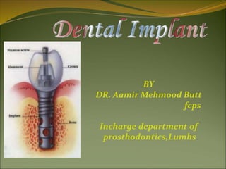
  • 2. WHAT IS A DENTAL IMPLANT? Dental implant is an artificial titanium fixture (similar to those used in orthopedics) which is placed surgically into the jaw bone to substitute for a missing tooth and its root(s). OR  A permucosal device which is biocompatible and biofunctional and is placed within mucosa or, on or within the bone associated with the oral cavity to provide support for fixed or removable prosthetics.
  • 3. Types of dental implant 1. Mucosal Insert 2. Endodontic Implant (Stabilizer) 3. Sub-periosteal implant 4. Endosteal or Endosseous implant  Plate-form implant  Ramus-frame implant  Root-form implant 5. Transosseous implant
  • 5. Endodontic Implant (Stabilizer)  Endodontic stabilizer implants are endosteal implants.  threaded post that passes at least 5 mm beyond the apex of the tooth root into available bone.  providing additional abutment support for restorative dentistry.  Five millimeters of available apical bone is the minimum that can increase the crown-root ratio to an extent sufficient to affect positively the prognosis of the tooth.
  • 6. Sub-periosteal implant  introduced in the 1940.  longest period of clinical application.  shaped to ride on the residual bony ridge of either the upper or lower.  have been used in completely edentulous as well as partially edentulous upper and lower jaws. However, the best results have been achieved in treatment of the edentulous lower jaw.
  • 7. Sub-periosteal implant… Indications  Usually a severely resorbed, completely edentulous, lower jaw bone which does not offer enough bone height to accommodate Root form Implants as anchoring devices.
  • 8. Endosteal or Endosseous implant  Plate-form implant  Ramus-frame implant  Root-form implant
  • 9. Plate-form implant  Their name is derived from their flat, blade-like (or plate-like) portion, which is the part that gets embedded into the bone.  Used where the residual bone ridge of the jaw is either too thin (due to resorption) & Difficult to place conventional Root form Implants or certain vital anatomical structures prevent conventional implants from being placed.  Bone grafting procedure, which re- establishes the lost bone have reduced the use of this form
  • 10. Ramus-frame implant  Ramus-frame Implants belong in the category of endosseous implants, although their appearance might not suggest that at first.  These implants are designed for the edentulous lower jaw only and are surgically inserted into the jaw bone in three different areas: the left and right back area of the jaw (the approximate area of the wisdom teeth), and the chin area in the front of the mouth.  The part of the implant that is visible in the mouth after the implant is placed looks similar to that of the Subperiosteal Implant.
  • 12. Dr,salah hegazy  Indications: Usually a severely resorbed, edentulous lower jaw bone, which does not offer enough bone height to accommodate Root form Implants as anchoring devices. These implants are usually indicated when the jaws are even resorbed to the point where Subperiosteal Implants will not suffice anymore. Ramus-frame implant
  • 13. Dr Aamir Butt An additional advantage that comes with this type of implant is a tripodial stabilization of the lower jaw. A jaw as thin as the one shown above can easily fracture at its thinnest part. The Ramus-frame Implant, once integrated (after a three month waiting period) will also stabilize and protect the jaw somewhat from fracturing.
  • 14. Dr Aamir Butt The Ramus-frame Implant usually comes in a standard pre-shaped form and needs to be custom-fitted to the patient's individual jaw dimension, as shown below:
  • 16. C.Root form implant Since the introduction of the Osseointegration concept and the Titanium Screw by Dr. Branemark, these implants have become the most popular implants in the world today.
  • 17. Root form Implants come in a variety of shapes, sizes, and materials and are being offered by many different companies worldwide. Some clinicians regard them to be the Standard of Care in Oral Implantology. These implants can be placed wherever a tooth or several teeth are missing, when enough bone is available to accommodate them. However, even if the bone volume is not sufficient to place Root form Implants, Bone grafting procedures within reasonable limits should be initiated, in order to benefit from these implants.
  • 18. Root form implant shape: Other variations dwell on the shape of the Root form implant. Some are screw-shaped, others are cylindrical, or even cone-shaped or any combination thereof.
  • 19. Today, the most accepted material for dental implants is high grade Titanium—either CP Titanium or an alloy thereof. The titanium alloy implants tend to be stronger than the CP titanium implants. The bone integration shows no difference to the two different types of titanium. Some implants have an outer coating of Hydroxyapatite (HA). Other implants have their surface altered through plasma spraying, or beading process. This was developed to increase the surface area of the titanium implant and, thus, in theory, give them more stability. These surface treatments were also offered as an alternative to the HA coatings, which on some implants have shown to break loose or even dissolve after a few years.
  • 20. 6. Transosseous implant These implants are not in use that much any more, because they necessitate an extraoral surgical approach to their placement, which again translates into general anesthesia, hospitalization and higher cost, but not necessarily higher benefits to the patient. In any case, these implants are used in mandibles only and are secured at the lower border of the chin via bone plates. These were originally designed to have a secure implant system, even for very resorbed lower jaws.
  • 21. A typical Transosseous Implant. The plate on the bottom is firmly pressed against the bottom part of the chin bone, whereas the long screw posts go through the chin bone, all the way to the top of the jaw ridge inside the mouth. The two attachments that will eventually protrude through the gums can be used to attach an overdenture-type prosthesis. The plate long screw posts The two attachments
  • 22. History and Present status of Implant Dentistry
  • 23. Ancient Implants  16 th Dark stone ( Egyptian-South American)  17 th Carved ivory teeth
  • 24. Early Implants  1809Gold implant  e.20th Lead, iridium, tantalum, stainless steel, and cobalt alloy  1913 hollow basket iridium + gold wires (Greenfield)
  • 25. Early Implants  1937Adams’s submergible threaded cylindrical implant with round bottom  1938 Strock’s (long term) threaded vitallium implant (cobalt+chrome+molybdenum)  The modern implants appear to be variants or composites of some of the designs of early implants
  • 26. Subperiosteal Implants  Placing implants on and around bone rather than in it  1943 Dahl of Sweden placed with 4 projecting posts  Direct bone impression  Cobalt-chrome-molybdenum casting  CT-generated CAD-CAM model
  • 27. One-stage pins and screws  Early 1960s pin, screw, and cylinder shaped implants  One piece and not submerged  Did not osseo-integration  Fibrous peri-implant membrane  Shock-absorbing claim
  • 28. Blade Implants  1967 Linkow blade implant-in narrow ridge  Required shared support with natural teeth  1970 Roberts and Roberts – Ramus blade implant (titanium)
  • 29. Transosteal Implants  1975Small introduced transosteal mandibular staple bone plate  Limited to mandible only
  • 30. Transosteal Implants  1970Cranin - single transosteal implant  1989 Bosker – transmandibular implant
  • 31.  The First Dental Implant Consensus Conference, sponsored by the National Institutes of Health (NIH) and Harvard University in 1978, was a landmark event. “ Dental Implants: Benefits and Risks”
  • 32.  The Toronto Conference opened the door to prompt widespread recognition of the Branemark implant.  The discovery of osseointegration has been one of the most significant scientific break throughs in dentistry.
  • 33. Endosteal root-form implants  1978Two-stage threaded titanium root-form implant was first presented in North America by Branemark (Toronto conference)  Terms “fixture”  First fixture was placed in 1965  Well-documented, long term prospective study
  • 34. Present Status Three Basic: “ In Bone” “Through Bone” “On Bone”
  • 35. “In Bone”  1. Ramus concepts (Harold and Ralph Roberts)  2. Pin concepts (J. Scialom Michelle Chercheve)  3. Disk concepts (Gerard Scorteci)  4. Plateform concepts (Harold + Roberts/Linkow)  5. Cylindrical or root form concepts
  • 36. Present Status  Many other root-forms have been introduced.  Body shaped competition  Surface competition – roughness  Varieties competition  Connection competition
  • 37.
  • 38. Dental Implants Implant material should have suitable:  Mechanical strength,  Biocompatibility,  Structural Biostability in physiologic environments.
  • 39. I. Modulus of elasticity II. Tensile strength III. Compressive strength IV. Elongation V. Metallurgy
  • 40. BIOCOMPATIBILITY  “The ability of an implanted material to undergo only a minimal amount of deterioration during service, to produce only a minimal change in the body environment, and to function satisfactorily in every other respect.”
  • 41. KEY FACTORS THAT INFLUENCE THE BENEFITS AND MAINTENANCE OF BIOCOMPATIBILITY  Corrosion resistance  Cytotoxicity of corrosion products  Metal contamination
  • 42. Biostability Based on tissue response and systemic toxicity effects of the implant:  Biotolerant  Bioinert  Bioactive
  • 43. Long term effects  Biotolerant materials, such as polymethylmethacrylate (PMMA), are usually characterized by thin fibrous tissue interface.  Chemical product irritate surrounding tissues.
  • 44. Long term effects  Bioinert materials, such as titanium and aluminum oxide, are characterized by direct bone contact, or osseointegration, at the interface under favorable mechanical conditions.  Non-reactive
  • 45. Long term effects  Bioactive materials, such as glass and calcium phosphate ceramics, have a bone-implant interface characterized by direct chemical bonding of the implant with surrounding bone.  Free calcium and phosphate compounds at the surface.
  • 46. Tissue response to implant materials  Most commonly used biomaterials:  Commercially pure (CP) titanium  Titanium-aluminum-vanadium alloy (Ti-6Al-4V)  Cobalt-chromium-molydenum (Co-Cr-Mo) alloy is most used for subperiosteal implants.
  • 47. Tissue response to implant materials  Calcium phosphate ceramics, Hydroxyapatite (HA), used for augmentation material or coating on surface.
  • 48. SUCCESS RATES % Subperiosteal 39 - 90 Staple 95 Vitreous carbon 50 Blade 65 - 90 Osseointegrated 80 - 100
  • 49. Advantages & disadvantages of implant over conventional treatment  Implants do not involve preparation of the adjacent teeth, they preserve the residual bone, and excellent aesthetics can be achieved.  However, it is expensive, the patient requires surgery, time consuming, and technically complex.
  • 50. INDICATIONS FOR TREATMENT Factors precluding wear of a removable prosthesis  Poor anatomy for denture support  Poor oral muscular coordination  Poor mucosal tissue tolerance  Parafunctional habits  Unrealistic expectations  Hyperactive gag reflex  Psychological inability to wear  Unfavourable number and location of abutments
  • 51. INDICATIONS  Fully edentulous  Partially edentulous  Single tooth
  • 52. INDICATIONS - FULLY EDENTULOUS  Poor retention  Functional disturbances  Psychological disturbances
  • 53. Dr,salah hegazy Diagnosis and Treatment Planning  The evaluation of a patient as a suitable candidate for implants should follow the same basic format as the standard patient evaluation, although some areas require additional emphasis and attention: I. Medical History. II. Psychological Status. III. Dental History.
  • 54. Dr,salah hegazy I. Medical History The patient’s medical history may reveal a number of conditions that could complicate or even contra- indicate implant therapy. These include: 1. Bleeding disorders; Paget’s disease; A history of radiation therapy in the maxilla or mandible region; Uncontrolled diabetes; Epilepsy that presents with more than one grand mal seizure per month; 2. In addition, there are a host of systemic medical conditions, including steroid therapy, hyperthyroidism, and adrenal gland dysfunction 3. Substance abuse including tobacco and alcohol
  • 55. Diabetes  7% population is affected  Type I (insulin depandent) & Type II which effects older age group & more common.  Blood glucose less than 150mg/dl with HbA1c value is 7. can be manage with normal protocols i:e Early morning appointment Stress reduction protocols Infection control measures Intravenous glucose for lengthy procedures Do not prescribe steroids For insulin controlled diabetes implant may be contraindicated . This may not be the case for diet
  • 56. Adrenal gland disorder  Epinephrine, nor epinephrine, corticosteroids & mineralocorticoids are affected.  Complicate the implant placement by: Inhibiting the response to inflammation ,pain & swelling Steroids reduced the protein synthesis & leukocytic activity that effects the healing process & incresed tendency to infection
  • 57. Thyroid disorders Large endocrine gland responsible for T3 & T4 hormones level in blood Sensitivity to Epinephrine in LA & retraction cords Stress related to implant surgery increase the catecholomine level that leads to thyrotoxicosis or thyroid storm symptoms includes: Fever Hypertension arrhythmias
  • 58. Hematological disorders  ANEMIA & POLYCYTHEMIA  Anemia characterized by reduced Hb level  Almost associated with every other blood disoder  Most common form is Iron deficiency anemia For implants special considerations are required including:  Suppressed bone marrow maturation  Increased trabecular pattern & reduced density of bone therefore more time for osseointegration is required
  • 59. LIVER DISEASES  Cirrhosis is the third leading cause of death  Alcohol, viruses are the common causes of liver damage.  Reduced formation of fibrinogen & clotting proteins  Vit: K  Qualitative & quantitative defect of platelet  1.5 times Increased PT contra indicate the implant placement. 
  • 60. OSTEOPOROSIS  Disease of bone metabolism.  Bone mineral density less than 2.5 standard deviation of the young healthy women.  Common in post menopausal women because of low estrogen level  Implant treatment need special considerations: Implant body with greater width & threads plus some surface coating to improve bone formation is selected More healing time Progressive loading of implant Hormonal therapy does not effect the prognosis
  • 61. OSTEOMALACIA  Vit: D deficiency  Oral findings are; Dec: trabecular bone Indistinct lamina dura Inc: chronic periodontitis  Treatment includes: supplement oral vit: D (50,000 IU)  Don’t Give implant during active phase of the disease
  • 62. Hyperparathyroidism  Hormonal problem  Sever skeletal depletion  Alveolar bone involvement is earlier than others bones  Ground glossy appearance b/c of altered trabecular pattern  Loose teeth  Loss of lamina dura  Implant is contraindicated in active disease
  • 63. Fibrous Dysplasia  Bone is replaced with the mass of fibrous connective tissue.  Twice as common in women as men.  May effect single or multiple bones  Ground glass appearance  Movement of teeth  Inc: in trabeculation  Implant is used following the excision & stabilization of bone in an affected area
  • 64. Osteitis Deformans (Paget's Disease)  Metabolic disease  Slow apposition & resorption of bone  Characterized by:  Lion face  Bone pain  Diastemas of teeth  High level of serum alkaline phosphatase level  Normal serum calcium level  Implant is contraindicated
  • 65. Multiple Myeloma  Plasma cell neoplasm originates in bone marrow.  Causes sever hypercalcemia, immune suppression, anemia, thrombocytopenia & widesprad bone destruction.  Found b/w 40 – 70 years of age.  Orally ( paresthesia, swelling, tooth mobility, gingival enlargement)  Plasma cell malignancy  Case report has described a successful placement of implants in this disease ( Sager RD 1990)
  • 66. Osteomyelitis  Acute or chronic inflammatory bone disease.  Bacterial in nature  Radiographically…… poorly defined radiolucent area with isolated segments of bone.  Caused by… odontogenic, periodontal infections, trauma, implants, immuno-compromised state & hypovascularized bone,  Common in mandible .  Treated by surgical drainage & I/V antibiotics  Relative contraindication to dental implants
  • 67. Osteogenesis imperfecta  Inherent bone disease,  characterized by poor bone quality & fragility.  Bone fractures with poor healing .  Thin cortical & trabecular pattern of bone .  Dental implants need prolong healing time.
  • 68. Cement-Osseous Dysplasia  Fibro-osseous lesion  Mandibular anterior region is affected.  Common in middle age women  Implants are only restricted in sclerotic phase of disease where bone is hypovascularized
  • 69. Prosthetic joints  450,000 joint arthroplasties are performed every year in USA.  Dental implants may be used with other prosthetic implants  The most critical period is up to 2 years after joints placement where hematogenous infection can spread b/c of dental implant placement.  Prophylactic antibiotic can prevent hematogenous infection .
  • 70. Ectodermal Dysplasia  Genetically inherent disorder affects 1 per 100,000 live births.  May be X= linked or Autosomal  Characterized by hypodontia, hypohydrosis & hypotrichosis.  Intra orally anodontia is common feature.  Conventional prosthodontics does not fullfill the functional, esthetic & psychological requirements b/c of anatomical variations.  success Rate of implants in Preadolescencents: Age 7 – 11 yrs, 87% …….. Age 12 – 17 yrs, 90%...... Age above 17 yrs 97%
  • 71. Ectodermal Dysplasia  Vertical growth results in submersion of implants need prosthetic revision or possible use of longer abutments
  • 72. Sjogren's Syndrome  Autoimmune disease  Xerostomia & xeropthalmia  Healing response & integration of implants is not affected.  non tissue borne prostheses reduced the prosthetic complications in these pt:
  • 73. Systemic Lupus Erythematosus  Autoimmune disorder  Dematological manifestation ( malar rash )  Oral vesicu lo-bulous lesions.  Treated with steroids & immunosuppresive drugs  No direct contraindications
  • 74. scleroderma  Chronic disease chracterized by deposits of collagen that cause musculoskeletal, pulmonary & GI involvement.  Detal implants with fixed prostheses is recommended since pt: is not be able to retrieve a removable prostheses.
  • 75. Rheumatoid Arthritis  Chronic inflammatory autoimmune disease  Affects muscles & joints  Loss of mobility & dexterity  Implants with fix restoration is indicated however special attention should be given to treatment medications as steroids may contraindicate the implant placement.
  • 76. Human Immunodeficiency Virus  HIV is a retrovirus resposible for AIDS.  Immune system get depressed  Pt: suffer life threatening opportunistic infections  Implant therapy is successful in HIV pts: however current status of immune system & medications toxic effects must be evaluated.
  • 77. Tobacco/smoking  Detrimental effects of tobacco 1. Vasoconstriction 2. Tissue hypoxia 3. Collagen deposition 4. Prostacyclin formation 5. Inc: platelate aggregation 6. Polymorphonuclear neutrophil dysfunction. 7. Inc: fibrinogen . 8. Blood viscosity 9. Systemic Inc: in epinephrine. Norepinephrine 10. Dec: calcium absorption & wound healing
  • 78. Tobacco/smoking  Pt should be informed abt detrimental effects of implants  Recommend ceasation of smoking 2 weeks before surgery & continue 8 weeks after the surgery.  Smoking is not the absolute contraindication
  • 79. Alcohol use  Ethyl alcohol most widely mood altering drug in world.  Associated with : dec: osteoblasts functions Dec bone formation Dec wound healing Inc parathyroid hormone secretion which lead to dec bone density
  • 80. pregnancy  Implants are contraindicated  Need medications & radiographs
  • 81. IRRADIATION  Radiotherapy results in: Progressive fibrosis of blood vessels & soft tissues Xerostomia Dec: bone healing quality. Tissues get hypovascularized, hypoxic, hypocellular These detrimental effects the wound repair & healing significantly .
  • 82. Implant Placement after Radiotherapy  The ability of implant to osseointegrate with the irradiated bone depends on:  Area of irradiation  Radiation dosage  Time elapsed since radiation exposure
  • 83. Radiation dosage…..  Dose > 50Gy …………..71% survival rate.  Dose <50Gy……………. 84% survival rate. Dose >120Gy……………very low success rate B/C ORN
  • 84. The time B/w radiotherapy & implant placement  Controversial…..?  More the time elapsed better will be the prognosis.  Different periods to initiate the implant treatment are recommended:  3 – 6 months ( King MA 1979)  12 months (Albrekttson T 1988)  & 24 months (Taylor TD 1993)
  • 85. II. Psychological Status  Perception of outcome  hypercritical  demanding  unrealistic expectation  Time and expense  Aesthetics  Maintenance
  • 86. III. Dental History It is also vital to evaluate the patient’s chief complaint, as it may have an equal bearing on treatment outcome. For example, the treatment plan recommended to the patient desiring a more secure lower denture will be quite different from the one proposed to the patient seeking a fixed and rigid appliance.
  • 87. Implant Guidelines Diagnostic phase  Problem list & treatment considerations -radiographic analysis  surgical analysis  esthetic analysis
  • 88. Implant Guidelines Diagnostic phase  radiographic analysis  periapical pathology  radiopaque/radiolucent regions  adequate vertical bone height  adequate space above inferior alveolar nerve or below maxillary sinus
  • 89. RADIOLOGY  Periapical  OPG  Lateral Cephalogram  CT Scan - axial - coronal - 3D reconstruction - Dentascan  MR
  • 94. CT SCANE  Give best cross sectional assessment of an object  Irradiated portion of the jaw can be determine  Height of the available bone can be assessed by a millimeter ruler.  Metallic restoration interferes the results.  Both quantity & density of bone can be assessed  Ideal for pre-maxillary region.
  • 96. MRI  Has limited use in implantology  Expensive  No radiations  Qulity of bone is difficult to assess.
  • 97. Implant Guidelines surgical analysis -  implant length/diameter  determined by quantity of bone apical to extraction site  use longest implant safely possible  diameter dictated by corresponding root anatomy at crest of bone
  • 98. Implant Guidelines surgical analysis  treatment options  immediate - place implant at time of tooth extraction  delayed immediate - 8-10 week delay  delayed - 9-10 months or longer  NOTE : immediate will not allow bone resorption, but delayed allows bone fill for stabilization
  • 99. Implant Guidelines  surgical analysis  proper surgical technique during implant placement is critical  minimal heat generation important
  • 100. esthetic analysis  implant emergence profile  restored implant should appear to “grow” or emerge from the gingiva  very natural & desirable in appearance
  • 101.
  • 102. A direct structural and functional connection between ordered living bone and the surface of a load carrying implant P-I Branemark
  • 103. IV. Osseointegration  Definition: A time-dependant healing process where by clinically symptomatic rigid fixation of alloplastic materials is achieved, and maintained, in bone during functional loading. (Zarb & Albrektson,1991)
  • 104. Factors affecting osseointegration 1. Implant biocompatibility 2. Implant design 3. Implant surface 4. Implant bed 5. Surgical technique 6. Loading condition
  • 105. 1. Implant biocompatibility  Materials used are:  Cp titanium (commercially pure titanium)  Titanium alloy (titanium-6aluminum- 4vanadium)  Zirconium  Hydroxyapatite (HA), one type of calcium phosphate ceramic material 5. Plasma sprayed coating
  • 106. Osseointegration (A) Hematoma occurs near screw threads (B) After 3 weeks – Osteoblasts begin forming spongy bone (C) After 4 months – spongy bone replaced by compact bone Lamellar bone – strongest type of bone, most desired next to implant (D) Osseointegration failure
  • 107. 2. Implant design (root-form)  Cylindrical Implant Some investigators explain the lack of bone steady state by overload due to micromovement of the cylindrical design, whereas others incriminates an inflammation/infection caused particularly by the very rough surfaces typical for these types of implant.  Threaded Implant In contrast, Threaded implants have demonstrated maintenance of a clear steady state bone response. To enhance initial stability and increase surface contact, most implant forms have
  • 108. 3. Implant surface The Pitch is the number of threads per unit length, is an important factor in implant osseointegration. Increased pitch and increased depth between individual threads allows for improved contact area between bone and implant. Moderately rough surfaces with 1.5µm also, improved contact area between bone and implant surface. Reactive implant surface by anodizing (Oxide layer) ,acid etching or HA coating enhanced osseointegration
  • 109.
  • 110. Bone Quality  Quality I Was composed of homogenous compact bone, usually found in the anterior lower jaw.  Quality II Had a thick layer of cortical bone surrounding dense trabecular bone, usually found in the posterior lower jaw.  Quality III Had a thin layer of cortical bone surrounding dense trabecular bone, normally found in the anterior upper jaw but can also be seen in the posterior lower jaw and the posterior upper jaw.  Quality IV Had a very thin layer of cortical bone surrounding a core of low-density trabecular bone, It is very soft bone and normally found in the posterior upper jaw. It can also be seen in the anterior upper jaw. According to Lekholm and Zarb.,1985
  • 113. 5. Surgical technique Minimal tissue violence at surgery is essential for proper osseointegration.  Careful cooling while surgical drilling is performed at low rotatory rates  Use of sharp drills  Use of graded series of drills  Proper drill geometry is important, as intermittent drilling.  The insertion torque should be of a moderate level because strong insertion torques may result in stress concentrations around the implant, with subsequent bone resorption.
  • 114. 6. Loading condition A. Delayed / conventional loading : B. Immediate loading: C. Early loading (prosthetic function within two months) D. Progressive loading
  • 115. Delayed / conventional loading : Restoration is placed from 3 – 6 months This is applicable for two stage protocol
  • 116. Immediate loading:  The biomechanical definition of immediate loading is also debated: For some researchers, The concept of immediate loading is satisfied as soon as the coronal portion of the prosthesis is inserted, even if it is kept out of occlusion.  • For others, The term immediate loading can be applied only if the prosthesis is subjected to occlusal forces as soon as it is inserted.  To qualify as an immediately loaded implant, the definitive prosthesis must be placed on the same day. Still others accept a delay in loading of 48 hours to 72 hours.
  • 117. Following are immediate-loading protocols • A. Immediate occlusal loading vs immediate functional loading B. Immediate functional loading vs immediate nonfunctional loading
  • 118. Progressive loading  Misch first proposed the concept of progressive or gradual bone loading during prosthetic reconstruction to decrease crestal bone loss and early implant failure of endosteal implants in 1980 based on empirical information.  98.9% survival at Stage II uncovery followed with a progressive loading format and found no early loading failures during the first year of function.
  • 120. Progressive loading protocol (TIME)  • The macroscopic coarse trabecular bone heals about 50% faster than dense cortical bone.  • The healing time between the initial and second-stage surgeries is kept similar for Dl and D2 bone and is 3 to 4 months.  A longer time is suggested for the initial healing phase of D3 and D4 bone (5 and 6 months, respectively) because of the lesser bone contact and decreased amount of cortical bone to allow for the maturation of the interface and the development of some lamellar bone.
  • 122. Progressive loading protocol (DIET)  • The patient is limited to a soft diet such as pasta and fish, from the initial transitional prosthesis delivery until the initial delivery of the final prosthesis.  • After the initial delivery of the final prosthesis, the patient may include meat in the diet, which requires about 21 psi in bite force.  The final restoration can bear the greater force without risk of fracture or uncementation. After the final evaluation appointment, the patient may include raw vegetables, which require an average 27 psi of force. A normal diet is permitted only after evaluation of the final prosthesis function, occlusion, and proper cementation.
  • 123. Progressive loading protocol (OCClUSAl. MATERIAL)  Using acrylic as the occlusal material, with the benefit of a lower impact force than metal or porcelain.  Either metal or porcelain can be used as the final occlusal material.  If parafunction or cantilever length cause concern relative to the amount of force on the early implant- bone interface, the dentist may extend the softer diet and acrylic restoration phase several months. In this way, the bone has a longer time to mineralize and organize to accommodate the higher forces.
  • 124. Progressive loading protocol (OCCLUSION) (step 1): No occlusal contacts are permitted during initial healing. (step 2). The first transitional prosthesis is left out of occlusion in partially edentulous patients The occlusal contacts then are similar to those of the final restoration for areas supported by implants. (step 3). However, no occlusal contacts are made on cantilevers The occlusal contacts of the final restoration follow the implant protective occlusion concepts.
  • 126. Progressive loading protocol (PROSTHESIS DESIGN)  Its purpose is to splint the implants together, to reduce stress by the mechanical advantage, and to have implants sustain masticatory forces solely from chewing.  In the second acrylic transitional restoration, occlusal contacts are placed on the implants with occlusal tables similar to the final restoration but with no cantilevers in nonesthetic regions.  In the final restoration, narrow occlusal tables and cantilevers are designed with occlusal contacts following implant-protective occlusion guidelines.
  • 127. implant-protective occlusion  Concept was developed by MISCH  Concept refers to an occlusal plane that is often unique & specifically designed for the restoration of endosteal implants, providing an environment for improved clinical longevity of both implant & prosthesis
  • 128. implant-protective occlusion The salient features are:  Using wider width of dental implant whenever possible.  Anterior teeth should disclude the posterior teeth.  Absence of lateral contacts in excursion.  All occlusal contacts more medial than the natural teeth.  A reduced width of occlusal table
  • 129. V. Biomechanics of osseointegrated implant. In all incidences of clinical loading, occlusal forces are first introduced to the prosthesis and then reach the bone implant interface via the implant. So far, many researchers have, therefore, focused on each of these steps of force transfer to gain insight into the biomechanical effect of several factors such as  Force directions and magnitudes,  Prosthesis type,  Prosthesis material,  Implant design,  Number and distribution of supporting implants,  Bone density, and  The mechanical properties of the bone-implant interface.
  • 131. Parts of Implant 1. Implant body or fixture The implant body is the component that is placed within the bone during first stage of surgery. It could be threaded or non threaded. The implant bodies may be coated with hydroxyapatite or titanium particles to incorporate microscopic component into them.
  • 132. Prosthetic Component Cont…. 2. First stage cover (Healing screw) During the healing phase ,this screw is normally placed in the superior surface of the body. The functions of this component are: • Facilitates the suturing of soft tissue. • Prevents the growth of tissue over the edge of the implant.
  • 133. Prosthetic Component Cont… 3. Second stage permucosal extension or healing abutment (healing cap) After a prescribed healing period, a second stage procedure may be performed to expose the implant and/or attach atransepithelial portion. This transepithelial portion is termed a permucosal extension because it extends the implant above the soft tissues and results in the development of a permucosal seal around the implant. This implant component is also called a healing abutment because stage II uncovery surgery often uses this device for initial soft tissue healing.
  • 134. Prosthetic Component Cont…. 4. Abutment It is the portion of the implant that supports or retains a prosthesis or implant superstructure.
  • 135. Three main categories of implant abutment are described according to the method by which the prosthesis or superstructure is retained to the abutment. A. An abutment for screw retention B. An abutment for cement retention C. An abutment for attachments
  • 136. Prosthetic Component Cont…. 5. Impression coping or impression posts It is a part of the implant that facilitates the transfer of the intraoral location (of the implant or abutment) to a similar position on the cast. Therefore it can also be called as implant body transfer coping or abutment transfer coping. It has two types:  Transfer impression  Pick type impression
  • 138. Pick up type impression
  • 139. Prosthetic Component Cont . . . 6. Laboratory analogues An analogue is something that is similar or analogous to something else. An implant analogue is used in the fabrication of the master cast to replicate the retentive portion of the implant body or abutment (implant body analogue, implant abutment analogue). After the master impression is obtained the corresponding analogue( e.g., implant body, abutment for screw) is attached to the transfer coping and the assembly is poured in stone to fabricate the master cast.
  • 140. Prosthetic Component Cont . . . 7. Prosthesis retaining screw A screw retained prosthesis or superstructure is secured to the implant body or abutment with a prosthetic screw.
  • 141. IMPLANT SUPER STRUCTURES A super structure is the prosthetic component fabricated over the implant after its placement. Commonly used super structures include 1. Overdentures, 2. Fixed partial dentures/bridges 3. Single Crown
  • 142. IMPLANT SUPER STRUCTURES 1 - Implant Supported Over Dentures They can be either a complete or a partial over denture. The implants are placed on suitable sites in the edentulous ridge. The implant abutments may either be present individually or be connected to one another with a bar.
  • 143. IMPLANT SUPER STRUCTURES 2 – Implant Supported Fixed Partial Dentures These may be either pure implant supported or a combination of implant and tooth supported fixed bridges.
  • 144. IMPLANT SUPER STRUCTURES 3 – Implant Supported Single Tooth Replacements
  • 146. C.Misch in 1989 reported five prosthetic options in implant dentistry. Of the five, the first three are fixed prosthesis (FP) that may be partial or complete replacements, which in turn may be cemented or screw retained. The remaining two are removable prosthesis (RP) that are classified on the support derived.
  • 147. PROSTHETIC OPTIONS IN IMPLANT DENTISTRY Cont. . . • FP-1: Fixed prosthesis; replaces only the crown; looks like a natural tooth.
  • 148. PROSTHETIC OPTIONS IN IMPLANT DENTISTRY Cont. . . • FP-2: Fixed prosthesis; replaces the crown and a portion of the root; crown contour appears normal in the occlusal half but it is elongated or hypercontoured in the gingival half.
  • 149. PROSTHETIC OPTIONS IN IMPLANT DENTISTRY Cont. . . • FP-3: Fixed prosthesis; replaces missing crowns and gingival color and the portion of the edentulous side; prosthesis most often uses denture teeth and acrylic gingiva, but may be made of porcelain.
  • 150. PROSTHETIC OPTIONS IN IMPLANT DENTISTRY Cont. . • RP-4: Removable prosthesis; overdenture supported completely by implants.
  • 151. PROSTHETIC OPTIONS IN IMPLANT DENTISTRY Cont. . • RP-5: Removable prosthesis; overdenture supported by both soft tissue and implants.
  • 152.  Ideal design of implant and tissue supported overdentures;  Internal of overdenture;
  • 154. 1. Number of implants abutment 2. Location of implants 3. Quality of bone 4. Amount of bone 5. Amount of circumoral activity
  • 155.  Quasi- general formula i:e • five implants b/w two mental foramina to support 10 – 12 units fixed mandibular prosthesis. •& six implants for maxilla  formula did not address the following considerations: Arch form ( Flat versus curvature) Length of implants Length of cantilever Occlusal forces
  • 156. Location of implants  Implant distribution is more favorable in curved arches since it provides : More occlusal units & optimal cantilever design  Flat arches are favorable for overdentures
  • 157. Quality of bone  D3 & D4 BONE
  • 158. Quantity of bone  10 mm of vertical height of the bone is minimally required  Bone grafting, sinus lift, frozen bone use to improves the quantity of bone
  • 159. Amount of circumoral activity  This would effect the choice of maxillary prosthesis  Where high lip line & advance residual ridge resorption will require the use of visible labial flange to compensate bone resorption .  This design demands high value for hygiene maintenance & preclude fixed option
  • 160. The most common line of treatment
  • 161. Treatment Planning Determinants 1. Changes in Oral Structures in Edentulism 2. Posterior Ridge Anatomy 3. Occlusal Forces 4. Quality, Location and Quantity of Bone 5. Implant Size 6. Implant Location 7. Arch configuration 8. "Mapping" the Mandible 9. Cantilevering
  • 162. 1. Changes in Oral Structures in Edentulism With successive denture treatments, it is common for the vertical dimension of occlusion to decrease as bone resorbs. This promotes an increased tendency toward a skeletal Class III relationship.
  • 163. Posteriorly, poor ridge height, inadequate attached gingiva and compromised ridge shape cause increased horizontal movement of the prosthesis. This increases the lateral forces that are brought to bear on the anterior implants, and will affect bar and prosthesis design. 2. Posterior Ridge Anatomy
  • 165. 3. Occlusal Forces The maximum bite force of subjects with a mandibular denture supported by implants is 60 to 200% higher than that of subjects with a conventional denture Edentulous patients that are predisposed to clenching and bruxing may be given the necessary "tools" to begin parafunctional habits once the implant bar is secured in place.
  • 166. The minimum buccal-lingual thickness of osseous tissue required to successfully place an implant is 5 mm. In order to achieve a 5.0 mm "flat" base, either the anterior ridge crest peak must be removed or a bone graft must be considered. 4. Quality, Location and Quantity of Bone
  • 167. 5. Implant Size. The greater the surface area of the implant- bone system, the less concentrated the force transmitted to the crest of bone at the implant interface. Similarly, the greater the surface area of the implant-bone system, the better the prognosis for the implant.  For each 0.25 mm increase in diameter, the surface area of a cylinder increases by more than 10 per cent;  For each 3.0 mm increase in length , the surface area of a cylinder increases by more than 10 per cent.
  • 168. Dr,salah hegazy Implant Size 0.25 mm diameter = 3.0 mm length
  • 169. 6. Implant Location  Ideally, occlusal forces should be directed along the long axis of the implants. Therefore ,The angle of the osseous ridge crest is a key determinant of implant angulation.  the distance between an implant and any adjacent "landmark" (natural tooth or another implant), which should be not less than 2.0 mm.
  • 170. The angle of the osseous ridge crest is a key determinant of implant angulation.
  • 171. 7. Arch configuration Mandibular arch forms may be classified as tapered or square.  With tapered arch forms, the most posterior right and left implants in a four-implant treatment are often placed well around the "turn" of the arch, creating a "U" shaped design that is well suited to cantilevering,  With a square arch, the four implants are usually placed in a relatively straight line. This "straight line" bar design is not well suited to cantilevering.
  • 172. 8. "Mapping" the Mandible The anterior symphysis can be divided into five geographic sites: A point, 6.0 mm anterior to each mental foramen, determines the most posterior boundaries, right and left. Another possible implant location occurs at the midline. Two additional sites are chosen on each side of the midline, spaced equidistantly between the midline and the respective distal sites.
  • 173. " Mapping" the Mandible
  • 174. Factors which helps in determining the appropriate cantilever than a suggested formula. The number of implants, Their respective lengths and locations,  The quality of bone support, The posterior ridge anatomy, Occlusal forces, And the opposing dentition
  • 175. 9. Cantilevering suggested formula.  One method is to draw a line through the most anterior implant, and another through the two most posterior implants. The distance between the two lines can then be measured. A suggested maximum cantilever would be 1.5 times this distance.
  • 176. Dr,salah hegazy The distance between the two lines can then be measured. A suggested maximum cantilever would be 1.5 times this distance.
  • 178. Treatment Planning When all the diagnostic information has been assembled, a variety of available treatment options must be assessed: 1. One-Implant Overdenture 2. Two-Implant Overdenture 3. Three-Implant Overdenture 4. Four-Implant Overdenture 5. Five-Implant Overdenture
  • 179. One-Implant Overdentures Indications:  The maladaptive or dissatisfied denture patient who demands greater stability and oral comfort,  Elderly patients desiring a more stable mandibular denture,  Or, as a minimal implant treatment objective for the partially edentulous patient with severely compromised teeth in which removal would convert a patient to a fully edentulous state
  • 181. In the two-implant over-denture, an attachment is used to greatly enhance the retentive potential of what is essentially a tissue-supported prosthesis. If only two implants are placed, which are 13mm long or longer, and they are in dense bone, they can be left as individual supporting units with little risk. . Two-Implant Solitary Overdenture
  • 184. 2. Two-Implant Bar Overdenture If the two implants are 10 mm long or shorter, or the bone quality is compromised, then ideally:  They should be splinted.  They should be at least 10 mm apart (in order to allow room for a clip or fastening mechanism)  They should be no further than 18 mm apart in order to limit bar flexure.
  • 187. 3. Three-Implant Overdenture The three-implant overdenture is still essentially a tissue- supported prosthesis with enhanced retention supplied by the attachment/bar complex.
  • 189. 4. Four-Implant Overdenture At this level, the prosthesis begins to derive a larger part of its support and retention from the implant/bar complex, and the importance of tissue support decreases. Also, the attachments selected for a four- implant bar over-denture can be more rigid, as the torquing forces generated by the prosthesis will be better tolerated. This number allows for some "insurance" in case one implant fails to integrate.
  • 192. 5. Five-Implant Overdenture At this level, a prosthesis can be fabricated that is completely implant supported and retained, if the AP spread of the implants is adequate. The decision to fabricate a bar over-denture over five implants, rather than a fixed detachable restoration, usually relates to the patients’ ability to maintain proper oral hygiene.
  • 195. PROSTHETIC PROTOCOL  Overdenture abutments were cemented or scrowed into the implants.  Pressure indicating paste was placed on each overdenture ball.  The denture was seated so that the pressure indicating paste could mark the exact location of the overdenture abutments. Then, a recess was cut into the denture at each abutment location  The resulting depressions in the mucosal aspect of the denture were lined with polyvinylsiloxane material and seated in the patient's mouth.  The denture was either lined with a lab-processed material or O-rings were used for retention.
  • 196. Overdenture abutments were cemented or scrowed into the implants.
  • 197. Pressure indicating paste was placed on each overdenture ball.
  • 198. Then, a recess was cut into the denture at each abutment location
  • 199. lined with polyvinylsiloxane material and seated in the patient's mouth.
  • 200. The denture was either lined with a lab-processed material or O-rings were used for retention
  • 201.
  • 202. MAINTENANCE & RECALL  Recall appointmens should be after every 3-4 months  Scaling and proper cleaning is done with only plastic disposible instruments  Use of steel probes and other instruments is prohibited  Home care aids like flosses interproximal brushes and water jets should be advised for patients to use at home
  • 203.
  • 204.
  • 205.
  • 206. COMPLICATIONS  Preoperative  Perioperative  Postoperative  Transient  Persistent  Permanent  Soft tissue  Hard tissue Time of operation Time Duration Tissue effected
  • 207. SERIOUS COMPLICATIONS  Jaw fracture  Haemorrhage  Ingestion  Inhalation  Neurological  Death
  • 208. COMPLICATIONS  Patient selection  Psyche  Anatomy  Systemic disease  Implant factors  Surgical  Prosthodontic  Errors in judgement  Deviation from established protocol
  • 209. ANATOMY  Unsuitable morphologically  Reduced bone density  Reduced bone volume  Attached tissue  Nerve position
  • 210. PREVENTION OF NERVE DAMAGE  CT  Bone density measurement  Drill sleeves  Discretion is better part of valour
  • 211. COMPLICATIONS Preoperative  Failure to obtain anaesthesia  Haemorrhage  Stuck implant  Loose implant  Lost implant
  • 212. SURGICAL FAILURE  Poor planning  Poor surgical technique  Lack of precision  Thermal injury  Faulty placement  Damage to adjacent structures
  • 213. SURGICAL  Haemorrhage  Stuck implant  Loose implant  Lost implant
  • 214.  Wound dehiscence  Infection  Mucosal perforation  Fistula formation  Anatomical - antral - nasal - neurological COMPLICATIONS
  • 215. STAGE ONE SURGERY  Failure to obtain anaesthesia  Faulty placement  Anatomical  Surgical
  • 216. SURGICAL  Stripped bone threads  Exposed implant threads  Fractured drill  Sheared implant hex  Excessive countersink  Eccentric drill
  • 217. Second stage  Loose implant  Excess bone coverage  Exposed threads  Coverscrew problems COMPLICATIONS
  • 218. STAGE TWO SURGERY  Wrong abutment length  Faulty abutment seating  Retained sutures  Gingival hyperplasia  Mobile tissue  Destroyed cover screw hex  Failure of integration
  • 219. FAULTY PLACEMENT  Labial / buccal  Lingual  Too close  Straight line in mandibular anteriors  Angulation  Divergence  Correct by use of a surgical template
  • 220. POSTOPERATIVE  Fascial space infections  Haematoma  Jaw fracture  Sinusitis  Wound dehiscence
  • 221. WOUND DEHISCENCE  Poor flap design  Poor surgical technique  Poor repair  Poor tissue quality  Previous surgery  Underlying medical condition  Superficial implant placement
  • 222. PERSISTENT  Neurological damage  Aesthetics  Speech  Function  Psychological
  • 223. Long term  Anatomical  Neurological  Deintegration  Progressive thread exposure  Gingivitis  Hyperplastic tissue  Fractured Implant COMPLICATIONS
  • 224. PROSTHODONTIC  Avoid premature loading  Passive fit  Good design  Good oral hygiene  Loss of integration  Soft tissue problems  Oral hygiene and maintenance  Retrievable vs cemented
  • 225. COMPONENT FAILURE  Fractured fixture  Fractured abutment screw  Fractured punch blade  Fractured screw driver tip  Fractured castings
  • 226. MANAGEMENT OF FAILURE  Failing implants FAIL  Removal  Abandon  Alternative site  Larger diameter  Replacement after healing
  • 227. PROSTHODONTIC PROBLEMS AND COMPLICATIONS TYPE  Structural  Cosmatic  Functional DESCRIPTION  Prosthesis fracture  Fracture of prosthesis rataining screw  Fracture of implant  Fracture of abutment  As perceived by patient and dentist  Speech problems  Transient muscle discomfort or TMJ disorders
  • 228. SURGICAL PROBLEMS AND COMPLICATIONS TIME  Stage 1 surgery  post- stage 1 surgery  Stage 2 surgery  Delayed complications DESCRIPTION  Unfavorable implant position/allignment  Swelling or echymosis. infection and neuroapthy  Failure of osseointegration  Unfavorable position or angle makes the implant unusable  Component fracture  Soft tissue complications