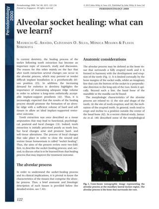
2. features of the alveolar process in the anterior maxilla
in humans. The authors included 250 periodontally
healthy subjects, 17–66 years of age. Cone beam com-
puted tomograms were obtained from the maxillary
front teeth. Measurements of the thickness of the
buccal bone plate of the alveolar process were per-
formed at three different positions in relation to the
buccal bone crest (i.e. at distances of 1, 3 and 5 mm
apical to the crest). The measurements demonstrated
that the buccal bone plate in most locations, in all
anterior tooth sites examined, was ≤1 mm thick (aver-
age thickness ~0.5 mm) and that close to 50% of sites
had a bone plate thickness that was ≤0.5 mm. In con-
clusion, tooth sites in the anterior maxilla have a thin
buccal bone wall (Fig. 2), which probably contributes
to its loss following tooth extraction.
Histologic considerations
The inner portion of socket walls is named “alveolar
bone proper” or bundle bone (a histological term)
and the remaining hard structure is called “alveolar
bone”. The bundle bone is a lamellar bone, 0.2–
0.4 mm wide (65), composed of circumferential
lamellae, whilst the alveolar bone is also of the lamel-
lar type but composed of concentric and interstitial
lamellae and of marrow. In the bundle bone, the
Sharpey’s fibers are invested in such way that they
connect the periodontal ligament to the alveolar bone
and skeleton. Likewise, on the contralateral side of
the periodontal ligament, the dental cementum
invested with Sharpey’s fibers connects the periodon-
tal ligament to the dentin. As with root cementum
and the periodontal ligament, the bundle bone is a
tooth-dependent structure. Overall, the bundle bone
and the buccal bone plate frequently exhibit a similar
thickness at the anterior front tooth region. Thus,
most of the thin buccal bone wall is a tooth-depen-
dent structure (Fig. 3).
Socket healing
Dimensional changes
The dimensional changes that occur in the alveolar
ridge following tooth extraction have been reported
in several human studies (14, 16, 47, 48, 62, 63, 66, 74)
and were determined using different methodologies,
including clinical, cast model and radiographic exam-
inations. After multiple tooth extractions and the use
of complete removable prostheses, the alveolar ridge
undergoes marked contraction in both vertical and
horizontal directions (13, 14, 32, 47, 48). Following
several years of full denture use, individuals may
undergo a wide variation in alveolar ridge reduction
and some may exhibit a fully resorbed alveolar ridge
(16). Following single-tooth extraction, the ridge
exhibits a limited reduction in its vertical dimension,
but the horizontal reduction is substantial (Fig. 4; 62,
63). It can be expected that: (i) up to 50% reduction of
the original ridge width will occur; (ii) the amount of
Fig. 2. Occlusal view of a dried skull specimen. Note the
limited thickness of the buccal bone wall at the central
incisor regions.
Fig. 3. Buccal–lingual section illustrating the most coronal
portion of the buccal bone wall. The buccal wall is made
mainly by bundle bone. Polarized light. Toluidine blue
stain; original magnification 3 50.
Alveolar socket healing
123
3. provisional matrix. Subsequently, the provisional
matrix is penetrated by several vessels and bone-
forming cells, and finger-like projections of woven
bone are laid down around the blood vessels. Eventu-
ally, the projections completely surround a vessel and
the primary osteon is thus formed (Fig. 6). The pri-
mary osteons may be occasionally reinforced by
parallel-fibered bone. Woven bone can be identified
in the healing socket as early as 2 weeks after tooth
extraction and remains in the wound for several
weeks. Woven bone is a provisional type of bone
without any load-bearing capacity and therefore
needs to be replaced with mature bone types (lamel-
lar bone and bone marrow).
Bone modeling and remodeling phase
Bone modeling and remodeling is the third and last
phase of the socket-healing process. Bone modeling
is defined as a change in the shape and architecture
of the bone, whereas bone remodeling is defined as a
change without concomitant change in the shape and
architecture of the bone. The replacement of woven
bone with lamellar bone or bone marrow is bone
remodeling, whereas the bone resorption that takes
place on the socket walls leading to a dimensional
alteration of the alveolar ridge is the result of bone
modeling. Bone remodeling in humans may take sev-
eral months and exhibits substantial variability
among individuals (32, 74). In a recent study, Lindhe
et al. (54) examined the tissue composition of biop-
sies from 36 individuals retrieved from previous
socket sites in the posterior maxilla after 16 weeks of
healing. The authors reported that about 60–65% of
the tissue volume was made up of lamellar bone and
bone marrow. Thus, the complete remodeling of the
woven bone into lamellar bone and bone marrow
may take several months or years.
The resorption of the socket walls was studied in
biopsies obtained from human samples (32) and from
a series of studies in dogs (3–6, 10). A few weeks after
tooth removal, osteoclasts could be found around the
crest of both buccal and lingual walls and on the
outer and inner (bundle bone) portions of the socket
(Fig. 7). Bone modeling takes place equally on buccal
and lingual walls, but because the lingual bone is usu-
ally wider than the buccal bone wall, modeling results
in greater vertical bone loss at the thin buccal plate
than at the wide lingual wall. In addition, bone mod-
eling takes place earlier than bone remodeling, in
such way that about two-thirds of the modeling pro-
cess occurs in the first 3 months of healing (66). In
summary, modeling and remodeling processes during
socket healing result in qualitative and quantitative
changes at the edentulous site, which culminate in a
reduction of the dimension of the ridge.
Fig. 6. Micrograph illustrating primary osteons in the
healing socket. The collagen fibers have a woven organiza-
tion. Toluidine blue stain; original magnification 3 100.
Fig. 7. Buccal–lingual section of the socket wall a few
months following tooth extraction. Note the intense mod-
eling and remodeling process characterized by the pres-
ence of bone multicellular units and reversal lines.
Ladewig fibrin stain; original magnification 3 20.
Alveolar socket healing
125
4. provisional matrix. Subsequently, the provisional
matrix is penetrated by several vessels and bone-
forming cells, and finger-like projections of woven
bone are laid down around the blood vessels. Eventu-
ally, the projections completely surround a vessel and
the primary osteon is thus formed (Fig. 6). The pri-
mary osteons may be occasionally reinforced by
parallel-fibered bone. Woven bone can be identified
in the healing socket as early as 2 weeks after tooth
extraction and remains in the wound for several
weeks. Woven bone is a provisional type of bone
without any load-bearing capacity and therefore
needs to be replaced with mature bone types (lamel-
lar bone and bone marrow).
Bone modeling and remodeling phase
Bone modeling and remodeling is the third and last
phase of the socket-healing process. Bone modeling
is defined as a change in the shape and architecture
of the bone, whereas bone remodeling is defined as a
change without concomitant change in the shape and
architecture of the bone. The replacement of woven
bone with lamellar bone or bone marrow is bone
remodeling, whereas the bone resorption that takes
place on the socket walls leading to a dimensional
alteration of the alveolar ridge is the result of bone
modeling. Bone remodeling in humans may take sev-
eral months and exhibits substantial variability
among individuals (32, 74). In a recent study, Lindhe
et al. (54) examined the tissue composition of biop-
sies from 36 individuals retrieved from previous
socket sites in the posterior maxilla after 16 weeks of
healing. The authors reported that about 60–65% of
the tissue volume was made up of lamellar bone and
bone marrow. Thus, the complete remodeling of the
woven bone into lamellar bone and bone marrow
may take several months or years.
The resorption of the socket walls was studied in
biopsies obtained from human samples (32) and from
a series of studies in dogs (3–6, 10). A few weeks after
tooth removal, osteoclasts could be found around the
crest of both buccal and lingual walls and on the
outer and inner (bundle bone) portions of the socket
(Fig. 7). Bone modeling takes place equally on buccal
and lingual walls, but because the lingual bone is usu-
ally wider than the buccal bone wall, modeling results
in greater vertical bone loss at the thin buccal plate
than at the wide lingual wall. In addition, bone mod-
eling takes place earlier than bone remodeling, in
such way that about two-thirds of the modeling pro-
cess occurs in the first 3 months of healing (66). In
summary, modeling and remodeling processes during
socket healing result in qualitative and quantitative
changes at the edentulous site, which culminate in a
reduction of the dimension of the ridge.
Fig. 6. Micrograph illustrating primary osteons in the
healing socket. The collagen fibers have a woven organiza-
tion. Toluidine blue stain; original magnification 3 100.
Fig. 7. Buccal–lingual section of the socket wall a few
months following tooth extraction. Note the intense mod-
eling and remodeling process characterized by the pres-
ence of bone multicellular units and reversal lines.
Ladewig fibrin stain; original magnification 3 20.
Alveolar socket healing
125
5. Stimulating factors
The initial healing responses in a wound are regulated
by signaling molecules (i.e. growth factors and cyto-
kines), such as platelet-derived growth factor, insulin-
like growth factors, transforming growth factor-beta
and fibroblastic growth factors. They initiate cell
migration, differentiation and proliferation as they
interact with each other in highly ordered temporal
and spatial sequences (53). These growth factors act
as mitogenic and angiogenic signals at the early stage
of bone healing. Once activated, growth factors insti-
gate a series of events via ligand–receptor interac-
tions, including signal transduction, gene
transcription, mRNA-directed protein biosynthesis
and secretion of post-translational proteins (44).
Few studies have examined the roles of growth fac-
tors and cytokines during socket healing (40, 74).
Fisher et al. (40) evaluated the expression of growth
factors during socket-healing events in a rabbit
model. The authors observed that: (i) fibroblast
growth factor-2 presented at higher levels at early
time points, before returning to lower levels; (ii) vas-
cular endothelial growth factor levels were main-
tained constant during healing; (iii) platelet-derived
growth factor-A levels increased during the first days
of socket healing; (iv) transforming growth factor-
beta1 presented a small elevation at early time points;
and (v) an increased expression of bone morphoge-
netic protein 2 was observed when osteoblast precur-
sors accumulated and began to proliferate. Trombelli
et al. (63) studied modeling and remodeling of
human extraction sockets and evaluated the expres-
sion of bone morphogenetic protein 7 during socket
healing. The results demonstrated that bone morpho-
genetic protein 7 increased during early and interme-
diate healing phases, and a period of increased bone
modeling and remodeling activity occurred, leading
to the deposition of woven bone from provisional
matrix. In summary, growth factors present multiple
activities, generally with overlapping actions, and a
simplistic characterization of their effects is not possi-
ble, or indeed appropriate.
What can we learn?
There are several lessons to be learned from the
various reports of local changes following tooth
extraction. The healed socket eventually fills with
newly formed bone and the alveolar ridge contracts.
The ridge reduction is larger in the molar region
(62), but it becomes more critical in the anterior
region as a result of esthetic demands. The anterior
maxillary region exhibits very thin socket walls (19,
46) that are frequently made up of only bundle
bone. As the bundle bone is a tooth-dependent
structure, it is gradually resorbed following exodon-
tia. Finally, the postextraction ridge reduction
appears to be related to several factors, including
surgical trauma, lack of a functional stimulus on the
bone walls, lack of bundle bone and periodontal lig-
ament and genetic information.
Tooth extraction is a traumatic procedure and, dur-
ing its course, the soft tissues are disrupted, the vas-
cular structures of the periodontal ligament are
damaged or destroyed and the principal fibers of the
periodontal ligament are severed (29). In addition, it
is well established in the dental literature that the ele-
vation of a full-thickness flap, in order to gain access
to the root, may cause resorption of thin bone walls
(50, 75–77; for reviews see 43, 70). However, different
animal and clinical studies have failed to support the
concept that tooth extraction without flap elevation
prevents ridge reduction (8, 17, 33, 39). These studies
indicate that the surgical trauma promoted by the
removal of the tooth itself overlaps with the surgical
trauma promoted by the elevation of a full-thickness
flap.
The surgical trauma caused by tooth extraction
may be limited by minimally invasive surgical proce-
dures (58). Such procedures aim to prevent expansion
of the socket housing, which otherwise may fracture
the thin adjacent bony walls. For this purpose, the
use of forceps to luxate the tooth by applying forces
toward the buccal palatal/lingual aspects of the
socket is not recommended. Likewise, the forceps
should not perform rotational movements, as the
cross-section shape of a root is seldom circular. Sev-
eral new surgical instruments, which promote mini-
mally invasive tooth extraction, are currently
commercially available. Periotomes and vertical
tooth-extraction systems are among the instruments
most frequently used for this purpose. Periotomes are
instruments designed to sever the periodontal liga-
ment fibers at the mesial and distal aspects of the
socket, in order to facilitate and improve the effi-
ciency of root elevators. Vertical tooth-extraction sys-
tems are, on the other hand, designed to pull roots in
a vertical direction and hence avoid any damage to
the socket walls. In both techniques described above,
no pressure is applied to the buccal socket wall; how-
ever, such techniques are efficient only for conical or
straight roots.
Ara
ujo et al.
126
10. Teeth provide support for very thin bone walls,
although fenestrations and dehiscence may occur
naturally when the bone thickness is below a certain
threshold (65). It is suggested, however, that implants
should be provided with bone walls about 1- to 2-
mm-wide on buccal and lingual aspects to allow a
stable bone height to be maintained (22, 42). The rea-
sons why teeth can support thin bone walls, and why
implants seem to fail to do so, remains obscure. It has
been suggested, however, that the presence of bundle
bone and periodontal ligament around teeth are
likely explanations. Bundle bone is capable of existing
in thinner dimensions than are alveolar or basal
bones because the periodontal ligament provides the
functional stimulus as well as the nutritional and cel-
lular source for its maintenance.
It is now well established that following tooth
extraction the ridge crest moves toward the long axis
of the basal bone (16, 63). The shape of the jawbone
appears to return to the shape that was present prior
to the development of the alveolar process during
tooth eruption. The lack of a functional stimulus on
the bone walls and the need for tissue adjustment to
meet “genetically” determined demands regarding
ridge geometry in the absence of teeth (2) may
explain this modification.
Grafting sockets with different materials, and the
use of mechanical barriers, have been proposed to
prevent alveolar ridge reduction, secondary to bone
modeling. Clinical studies have been performed to
evaluate the outcome of such surgical protocols
(Table 1). The results from these studies indicate
that ridge contraction following tooth extraction
can be diminished when combined with socket
grafts and/or the use of mechanical barriers. Exper-
imental studies in a dog model (6, 9) have demon-
strated that placement of bone substitutes in the
fresh extraction socket failed to inhibit the pro-
cesses of modeling and remodeling that took place
in the socket walls following tooth extraction. The
authors observed, however, that the graft supported
de novo hard-tissue formation, in particular in the
cortical region of the extraction site, and the
dimension and profile of the alveolar ridge was bet-
ter preserved. The authors concluded that the
placement of a biomaterial in an extraction socket
may modify modeling and compensate for the buc-
cal bone loss. The histological observations
described above were confirmed by a recent ran-
domized clinical trial (12) that evaluated radio-
graphically the dimensional alterations of the
alveolar ridge at socket sites grafted with anorganic
bovine bone. The authors observed that after
4 months of healing, the buccal bone wall at the
grafted socket sites was markedly reduced in
height. On the other hand, the cross-sectional area
of the grafted sites exhibited a reduction of only
3% of their initial dimensions, whilst in the non-
grafted sites, the corresponding reduction was 25%.
It has been well established in the literature that
immediate implant placement in fresh extraction
sockets fails to prevent bone modeling and thus
maintains the original shape of the ridge (3–5, 18, 33,
36, 73). The use of hard- or soft-tissue grafts with
immediate implant placement to prevent ridge
reduction has been evaluated in various clinical and
experimental studies (11, 25–28, 37, 64, 78). In these
studies, the hard-tissue graft, mainly a bone substi-
tute, was placed in the space between the implant
surface and the inner surface of the buccal bone wall,
whilst the soft-tissue graft was adapted to the outer
surface of the bone wall. The findings from these
reports demonstrate that graft procedures, combined
with implant placement, may counteract ridge altera-
tions following tooth extraction.
In summary, there are four fundamental learnings
from current knowledge of the socket-healing pro-
cess. First, a relatively thin buccal bone wall at the
anterior maxillary region characterizes the alveolar
socket. Such a thin bony wall provides the framework
for the outline of the buccal aspect of the alveolar
process. Second, the buccal bone wall will eventually
be resorbed following tooth extraction. Following
buccal bone resorption, the soft tissue collapses into
the socket, creating a ridge defect. Third, the immedi-
ate placement of an implant does not prevent buccal
bone loss, nor, indeed, does a socket graft with vari-
ous biomaterials. In contrast, grafting sockets limits
the collapse of the soft tissues into the healing alveo-
lar socket and, at the same time, supports bone for-
mation. Thus, the preservation of the ridge
dimension occurs as a compensatory mechanism for
the buccal bone loss. Finally, tooth extraction, once
considered a simple and straightforward surgical pro-
cedure, should be performed with the understanding
that ridge reduction will follow and thus further clini-
cal steps should be considered to compensate for
such a change when considering future reconstruc-
tion or replacement of the extracted tooth.
References
1. Amler MH. The time sequence of tissue regeneration in
human extraction wounds. Oral Surg Oral Med Oral Pathol
1969: 27: 309–318.
Alveolar socket healing
131
11. 2. Ara
ujo MG, Lindhe J. Dimensional ridge alterations follow-
ing tooth extraction. An experimental study in the dog. J
Clin Periodontol 2005: 32: 212–218.
3. Ara
ujo MG, Sukekava F, Wennstr€
om JL, Lindhe J. Ridge
alterations following implant placement in fresh extraction
sockets: an experimental study in the dog. J Clin Periodon-
tol 2005: 32: 645–652.
4. Ara
ujo MG, Wennstr€
om JL, Lindhe J. Modeling of the lin-
gual bone walls of fresh extraction sites following implant
installation. Clin Oral Impl Res 2006: 17: 606–614.
5. Ara
ujo MG, Sukekava F, Wennstr€
om JL, Lindhe J. Tissue
modeling following implant placement in fresh extaction
sockets. Clin Oral Impl Res 2006: 17: 615–624.
6. Ara
ujo MG, Linder E, Wennstr€
om JL, Lindhe J. The influ-
ence of Bio-Oss collagen on healing of an extraction socket:
an experimental study in the dog. Int J Periodontics Restor-
ative Dent 2008: 28: 123–135.
7. Ara
ujo MG, Lindhe J. The edentulous alveolar ridge. In:
Lindhe J, Lang NP, Thorkild K, editors. Clinical periodontol-
ogy and implant dentistry. Oxford: Blackwell Munksgaard,
2008: 50–68.
8. Ara
ujo MG, Lindhe J. Ridge alterations following tooth
extraction with and without flap elevation. An experimental
study in the dog. Clin Oral Impl Res 2009: 20: 545–549.
9. Ara
ujo MG, Lindhe J. Ridge preservation with the use of
Bio-Oss collagen: a 6-month study in the dog. Clin Oral
Impl Res 2009: 20: 433–440.
10. Ara
ujo MG, Linder E, Lindhe J. Effect of a xenograft on early
bone formation in extraction sockets: an experimental
study in dog. Clin Oral Impl Res 2009: 20: 1–6.
11. Ara
ujo MG, Linder E, Lindhe J. Bio-Oss collagen in the buc-
cal gap at immediate implants: a 6-month study in the dog.
Clin Oral Implants Res. 2011: 22: 1–8.
12. Ara
ujo MG, da Silva JC, de Mendonc
ßa AF, Lindhe J. Ridge
alterations following grafting of fresh extraction sockets in
man. A randomized clinical trial. Clin Oral Implants Res
2014. doi: 10.1111/clr.12366 [Epub ahead of print].
13. Atwood DA. Some clinical factors related to the rate of
resorption of residual ridges. J Prosthet Dent 1962: 12: 441–
450.
14. Atwood DA. Postextraction changes in the adult mandible
as illustrate by microradiographs of midsagittal section and
serial cephalometric roentgenograms. J Prosthet Dent 1963:
13: 810–824.
15. Barone A, Aldini NN, Fini M, Giardino R, Calvo Guirado JL,
Covani U. Xenograft versus extraction alone for ridge pres-
ervation after tooth removal: a clinical and histomorpho-
metric study. J Periodontol 2008: 79: 1370–1377.
16. Bergman B, Carlsson GE. Clinical long-term study of com-
plete denture wearers. J Prosthet Dent 1985: 53: 56–61.
17. Blanco J, Nunez V, Aracil L, Munoz F, Ramos I. Ridge altera-
tions following immediate implant placement in the dog:
flap versus flapless surgery. J Clin Periodontol 2008: 35:
640–648.
18. Botticelli D, Berglundh T, Lindhe J. Hard-tissue alterations
following immediate implant placement in extraction sites.
J Clin Periodontol 2004: 31: 820–828.
19. Braut V, Bornstein MM, Belser U, Buser D. Thickness of
the anterior maxillary facial bone wall – a retrospective
radiographic study using cone beam computed tomogra-
phy. Int J Periodontics Restorative Dent 2011: 31: 125–
131.
20. Brkovic BM, Prasad HS, Rohrer MD, Konandreas G, Agro-
giannis G, Antunovic D, S
andor GK. Beta-tricalcium phos-
phate/type I collagen cones with or without a barrier
membrane in human extraction socket healing: clinical,
histologic, histomorphometric, and immunohistochemical
evaluation. Clin Oral Investig 2012: 16: 581–590.
21. Brownfield LA, Weltman RL. Ridge preservation with or
without an osteoinductive allograft: a clinical, radiographic,
micro-computed tomography, and histologic study evaluat-
ing dimensional changes and new bone formation of the
alveolar ridge. J Periodontol 2012: 83: 581–589.
22. Buser D, von Arx T, ten Bruggenkate C, Weingart D. Basic
surgical principles with ITI implants. Clin Oral Impl Res
2000: 11(Suppl): 59–68.
23. Buser D, Martin W, Belser UC. Optimizing esthetics for
implant restorations in the anterior maxilla: anatomic and
surgical considerations. Int J Oral Maxillofac Implants 2004:
19(Suppl): 43–61.
24. Camargo PM, Lekovic V, Weilaender M, Klokkevold PR,
Kenney EB, Dimitrijevic B, Nedic M, Jancovic S, Orsini M.
Influence of bioactive glass on chages in alveolar process
dimensions after exodontias. Oral Surg Oral Med Oral
Pathol Oral Radiol Endod 2000: 90: 581–586.
25. Caneva M, Botticelli D, Stellini E, Souza SL, Salata LA, Lang
NP. Magnesium-enriched hydroxyapatite at immediate
implants: a histomorphometric study in dogs. Clin Oral
Implants Res 2011: 22: 512–517.
26. Caneva M, Botticelli D, Morelli F, Cesaretti G, Beolchini M,
Lang NP. Alveolar process preservation at implants installed
immediately into extraction sockets using deproteinized
bovine bone mineral – an experimental study in dogs. Clin
Oral Implants Res 2012: 23: 789–796.
27. Caneva M, Botticelli D, Pantani F, Baffone GM, Rangel IG
Jr, Lang NP. Deproteinized bovine bone mineral in mar-
ginal defects at implants installed immediately into extrac-
tion sockets: an experimental study in dogs. Clin Oral
Implants Res 2012: 23: 106–112.
28. Caneva M, Botticelli D, Vigan
o P, Morelli F, Rea M, Lang
NP. Connective tissue grafts in conjunction with implants
installed immediately into extraction sockets. An experi-
mental study in dogs. Clin Oral Implants Res 2013: 24: 50–
56.
29. Cardaropoli G, Ara
ujo MG, Lindhe J. Dynamics of bone tis-
sue formation in tooth extraction sites. An experimental
study in dogs. J Clin Periodontol 2003: 30: 809–818.
30. Cardaropoli D, Cardaropoli G. Preservation of the postex-
traction alveolar ridge: a clinical and histologic study. Int J
Periodontics Restorative Dent 2008: 28: 469–477.
31. Cardaropoli D1, Tamagnone L, Roffredo A, Gaveglio L,
Cardaropoli G. Socket preservation using bovine bone min-
eral and collagen membrane: a randomized controlled clin-
ical trial with histologic analysis. Int J Periodontics
Restorative Dent 2012: 4: 421–430.
32. Carlsson GE, Persson G. Morphologic changes of the man-
dible after extraction and wearing of dentures. A longitudi-
nal, clinical, and X-ray cephalometric study covering
5 years. Odontologisk Revy 1967: 18: 27–54.
33. Chen ST, Darby IB, Reynolds EC, Clement JG. Immediate
implant placement postextraction without flap elevation. J
Periodontol 2009: 80: 163–172.
34. Clozza E, Biasotto M, Cavalli F, Moimas L, Di Lenarda R.
Three-dimensional evaluation of bone changes following
Ara
ujo et al.
132
12. ridge preservation procedures. Int J Oral Maxillofac
Implants 2012: 4: 770–775.
35. Cook DC, Mealey BL. Histologic comparison of healing fol-
lowing tooth extraction with ridge preservation using two
different xenograft protocols. J Periodontol 2013: 5: 585–
594.
36. Evans CD, Chen ST. Esthetic outcomes of immediate
implant placements. Clin Oral Impl Res 2008: 19: 73–80.
37. Favero G, Lang NP, Romanelli P, Pantani F, Caneva M, Bot-
ticelli D. A digital evaluation of alveolar ridge preservation
at implants placed immediately into extraction sockets: an
experimental study in the dog. Clin Oral Implants Res 2015:
26: 102–108.
38. Fernandes PG, Novaes AB Jr, de Queiroz AC, de Souza SL,
Taba M Jr, Palioto DB, Grisi MF. Ridge preservation with
acellular dermal matrix and anorganic bone matrix cell-
binding peptide P-15 after tooth extraction in humans. J Pe-
riodontol 2011: 82: 72–79.
39. Fickl S, Zuhr O, Wachtel H, Bolz W, Huerzeler M. Tissue
alterations after tooth extraction with and without surgical
trauma: a volumetric study in the beagle dog. J Clin Period-
ontol 2008: 35: 356–363.
40. Fisher JP, Lalani Z, Bossano CM, Brey EM, Demian N, John-
ston CM, Dean D, Jansen JA, Wong MEK, Mikos AG. Effect
of biomaterial properties on bone healing in a rabbit tooth
extraction socket model. Part A. J Biomed Mater Res 2004:
68: 428–438.
41. Gholami GA, Najafi B, Mashhadiabbas F, Goetz W, Najafi S.
Clinical, histologic and histomorphometric evaluation of
socket preservation using a synthetic nanocrystalline
hydroxyapatite in comparison with a bovine xenograft: a
randomized clinical trial. Clin Oral Implants Res 2012: 23:
1198–1204.
42. Grunder U, Gracis S, Capelli M. Influence of the 3-D bone-
to-implant relationship on esthetics. Int J Periodontics
Restorative Dent 2005: 25: 113–119.
43. Heitz-Mayfield LJA, Trombelli L, Heitz F, Needlemann IG,
Moles DR. A systematic review of the effect of surgical de-
bridment vs. non-surgical debridment for the treatment of
chronic periodontitis. J Clin Periodontol 2002: 29: 92–102.
44. Hollinger J, Wong ME. The integrated processes of hard tis-
sue regeneration with special emphasis on fracture healing.
Oral Surg Oral Med Oral Pathol Oral Radiol Endod 1996:
82: 594–606.
45. Iasella JM, Greenwell H, Miller RL, Hill M, Drisko C, Bohra
AA, Scheetz JP. Ridge preservation with freeze-dried bone
allograft and a collagen membrane compared to extraction
alone for implant development: a clinical and histologic
study in humans. J Periodontol 2003: 74: 990–999.
46. Januario AL, Duarte WR, Barriviera M, Mesti JC, Ara
ujo MG,
Lindhe J. Dimension of the facial bone wall in the anterior
maxilla: a cone-beam computed tomography study. Clin
Oral Impl Res 2011: 10: 1168–1171.
47. Johnson K. A study of the dimensional changes occurring in
the maxilla after tooth extraction. Part I. Normal healing.
Aust Dent J 1963: 8: 428–433.
48. Johnson K. A study of the dimensional changes occurring in
the maxilla following tooth extraction. Aust Dent J 1969: 14:
241–244.
49. Jung RE, Philipp A, Annen BM, Signorelli L, Thoma DS,
H€
ammerle CH, Attin T, Schmidlin P. Radiographic evalua-
tion of different techniques for ridge preservation after
tooth extraction: a randomized controlled clinical trial. J
Clin Periodontol 2013: 1: 90–98.
50. Karring T, Cumming BR, Oliver RC, L€
oe H. The origin of
granulation tissue and its impact on postoperative results
of mucogingival surgery. J Periodontol 1975: 46: 577–585.
51. Kim YK, Yun PY, Lee HJ, Ahn JY, Kim SG. Ridge preservation
of the molar extraction socket using collagen sponge and
xenogeneic bone grafts. Implant Dent 2011: 4: 267–272.
52. Kutkut A, Andreana S, Kim HL, Monaco E Jr. Extraction
socket preservation graft before implant placement with
calcium sulfate hemihydrate and platelet-rich plasma: a
clinical and histomorphometric study in humans. J Period-
ontol 2012: 4: 401–409.
53. Lalani Z, Wong M, Brey EM, Mikos AG, Duke PJ. Spatial and
temporal localization of transforming growth factor-beta1,
bone morphogenetic protein-2, and platelet-derived
growth factor-A in healing tooth extraction sockets in a rab-
bit model. J Oral Maxillofac Surg 2003: 61: 1061–1072.
54. Lindhe J, Cecchinato D, Bressan EA, Toia M, Ara
ujo MG, Lil-
jenberg B. The alveolar process of the edentulous maxilla in
periodontitis and non-periodontitis subjects. Clin Oral
Impl Res 2012: 23: 5–11.
55. Luczyszyn SM, Papalexiou V, Novaes AB Jr, Grisi MFM, Sou-
za SLS, Taba M Jr. Acellular dermal matrix and hydroxyapa-
tite in prevention of ridge deformities after tooth
extraction. Implant Dent 2005: 14: 176–184.
56. Mardas N, Chadha V, Donos N. Alveolar ridge preservation
with guided bone regeneration and a synthetic bone substi-
tute or a bovine-derived xenograft: a randomized, con-
trolled clinical trial. Clin Oral Impl Res 2010: 21: 688–698.
57. Mardas N, D’Aiuto F, Mezzomo L, Arzoumanidi M, Donos
N. Radiographic alveolar bone changes following ridge
preservation with two different biomaterials. Clin Oral
Implants Res 2011: 22: 416–423.
58. Muska E, Walter C, Knight A, Taneja P, Bulsara Y, Hahn M,
Desai M, Dietrich T. Atraumatic vertical tooth extraction: a
proof of principle clinical study of a novel system. Oral Surg
Oral Med Oral Pathol Oral Radiol 2013: 116: e303–e310.
59. Nam HW, Park JB, Lee JY, Rhee SH, Lee SC, Koo KT, Kim
TI, Seol YJ, Lee YM, Ku Y, Rhyu IC, Park YJ, Chung CP.
Enhanced ridge preservation by bone mineral bound
with collagen-binding synthetic oligopeptide: a clinical
and histologic study in humans. J Periodontol 2011: 82:
471–480.
60. National Research Council. Committee on animal models
for research on aging. Mammalian models for research on
aging, 1st edn. Washington, DC: National Academy Press,
1980.
61. Neiva RF, Tsao YP, Eber R, Shotwell J, Billy E, Wang HW.
Effects of a putty-form hydroxyapatite matrix combined
with the synthetic cell-binding peptide P-15 on alveolar
ridge preservation. J Periodontol 2008: 79: 291–299.
62. Pietrokovski J, Massler M. Alveolar ridge resorption follow-
ing tooth extraction. J Prosthet Dent 1967: 17: 21–27.
63. Pietrokovski J, Starinsky R, Arensburg B, Kaffe I. Morpho-
logic characteristics of bony edentulous jaws. J Prosthodont
2007: 16: 141–147.
64. Rungcharassaeng K, Kan JY, Yoshino S, Morimoto T, Zimm-
erman G. Immediate implant placement and provisional-
ization with and without a connective tissue graft: an
analysis of facial gingival tissue thickness. Int J Periodontics
Restorative Dent 2012: 6: 657–663.
Alveolar socket healing
133
13. 65. Schroeder HE. The periodontium. Berlin Heidelberg:
Springer-Verlag, 1986.
66. Schropp L, Wenzel A, Kostopoulos L, Karring T. Bone heal-
ing and soft tissue contour changes following single-tooth
extraction: a clinical and radiographic 12-month prospec-
tive study. Int J Periodontics Restorative Dent 2003: 23: 313–
323.
67. Serino G, Biancu S, Iezzi G, Piatelli A. Ridge preservation
following tooth extraction using a polylactide and polygly-
colide sponge as space filler: a clinical and histological
study in humans. Clin Oral Implants Res 2003: 14: 651–658.
68. Shakibaie-M B. Comparison of the effectiveness of two dif-
ferent bone substitute materials for socket preservation
after tooth extraction: a controlled clinical study. Int J Peri-
odontics Restorative Dent 2013: 2: 223–228.
69. Tal H. Autogenous masticatory mucosal grafts in extraction
socket seal procedures: a comparison between sockets
grafted with demineralized freeze-dried bone and deprotei-
nized bovine bone mineral. Clin Oral Impl Res 1999: 10:
289–296.
70. Tavtigian R. The height of the facial radicular alveolar crest
following apically positioned flap operations. J Periodontol
1970: 41: 412–418.
71. Thalmair T, Fickl S, Schneider D, Hinze M, Wachtel H.
Dimensional alterations of extraction sites after different
alveolar ridge preservation techniques – a volumetric study.
J Clin Periodontol 2013: 40: 721–727.
72. Toloue SM, Chesnoiu-Matei I, Blanchard SB. A clinical and
histomorphometric study of calcium sulfate compared with
freeze-dried bone allograft for alveolar ridge preservation. J
Periodontol 2012: 83: 847–855.
73. Tomasi C, Sanz M, Cecchinato D, Pjetursson B, Ferrus J,
Lang NP, Lindhe J. Bone dimensional variations at implants
placed in fresh extraction sockets: a multilevel multivariate
analysis. Clin Oral Impl Res 2010: 21: 30–36.
74. Trombelli L, Farina R, Marzola A, Bozzi L, Liljenberg B,
Lindhe J. Modeling and remodeling of human extraction
sockets. J Clin Periodontol 2008: 35: 630–639.
75. Wilderman MN. Repair after a periosteal retention proce-
dure. J Periodontol 1963: 34: 487–503.
76. Wood DL, Hoag PM, Donnenfeld OW, Rosenberg DL. Alve-
olar crest reduction following full and partial thickness
flaps. J Periodontol 1972: 43: 141–144.
77. Yaffe A, Fine N, Binderman I. Regional accelerated phe-
nomena in the mandible following mucoperiosteal flap sur-
gery. J Periodontol 1994: 65: 79–83.
78. Yoshino S, Kan JY, Rungcharassaeng K, Roe P, Lozada JL.
Effects of connective tissue grafting on the facial gingival
level following single immediate implant placement and
provisionalization in the esthetic zone: a 1-year randomized
controlled prospective study. Int J Oral Maxillofac Implants
2014: 29: 432–440.
Ara
ujo et al.
134