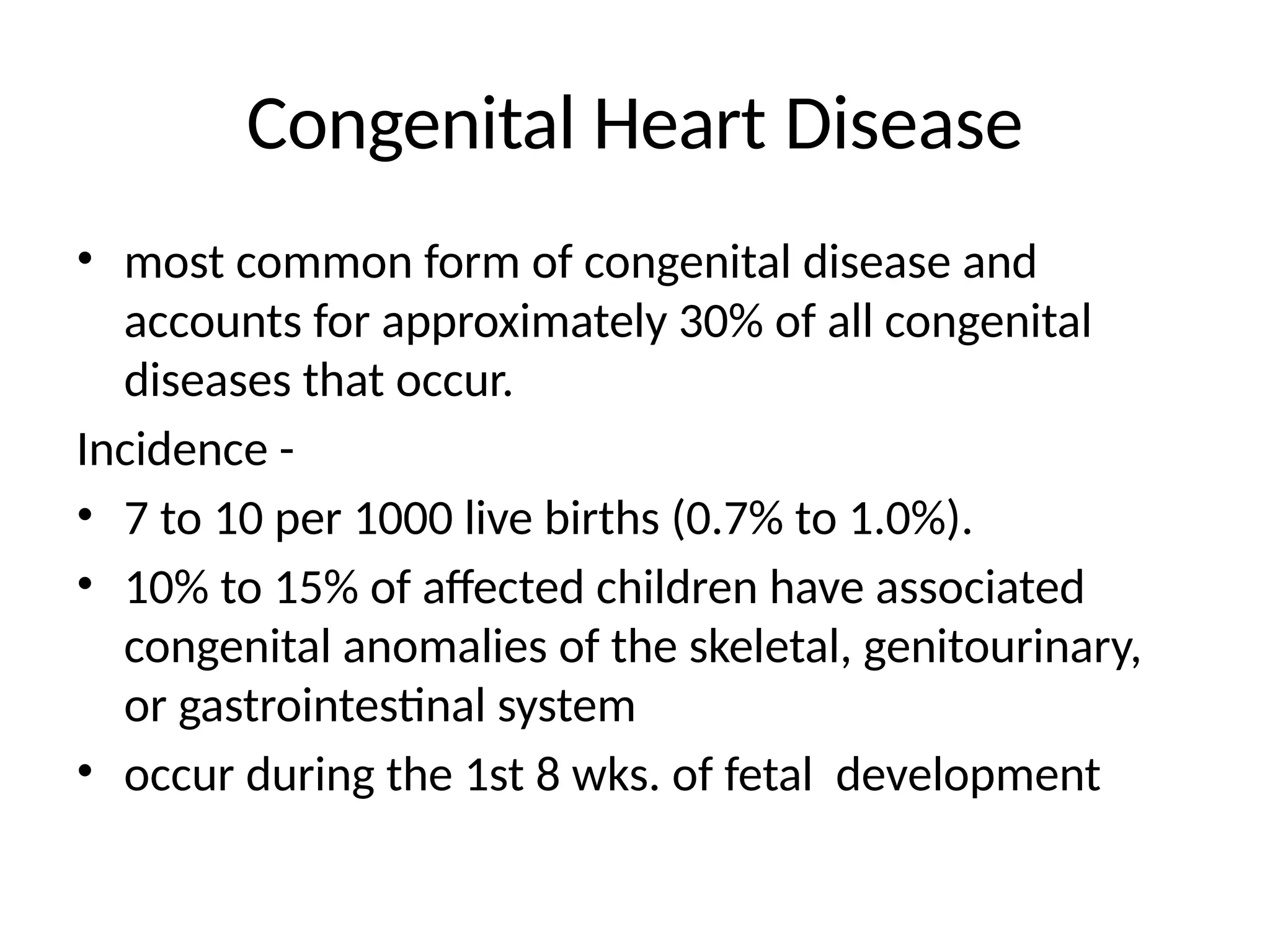
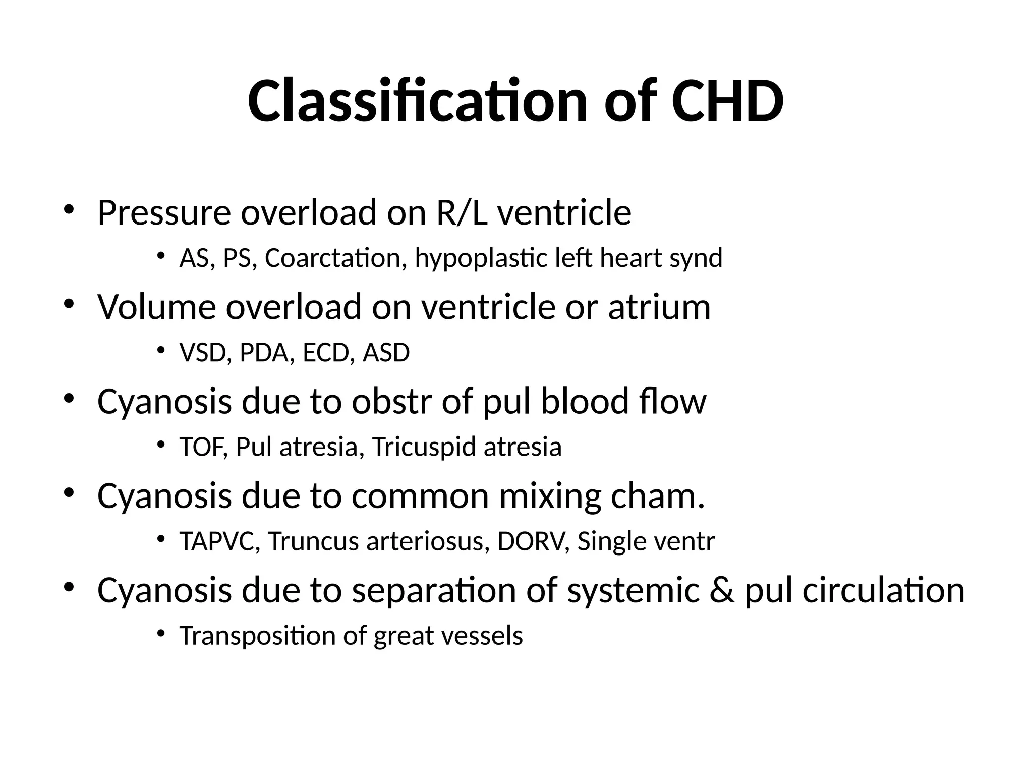
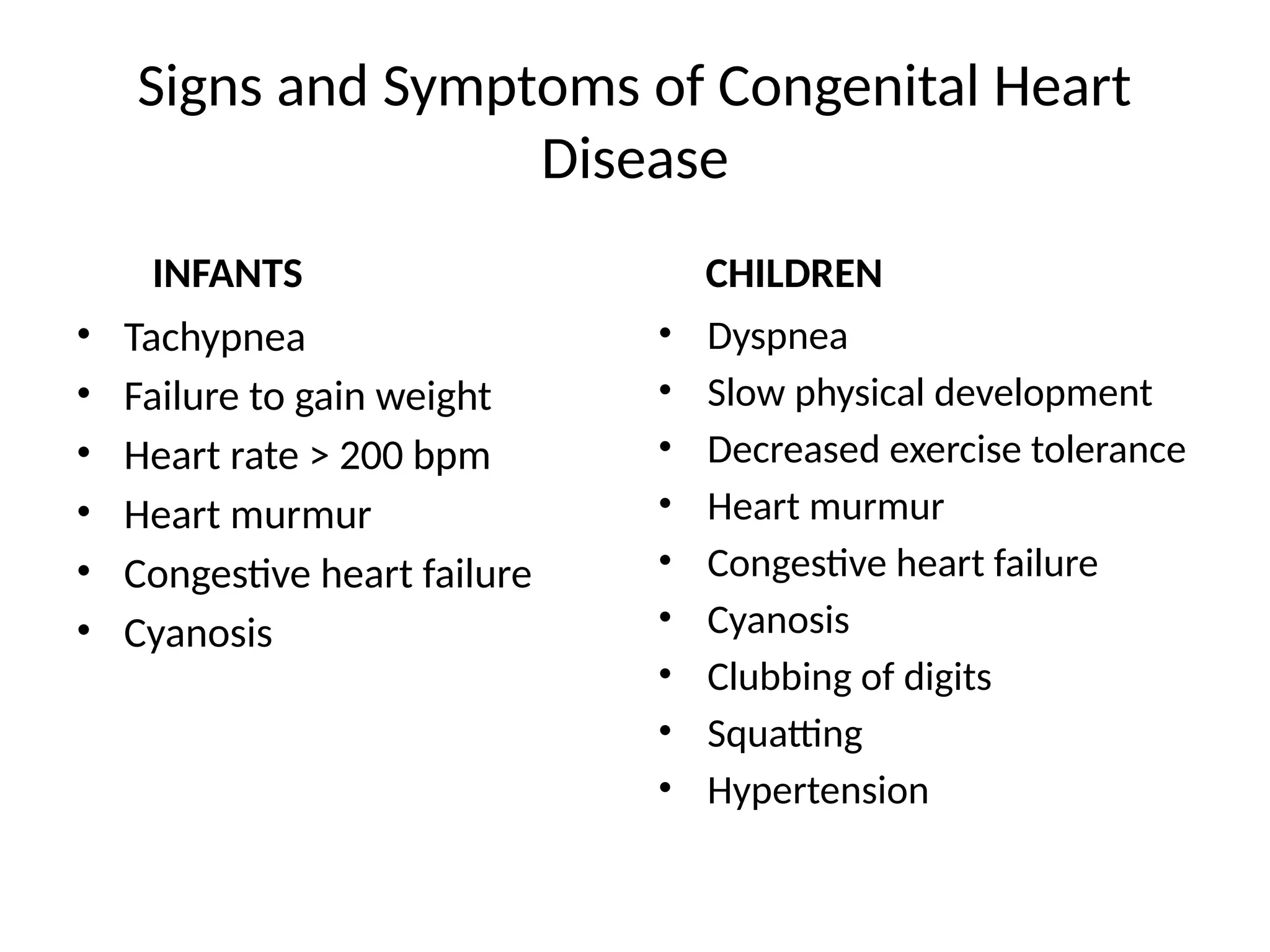
Congenital Heart Diseases
Moderator – Dr. Kirti
Presenters - Dr. Pushpa
Dr. Jyotsna
Dr. Yatender
Congenital Heart Disease
• most common form of congenital disease and
accounts for approximately 30% of all congenital
diseases that occur.
Incidence -
• 7 to 10 per 1000 live births (0.7% to 1.0%).
• 10% to 15% of affected children have associated
congenital anomalies of the skeletal, genitourinary,
or gastrointestinal system
• occur during the 1st 8 wks. of fetal development
Factors Contributing to CHD
85-90% -no identifiable cause,
multifactorial inheritance - Factors are usually both genetic and
environmental
Maternal Factors:
• seizure disorders w/ intake of anti-seizure medications
• intake of lithium for depression
• uncontrolled IDDM
• lupus
• german measles (rubella) – 1st trimester of pregnancy
Chromosome abnormalities:
• 5 to 8 % of all babies with CHD have a chromosome abnormality
• includes Down syndrome, trisomy 18 and trisomy 13, Turner’s syndrome,
Cri-du-chat syndrome
Blood circulation after birth:
The transformation from fetal to neonatal circulation involves two major
changes:
• A marked increase in systemic resistance. caused by loss of the low-
resistance placenta.
• A marked decrease in pulmonary resistance. caused by pulmonary
artery dilation with the neonate’s first breaths.
Effect of anaesthetics on PVR and SVR
Barbiturates: vasodilatingeffect, lowering both SVR and PVR.
Ketaminedecrease PVR in adults but not in children. Its
sympathomimetic effect helps in maintaining SVR, and it is
considered one of the safest induction techniques for a cyanotic
patient.
Narcotics provide good hemodynamic stability without altering
PVR. Fentanyl and sufentanil are used for maintenance of
anesthesia because they blunt the sympathetic stress response.
Volatile anaesthetics: decrease SVR in a dose-dependent manner
and are myocardial depressants.The use of potent inhalation
agents as primary anesthetics should be reserved for the child
with adequate cardiovascular reserve who is a candidate for
early postoperative extubation. In these patients, the myocardial
depression and hypotension associated with the use of
inhalation agents are well tolerated.
Nitrous oxide does not increase PVR in infants, but 100% oxygen is
preferred to avoid hypoxemia
Inflation of alveoli →↑PAO2→ vasodilatation→↓PVR→
↑PBF →↑LA Pressure exceeding RA pressure→foramen
ovale close
More heavily oxygenated blood passing by the ductus
arteriosus causes it to constrict
Functional closure of the foramen ovale and ductus
arteriosus occurs soon after birth
Overall anatomic changes are not complete for weeks
Transform of Fetal circulation to Adult
(parallel to Series)
Classification of CHD
• Pressure overload on R/L ventricle
• AS, PS, Coarctation, hypoplastic left heart synd
• Volume overload on ventricle or atrium
• VSD, PDA, ECD, ASD
• Cyanosis due to obstr of pul blood flow
• TOF, Pul atresia, Tricuspid atresia
• Cyanosis due to common mixing cham.
• TAPVC, Truncus arteriosus, DORV, Single ventr
• Cyanosis due to separation of systemic & pul circulation
• Transposition of great vessels
Signs and Symptoms of Congenital Heart
Disease
INFANTS
• Tachypnea
• Failure to gain weight
• Heart rate > 200 bpm
• Heart murmur
• Congestive heart failure
• Cyanosis
CHILDREN
• Dyspnea
• Slow physical development
• Decreased exercise tolerance
• Heart murmur
• Congestive heart failure
• Cyanosis
• Clubbing of digits
• Squatting
• Hypertension
Common Problems Associated with Congenital Heart Disease
• Infective endocarditis
• Cardiac dysrhythmias
• Complete heart block
• Hypertension (systemic or pulmonary)
• Erythrocytosis
• Thromboembolism
• Coagulopathy
• Brain abscess
• Increased plasma uric acid concentration
• Sudden death
Acyanotic Congenital Heart Defects
• L → R shunts cause CHF and pulmonary hypertension.
This leads to RV enlargement, RV failure
• These babies present with CHF and respiratory
distress.
• They are not typically cyanotic
• Examples: Patent Ductus Arteriosus (PDA)
Ventricular Septal Defect (VSD)
Atrial Septal Defect (ASD)
Coarctation of the Aorta
Atrial Septal Defect (ASD)
• an opening in the atrial septum
• occur in 4-10% of all infants with CHD
• ASD may take the form of ostium secundum in the region
of the fossa ovalis (often located near the center of the
interatrial septum and varying from a single opening to a
fenestrated septum)
• ostium primum(endocardial cushion defect characterized
by a large opening in the interatrial septum)
• sinus venosus located in the upper atrial septum.
• Secundum ASDs account for 75% of all ASDs.
• Additional cardiac abnormalities may occur with each
type of defect and include mitral valve prolapse (ostium
secundum) and mitral regurgitation resulting from a
cleft in the anterior mitral valve leaflet (ostium primum).
• EFFECTS: When blood passes through the ASD from the
left atrium to the right atrium → a larger volume of
blood than normal must be handled by the right side of
the heart → extra blood then passes through the
pulmonary artery into the lungs → pulmonary
hypertension and pulmonary congestion
Sign and symptoms
• dyspnea on exertion,
• supraventricular dysrhythmias,
• right heart failure,
• Paradoxical embolism, and recurrent
pulmonary infections.
• Abdominal viscera may become congested
because of increased right sided pressures and
circulating volume.
Diagnostic tests:
• CXR – prominent pulmonary arteries and mild to
moderate cardiomegaly
• ECG-right axis deviation and incomplete right bundle
branch block. Atrial fibrillation and supraventricular
tachycardia may accompany an ASD
• Transesophageal and color flow Doppler
echocardiography are both useful for detecting and
determining the location of ASDs.
Treatment
• A small defect with minimal right-to-left shunting (ratio of
pulmonary flow to systemic flow is <1.5) usually causes no
symptoms and therefore does not require closure.
• When pulmonary blood flow is 1.5 times the systemic blood
flow, the ASD should be closed either percutaneously via
cardiac catheterization or surgically with open sternotomy using
pericardial patch or dacron patch and cardiopulmonary bypass
to prevent right ventricular dysfunction and irreversible
pulmonary hypertension.
• Prophylaxis against infective endocarditis is not recommended
for patients with ASDs unless a concomitant valvular
abnormality is present.
MANAGEMENT OF ANESTHESIA
• Aims – ↓ SVR
• ↑ PVR
• Bubble avoidance: prevent injection of air bubble
• Speed of induction: L to R, speeds equilibration of inhalational anaesthetics,
specially for more soluble agents. increased pulmonary blood flow can dilute
drugs injected intravenously. It is unlikely, however, that this potential dilution
will alter the clinical response to these drugs because the pulmonary circulation
time is brief.
• drugs or events that produce prolonged increases in systemic vascular
resistance should be avoided, because this change favors an increase in the
magnitude of the left-to-right shunt at the atrial level.
• decreases in systemic vascular resistance, as produced by volatile anesthetics or
increases in pulmonary vascular resistance due to positive pressure ventilation
of the lungs, tend to decrease the magnitude of the left-to-right shunt.
Ventricular Septal Defect (VSD)
• an opening in the ventricular septum
• VSD is the most common congenital cardiac abnormality in
infants and children occurring in 50% of all children with
congenital heart disease and in 20% as an isolated lesion.
• allows oxygenated blood to pass from the left ventricle, through
the opening in the septum, and then mix with unoxygenated
blood in the right ventricle.
• 70% of these defects are located in the membranous portion of
the intraventricular septum; 20% in the muscular portion of the
septum; 5% just below the aortic valve, causing aortic
regurgitation; and 5% near the junction of the mitral and
tricuspid valves (atrioventricular canal defect).
Pathophysiology
• Initially, systemic vascular resistance exceeds pulmonary
vascular resistance, and left-to-right intracardiac shunting
predominates, with corresponding pulmonary artery, left
atrial, and left ventricular volume overload.
• Over time, the pulmonary vascular resistance increases,
and the magnitude of the left-to-right intracardiac
shunting decreases; eventually, the shunt may become
right to left with the development of arterial hypoxemia
(cyanosis).
• The murmur of a moderate to large VSD is holosystolic
and is loudest at the lower left sternal border.
Diagnostic tests
• ECG - When the VSD is large, there is evidence
of left atrial and ventricular enlargement on
the ECG. If pulmonary hypertension develops,
signs of right atrial and ventricular
enlargement are noted
• chest radiographic findings – chamber
enlargement to varying degrees, depending on
the volume of the shunt, and increased
pulmonary vascularity.
Treatment
Adequate nutrition
• high-calorie formula or breast milk
• supplemental tube feedings
Prophylactic antibiotics to prevent bacterial endocarditis
Surgical repair –primary closure or with special patch like
dacron patch
Interventional cardiac catheterization
Once the pulmonary/systemic vascular resistance ratio
exceeds 0.7, the risk of surgical closure becomes
prohibitive.
MANAGEMENT OF ANESTHESIA
• volatile anesthetics (which decrease systemic vascular resistance) and
positive pressure ventilation (which increases pulmonary vascular
resistance) are well tolerated.
• However, there may be increased delivery of depressant drugs to the
heart if coronary blood flow is increased to supply the hypertrophied
ventricles. So, the technique of increasing the inspired concentrations of
volatile anesthetics to achieve rapid induction of anesthesia, as is often
done in children without cardiac defects, could result in excessive
depression of the heart before central nervous system depression is
achieved in children with VSD.
• Right ventricular infundibular hypertrophy may be present in patients with
VSDs. Perioperative events that exaggerate this obstruction to right
ventricular outflow, such as increased myocardial contractility or
hypovolemia, must be minimized.
Patent Ductus Arteriosus
PDA is present when the ductus arteriosus (which arises just distal to the
left subclavian artery and connects the descending aorta to the left
pulmonary artery) fails to close spontaneously shortly after birth.
• In fullterm newborns, the ductus arteriosus closes within 24 to 48 hours
after delivery, but in preterm newborns, the ductus arteriosus frequently
fails to close.
PDA is seen more often in the following:
• premature infants
• infants born to a mother who had rubella during the first trimester of
pregnancy
The pulmonary/systemic blood flow ratio depends on the pressure gradient
from the aorta to the pulmonary artery, the pulmonary/ systemic vascular
resistance ratio, and the diameter and length of the ductus arteriosus.
SIGNS AND SYMPTOMS
• characteristic continuous systolic an diastolic murmur best
heard at the left infraclavicular area or left upper sternal border.
• ECG and CXR - If the left-to-right shunt is large, left ventricular
hypertrophy. If pulmonary hypertension develops, right
ventricular hypertrophy is apparent.
• The potential adverse effects of an untreated PDA include
ventricular hypertrophy with congestive heart failure,
pulmonary vascular disease including Eisenmenger's syndrome
with shunt flow reversal, poor physical growth, infective
endocarditis, aneurysmal dilatation of the ductus, and ductal
calcification.
Treatment
Medical Management
• Indomethacin IV (prostaglandin inhibitor) may help close a PDA.
works by stimulating the muscles inside the PDA to constrict, thereby closing the
connection
Digoxin
Diuretics
adequate nutrition
(premature infants or those infants with a large PDA may become tired when
feeding, and are not able to eat enough to gain weight)
– high-calorie formula or breast milk
PDA surgical repair or closure
-Repair is usually indicated in infants younger than 6 months of age who have large
defects that are causing symptoms, such as poor weight gain and rapid breathing
-PDA ligation
MANAGEMENT OF ANESTHESIA
• increases in systemic vascular resistance or decreases in
pulmonary vascular resistance should be avoided, because
these changes will increase the magnitude of the left-to-
right shunt.
• Appropriate preparations must be made in anticipation of
the possibility of large blood loss should control of the PDA
be lost during attempted ligation.
• Ligation of the PDA is often associated with significant
systemic hypertension during the postoperative period.
This hypertension can be managed with continuous
infusion of vasodilating drugs such as nitroprusside.
Coarctation of the Aorta
• typically consists of a discrete, diaphragm-like ridge extending
into the aortic lumen.
• described by its relationship to the ductus arteriosus
(preductal, juxtaductal, postductal).
• Postductal - coarctation extends just distal to the left
subclavian artery at the site of the aortic ductal attachment
(ligamentum arteriosum) and is most likely to manifest in
young adults.
• Preductal - coarctation is immediately proximal to the left
subclavian artery. This situation is most likely to present in
infants.
• A/w - bicuspid aortic valve, PDA, mitral stenosis or
regurgitation, aneurysms of the circle of Willis, and gonadal
dysgenesis (Turner's syndrome).
SIGNS AND SYMPTOMS
• headache,
• dizziness, epistaxis, and palpitations.
• diminished blood flow to the legs causes claudication.
• systolic blood pressure is higher in arms than in the legs, but the
diastolic pressure is similar, which results in widened pulse pressure
in the arms.
• The femoral arterial pulses are weak and delayed.
• Systemic hypertension presumably reflects ejection of the left
ventricular stroke volume into the fixed resistance created by the
narrowed aorta.
• A harsh systolic ejection murmur is present along the left sternal
border and in the back, particularly over the area of the coarctation.
Diagnosis –
• ECG – left ventricular hypertrophy
• CXR - symmetrical notching of the posterior third of the third
through eighth ribs. Indentation of the aorta with prestenotic or
poststenotic dilation of the aorta, producing the "reversed E," or
"3," sign.
• Echo - estimate the transcoarctation pressure gradient.
Treatment
• Surgical resection of the coarctation of the aorta
• balloon dilation is a therapeutic alternative, the procedure is
associated with a higher incidence of subsequent aortic aneurysm
and recurrent coarctation than is surgical resection.
MANAGEMENT OF ANESTHESIA
• Management of anesthesia for surgical resection of coarctation of
the aorta must consider
1. the adequacy of perfusion to the lower portion of the body during
cross-clamping of the aorta,
2. the propensity for systemic hypertension during crossclamping of
the aorta, and
3. the risk of neurologic sequelae due to ischemia of the spinal cord.
• Continuous monitoring of systemic blood pressure above and below
the coarctation is achieved by placing a catheter in the right radial
artery and in a femoral artery.
• Mean arterial pressures in the lower extremities should be
at least 40 mm Hg to ensure adequate blood flow to the
kidneys and spinal cord.
• Somatosensory evoked potentials are useful for
monitoring spinal cord function and the adequacy of its
blood flow during cross-clamping of the aorta.
• Excessive increases in systolic blood pressure during cross-
clamping of the aorta may adversely increase the work of
the heart and make surgical repair more difficult. In this
situation, the use of volatile anesthetics is helpful for
maintaining normal systemic blood pressures.
Blue Baby (R → L shunt)
• R → L shunts cause hypoxia and central cyanosis.
• Venous blood is shunted from the R to the L side of the
heart w/o passing through the lungs to be oxygenated
• Unoxygenated blood circulates in arteries
• cyanosis
Examples:
 Tetralogy of Fallot (TOF)
 Transposition of the Great Arteries (TGA)
 Truncus Arteriosus (TA)
Cyanotic Congenital Heart Defects
Tetralogy of Fallot
• the most common cyanotic congenital heart defect
• 10% of all congenital heart disease cases
characterized by a
• large single VSD,
• an aorta that overrides the right and left ventricles,
• obstruction to right ventricular outflow (subvalvular,
valvular, supravalvular, pulmonary arterial
branches), and
• right ventricular hypertrophy
Pathophysiology
• Right ventricular hypertrophy occurs because the VSD permits
continuous exposure of the right ventricle to the high
pressures present in the left ventricle.
• Right-to-left intracardiac shunting occurs because of increased
resistance to flow in the right ventricular outflow tract, the
severity of which determines the magnitude of the shunt.
• Because the resistance to flow across the right ventricular
outflow tract is relatively fixed, changes in systemic vascular
resistance (drug induced) may affect the magnitude of the
shunt. Decreases in systemic vascular resistance increase
right-to-left intracardiac shunting and accentuate arterial
hypoxemia, whereas increases in systemic vascular resistance
(squatting) decrease left-to-right intracardiac shunting with
resultant increases in pulmonary blood flow.
SIGNS AND SYMPTOMS
The main characteristic of TOF is cyanosis
• Cyanosis can result from three separate mechanisms:
(a) inadequate PBF
(b) right-to-left shunting
(c) intrinsic pulmonary disease.
• Because of the right ventricular outflow obstruction, blood ejected from the
RV crosses the VSD and enters the overriding aorta.
• This reduces the amount of pulmonic blood flow available for oxygenation and
adds desaturated blood to the systemic circulation leading to cyanosis.
• Decreases in SVR or increases in PVR will worsen the degree of cyanosis
The most common auscultatory finding is a systolic ejection murmur heard
along the left sternal border resulting from blood flow across the stenotic
pulmonic valve.
What are hypercyanotic spells? How are they
treated?
• Hypercyanotic spells or "tet spells" are paroxysmal episodes
in which the cyanosis acutely worsens. Crying, feeding, or
defecating can bring on these episodes. The common
pathway for all these activities is an increase in right-to-left
shunting. Three mechanisms can explain the increase in
shunt:
1. Increase in PVR This is associated with a reduction in PBF
and increase in R→L shunt. Treatment is to lower PVR
through hyperventilation with 100% O2 and bicarbonate
administration to temper the effects of acidosis on PVR.
What are hypercyanotic…...
2 Dynamic outflow obstruction Tachycardia, hypovolemia and
increased myocardial contractility can cause infundibular
spasm and worsens the R→L shunt. The spasm can be
treated with β-blockers, with volume, and by deepening the
level of anesthesia to decrease catecholamine levels.
Morphine can be given to diminish the hyperpneic response.
3 Decrease in SVR This will favor right-to-left shunting through
the VSD. Treatment is volume administration to ensure
adequate filling of the right ventricle and an -adrenergic
agonist eg. Phenylephrine to increase the SVR. SVR can also
be increased by flexing the legs or by compressing the
abdominal aorta directly. Children will squat during a
hypercyanotic spell to increase their SVR by kinking the large
arteries in the inguinal area and cause a decrease in the
right-to-left shunt.
TREATMENT
• Treatment of tetralogy of Fallot is complete surgical correction when
patients are extremely young (closure of the VSD with a Dacron patch and
relief of right ventricular outflow obstruction by placement of a synthetic
graft).
Palliative procedures to increase pulmonary blood flow.
- involved anastomosis of a systemic artery to a pulmonary artery in an
effort to increase pulmonary blood flow and improve arterial oxygenation.
These are -
• Waterston's operation (side-to-side anastomosis of the ascending aorta
and the right pulmonary artery),
• Potts's operation (side-to-side anastomosis of the descending aorta to the
left pulmonary artery), and
• Blalock-Taussig operation (end-to-side anastomosis of the subclavian
artery to the pulmonary artery).
Pre operative preparation
• General NPO guidelines can be followed
• avoid dehydration
• Crying associated with im administration of drugs used for
preoperative medication can lead to hypercyanotic attacks.
• Midazolam 0.5 to 1.0 mg/kg can be given by mouth 10 to
20 minutes before induction in children older than 9
months
• β-adrenergic antagonists should be continued until the
induction of anesthesia in patients receiving these drugs
for prophylaxis against hypercyanotic attacks.
Anaesthetic considerations
• The onset of action of IV drugs may be more rapid in the
presence of R→L shunts because the dilutional effect in the
lungs is decreased. For this reason, it may be prudent to
decrease the rate of intravenous injection of depressant drugs
in these patients.
• Ketamine causes ↑SVR, leading to a decrease in the
magnitude of the R→L intracardiac shunt, thereby
↑PBF,hence a/w improved arterial oxygenation
• Tracheal intubation is facilitated by administration of muscle
relaxants.
• Halothane is the preferred inhalational anesthetic as it
decreases contractility and maintains SVR.
• hypercyanotic attacks can occur during administration of low
concentrations of volatile anesthetics.
Maintenance of Anesthesia
• Maintenance of anesthesia is often achieved with nitrous oxide
combined with ketamine. The advantage of this combination is
preservation of the systemic vascular resistance.
• Nitrous oxide may also increase pulmonary vascular resistance,
but this potentially adverse effect is more than offset by its
beneficial effects on systemic vascular resistance.
• The use of an opioid or benzodiazepine may also be considered
during maintenance of anesthesia, but the dose and rate of
administration must be adjusted to minimize decreased
systemic blood pressure and systemic vascular resistance.
• Intraoperative skeletal muscle paralysis may be provided
with pancuronium in view of its ability to maintain
systemic blood pressure and systemic vascular resistance.
An increase in heart rate associated with pancuronium is
helpful for maintaining left ventricular cardiac output.
• Controlled ventilation, but avoid excessive positive airway
pressure may adversely increase the resistance to blood
flow through the lungs.
• Intravascular fluid volume must be maintained with ivf
administration bc acute hypovolemia tends to increase the
magnitude of shunt.
• predictable erythrocytosis, it is probably not
necessary to consider blood replacement until
approximately 20% of the patient's blood volume
has been lost.
• meticulous care to avoid infusion of air through the
tubing used to deliver intravenous solutions because
it could lead to systemic air embolization.
• α-Adrenergic agonist drugs such as phenylephrine
must be available to treat undesirable decreases in
systemic blood pressure caused by decreased SVR.
Transposition of the Great Arteries
• Results from failure of the truncus arteriosus to spiral, so that
the aorta arises from the anterior portion of the right ventricle
and the pulmonary artery arises from the left ventricle
• There is complete separation of the pulmonary and systemic
circulations such that systemic venous blood traverses the right
atrium, right ventricle, aorta, and systemic circulation; and
pulmonary venous blood traverses the left atrium, left ventricle,
pulmonary artery, and lungs.
• Thus, the circulation is parallel instead of normal in-series
circulation.
• Survival is possible only if there is communication between the
two circulations in the form of a VSD, ASD, or PDA.
Types –
• group I, who have transposition of the great arteries with intact
ventricular septum
• group II, who have transposition of the great arteries with VSD, show
symptoms of congestive heart failure (tachypnea, tachycardia, sweating,
and poor feeding)
• group III have transposition of the great arteries with VSD and
pulmonary stenosis, and presentation varies based on the severity of
the stenosis.
Diagnostic tests –
• ECG - right axis deviation and right ventricular hypertrophy
• CXR - cardiac silhouette described as being "egg shaped with a narrow
stalk."
TREATMENT
• The immediate management involves creating
intracardiac mixing or increasing the degree of mixing.
This goal is accomplished with infusions of prostaglandin
E1 to maintain patency of the ductus arteriosus
• and/or balloon atrial septostomy (Rashkind's
procedure).
• Administration of oxygen may decrease pulmonary
vascular resistance and increase pulmonary blood flow.
Diuretics and digoxin are administered to treat
congestive heart failure.
Treatment
Two surgical switch procedures have been used to treat
complete transition of the great arteries.
• Mustard's or Senning's operation, generally involved
resection of the atrial septum.
• arterial switch operation in which the pulmonary artery
and ascending aorta are transected above the semilunar
valves and reanastomosed with the right and left
ventricles; the coronary arteries are then reimplanted so
that the aorta is connected to the left ventricle and the
pulmonary artery is connected to the right ventricle.
MANAGEMENT OF ANESTHESIA
• Drugs administered intravenously are distributed with
minimal dilution to organs such as the heart and brain.
Therefore, doses and rates of injection of intravenously
administered drugs may have to be decreased.
• Conversely, the onset of anesthesia produced by inhaled
drugs is delayed because only small amounts of the inhaled
drug reach the systemic circulation.
• induction and maintenance of anesthesia are often
accomplished with ketamine combined with muscle relaxants
to facilitate tracheal intubation.
• Ketamine can be supplemented with opioids or
benzodiazepines for maintenance of anesthesia.
• Dehydration must be avoided during the perioperative period.
Truncus Arteriosus
• congenital cardiac defect in which a single arterial trunk
serves as the origin of the aorta and pulmonary artery.
• This single arterial trunk overrides both ventricles, which are
connected through a VSD.
• Types –
• type I - A main pulmonary artery may arise from the truncal
artery (truncus arteriosus),
• type II - the branch pulmonary arteries may arise separately
but in close proximity,
• type III - the branch pulmonary arteries may arise widely
separate from the lateral aspects of the truncal artery.
SIGNS AND SYMPTOMS
• cyanosis
• arterial hypoxemia,
• failure to thrive, and
• congestive heart failure early in life.
Diagnosis –
• confirmed by angiocardiography performed during
cardiac catheterization.
• CXR – cardiomegaly and increased vascularity of the
lung fields.
Treatment
• Truncus arteriosus must be treated by surgical repair of the defects.
• The pulmonary arteries are detached from the common artery
(truncus arteriosus) and connected to the right ventricle using a
homograft (a section of pulmonary artery with its valves intact
from a tissue donor).
• The ventricular septal defect is closed with a patch.
• Banding of the right and left pulmonary arteries if pulmonary blood
flow is excessive. In addition, an associated VSD can be closed so
only left ventricular output enters the truncus arteriosus.
• When this is done, a Dacron conduit with a valve is also placed
between the right ventricle and pulmonary artery.
MANAGEMENT OF ANESTHESIA
• When pulmonary blood flow is increased, the use of
positive end-expiratory pressure is beneficial and may serve
to decrease the symptoms of congestive heart failure.
• Increased pulmonary blood flow may be associated with
evidence of myocardial ischemia on the ECG.
• When myocardial ischemia that occurs intraoperatively
does not respond to intravenous administration of
phenylephrine or fluids, or the use of positive end-
expiratory pressure, consideration may be given to
temporary banding of the pulmonary artery to increase
systemic and coronary blood flow.

Congenital Heart Diseases classificayion.pptx

  • 1.
    Congenital Heart Diseases Moderator– Dr. Kirti Presenters - Dr. Pushpa Dr. Jyotsna Dr. Yatender
  • 2.
    Congenital Heart Disease •most common form of congenital disease and accounts for approximately 30% of all congenital diseases that occur. Incidence - • 7 to 10 per 1000 live births (0.7% to 1.0%). • 10% to 15% of affected children have associated congenital anomalies of the skeletal, genitourinary, or gastrointestinal system • occur during the 1st 8 wks. of fetal development
  • 3.
    Factors Contributing toCHD 85-90% -no identifiable cause, multifactorial inheritance - Factors are usually both genetic and environmental Maternal Factors: • seizure disorders w/ intake of anti-seizure medications • intake of lithium for depression • uncontrolled IDDM • lupus • german measles (rubella) – 1st trimester of pregnancy Chromosome abnormalities: • 5 to 8 % of all babies with CHD have a chromosome abnormality • includes Down syndrome, trisomy 18 and trisomy 13, Turner’s syndrome, Cri-du-chat syndrome
  • 5.
    Blood circulation afterbirth: The transformation from fetal to neonatal circulation involves two major changes: • A marked increase in systemic resistance. caused by loss of the low- resistance placenta. • A marked decrease in pulmonary resistance. caused by pulmonary artery dilation with the neonate’s first breaths. Effect of anaesthetics on PVR and SVR Barbiturates: vasodilatingeffect, lowering both SVR and PVR. Ketaminedecrease PVR in adults but not in children. Its sympathomimetic effect helps in maintaining SVR, and it is considered one of the safest induction techniques for a cyanotic patient.
  • 6.
    Narcotics provide goodhemodynamic stability without altering PVR. Fentanyl and sufentanil are used for maintenance of anesthesia because they blunt the sympathetic stress response. Volatile anaesthetics: decrease SVR in a dose-dependent manner and are myocardial depressants.The use of potent inhalation agents as primary anesthetics should be reserved for the child with adequate cardiovascular reserve who is a candidate for early postoperative extubation. In these patients, the myocardial depression and hypotension associated with the use of inhalation agents are well tolerated. Nitrous oxide does not increase PVR in infants, but 100% oxygen is preferred to avoid hypoxemia
  • 7.
    Inflation of alveoli→↑PAO2→ vasodilatation→↓PVR→ ↑PBF →↑LA Pressure exceeding RA pressure→foramen ovale close More heavily oxygenated blood passing by the ductus arteriosus causes it to constrict Functional closure of the foramen ovale and ductus arteriosus occurs soon after birth Overall anatomic changes are not complete for weeks
  • 8.
    Transform of Fetalcirculation to Adult (parallel to Series)
  • 9.
    Classification of CHD •Pressure overload on R/L ventricle • AS, PS, Coarctation, hypoplastic left heart synd • Volume overload on ventricle or atrium • VSD, PDA, ECD, ASD • Cyanosis due to obstr of pul blood flow • TOF, Pul atresia, Tricuspid atresia • Cyanosis due to common mixing cham. • TAPVC, Truncus arteriosus, DORV, Single ventr • Cyanosis due to separation of systemic & pul circulation • Transposition of great vessels
  • 10.
    Signs and Symptomsof Congenital Heart Disease INFANTS • Tachypnea • Failure to gain weight • Heart rate > 200 bpm • Heart murmur • Congestive heart failure • Cyanosis CHILDREN • Dyspnea • Slow physical development • Decreased exercise tolerance • Heart murmur • Congestive heart failure • Cyanosis • Clubbing of digits • Squatting • Hypertension
  • 11.
    Common Problems Associatedwith Congenital Heart Disease • Infective endocarditis • Cardiac dysrhythmias • Complete heart block • Hypertension (systemic or pulmonary) • Erythrocytosis • Thromboembolism • Coagulopathy • Brain abscess • Increased plasma uric acid concentration • Sudden death
  • 12.
    Acyanotic Congenital HeartDefects • L → R shunts cause CHF and pulmonary hypertension. This leads to RV enlargement, RV failure • These babies present with CHF and respiratory distress. • They are not typically cyanotic • Examples: Patent Ductus Arteriosus (PDA) Ventricular Septal Defect (VSD) Atrial Septal Defect (ASD) Coarctation of the Aorta
  • 13.
    Atrial Septal Defect(ASD) • an opening in the atrial septum • occur in 4-10% of all infants with CHD • ASD may take the form of ostium secundum in the region of the fossa ovalis (often located near the center of the interatrial septum and varying from a single opening to a fenestrated septum) • ostium primum(endocardial cushion defect characterized by a large opening in the interatrial septum) • sinus venosus located in the upper atrial septum. • Secundum ASDs account for 75% of all ASDs.
  • 15.
    • Additional cardiacabnormalities may occur with each type of defect and include mitral valve prolapse (ostium secundum) and mitral regurgitation resulting from a cleft in the anterior mitral valve leaflet (ostium primum). • EFFECTS: When blood passes through the ASD from the left atrium to the right atrium → a larger volume of blood than normal must be handled by the right side of the heart → extra blood then passes through the pulmonary artery into the lungs → pulmonary hypertension and pulmonary congestion
  • 16.
    Sign and symptoms •dyspnea on exertion, • supraventricular dysrhythmias, • right heart failure, • Paradoxical embolism, and recurrent pulmonary infections. • Abdominal viscera may become congested because of increased right sided pressures and circulating volume.
  • 17.
    Diagnostic tests: • CXR– prominent pulmonary arteries and mild to moderate cardiomegaly • ECG-right axis deviation and incomplete right bundle branch block. Atrial fibrillation and supraventricular tachycardia may accompany an ASD • Transesophageal and color flow Doppler echocardiography are both useful for detecting and determining the location of ASDs.
  • 18.
    Treatment • A smalldefect with minimal right-to-left shunting (ratio of pulmonary flow to systemic flow is <1.5) usually causes no symptoms and therefore does not require closure. • When pulmonary blood flow is 1.5 times the systemic blood flow, the ASD should be closed either percutaneously via cardiac catheterization or surgically with open sternotomy using pericardial patch or dacron patch and cardiopulmonary bypass to prevent right ventricular dysfunction and irreversible pulmonary hypertension. • Prophylaxis against infective endocarditis is not recommended for patients with ASDs unless a concomitant valvular abnormality is present.
  • 19.
    MANAGEMENT OF ANESTHESIA •Aims – ↓ SVR • ↑ PVR • Bubble avoidance: prevent injection of air bubble • Speed of induction: L to R, speeds equilibration of inhalational anaesthetics, specially for more soluble agents. increased pulmonary blood flow can dilute drugs injected intravenously. It is unlikely, however, that this potential dilution will alter the clinical response to these drugs because the pulmonary circulation time is brief. • drugs or events that produce prolonged increases in systemic vascular resistance should be avoided, because this change favors an increase in the magnitude of the left-to-right shunt at the atrial level. • decreases in systemic vascular resistance, as produced by volatile anesthetics or increases in pulmonary vascular resistance due to positive pressure ventilation of the lungs, tend to decrease the magnitude of the left-to-right shunt.
  • 20.
    Ventricular Septal Defect(VSD) • an opening in the ventricular septum • VSD is the most common congenital cardiac abnormality in infants and children occurring in 50% of all children with congenital heart disease and in 20% as an isolated lesion. • allows oxygenated blood to pass from the left ventricle, through the opening in the septum, and then mix with unoxygenated blood in the right ventricle. • 70% of these defects are located in the membranous portion of the intraventricular septum; 20% in the muscular portion of the septum; 5% just below the aortic valve, causing aortic regurgitation; and 5% near the junction of the mitral and tricuspid valves (atrioventricular canal defect).
  • 22.
    Pathophysiology • Initially, systemicvascular resistance exceeds pulmonary vascular resistance, and left-to-right intracardiac shunting predominates, with corresponding pulmonary artery, left atrial, and left ventricular volume overload. • Over time, the pulmonary vascular resistance increases, and the magnitude of the left-to-right intracardiac shunting decreases; eventually, the shunt may become right to left with the development of arterial hypoxemia (cyanosis). • The murmur of a moderate to large VSD is holosystolic and is loudest at the lower left sternal border.
  • 23.
    Diagnostic tests • ECG- When the VSD is large, there is evidence of left atrial and ventricular enlargement on the ECG. If pulmonary hypertension develops, signs of right atrial and ventricular enlargement are noted • chest radiographic findings – chamber enlargement to varying degrees, depending on the volume of the shunt, and increased pulmonary vascularity.
  • 24.
    Treatment Adequate nutrition • high-calorieformula or breast milk • supplemental tube feedings Prophylactic antibiotics to prevent bacterial endocarditis Surgical repair –primary closure or with special patch like dacron patch Interventional cardiac catheterization Once the pulmonary/systemic vascular resistance ratio exceeds 0.7, the risk of surgical closure becomes prohibitive.
  • 25.
    MANAGEMENT OF ANESTHESIA •volatile anesthetics (which decrease systemic vascular resistance) and positive pressure ventilation (which increases pulmonary vascular resistance) are well tolerated. • However, there may be increased delivery of depressant drugs to the heart if coronary blood flow is increased to supply the hypertrophied ventricles. So, the technique of increasing the inspired concentrations of volatile anesthetics to achieve rapid induction of anesthesia, as is often done in children without cardiac defects, could result in excessive depression of the heart before central nervous system depression is achieved in children with VSD. • Right ventricular infundibular hypertrophy may be present in patients with VSDs. Perioperative events that exaggerate this obstruction to right ventricular outflow, such as increased myocardial contractility or hypovolemia, must be minimized.
  • 26.
    Patent Ductus Arteriosus PDAis present when the ductus arteriosus (which arises just distal to the left subclavian artery and connects the descending aorta to the left pulmonary artery) fails to close spontaneously shortly after birth. • In fullterm newborns, the ductus arteriosus closes within 24 to 48 hours after delivery, but in preterm newborns, the ductus arteriosus frequently fails to close. PDA is seen more often in the following: • premature infants • infants born to a mother who had rubella during the first trimester of pregnancy The pulmonary/systemic blood flow ratio depends on the pressure gradient from the aorta to the pulmonary artery, the pulmonary/ systemic vascular resistance ratio, and the diameter and length of the ductus arteriosus.
  • 28.
    SIGNS AND SYMPTOMS •characteristic continuous systolic an diastolic murmur best heard at the left infraclavicular area or left upper sternal border. • ECG and CXR - If the left-to-right shunt is large, left ventricular hypertrophy. If pulmonary hypertension develops, right ventricular hypertrophy is apparent. • The potential adverse effects of an untreated PDA include ventricular hypertrophy with congestive heart failure, pulmonary vascular disease including Eisenmenger's syndrome with shunt flow reversal, poor physical growth, infective endocarditis, aneurysmal dilatation of the ductus, and ductal calcification.
  • 29.
    Treatment Medical Management • IndomethacinIV (prostaglandin inhibitor) may help close a PDA. works by stimulating the muscles inside the PDA to constrict, thereby closing the connection Digoxin Diuretics adequate nutrition (premature infants or those infants with a large PDA may become tired when feeding, and are not able to eat enough to gain weight) – high-calorie formula or breast milk PDA surgical repair or closure -Repair is usually indicated in infants younger than 6 months of age who have large defects that are causing symptoms, such as poor weight gain and rapid breathing -PDA ligation
  • 30.
    MANAGEMENT OF ANESTHESIA •increases in systemic vascular resistance or decreases in pulmonary vascular resistance should be avoided, because these changes will increase the magnitude of the left-to- right shunt. • Appropriate preparations must be made in anticipation of the possibility of large blood loss should control of the PDA be lost during attempted ligation. • Ligation of the PDA is often associated with significant systemic hypertension during the postoperative period. This hypertension can be managed with continuous infusion of vasodilating drugs such as nitroprusside.
  • 31.
    Coarctation of theAorta • typically consists of a discrete, diaphragm-like ridge extending into the aortic lumen. • described by its relationship to the ductus arteriosus (preductal, juxtaductal, postductal). • Postductal - coarctation extends just distal to the left subclavian artery at the site of the aortic ductal attachment (ligamentum arteriosum) and is most likely to manifest in young adults. • Preductal - coarctation is immediately proximal to the left subclavian artery. This situation is most likely to present in infants. • A/w - bicuspid aortic valve, PDA, mitral stenosis or regurgitation, aneurysms of the circle of Willis, and gonadal dysgenesis (Turner's syndrome).
  • 33.
    SIGNS AND SYMPTOMS •headache, • dizziness, epistaxis, and palpitations. • diminished blood flow to the legs causes claudication. • systolic blood pressure is higher in arms than in the legs, but the diastolic pressure is similar, which results in widened pulse pressure in the arms. • The femoral arterial pulses are weak and delayed. • Systemic hypertension presumably reflects ejection of the left ventricular stroke volume into the fixed resistance created by the narrowed aorta. • A harsh systolic ejection murmur is present along the left sternal border and in the back, particularly over the area of the coarctation.
  • 34.
    Diagnosis – • ECG– left ventricular hypertrophy • CXR - symmetrical notching of the posterior third of the third through eighth ribs. Indentation of the aorta with prestenotic or poststenotic dilation of the aorta, producing the "reversed E," or "3," sign. • Echo - estimate the transcoarctation pressure gradient. Treatment • Surgical resection of the coarctation of the aorta • balloon dilation is a therapeutic alternative, the procedure is associated with a higher incidence of subsequent aortic aneurysm and recurrent coarctation than is surgical resection.
  • 35.
    MANAGEMENT OF ANESTHESIA •Management of anesthesia for surgical resection of coarctation of the aorta must consider 1. the adequacy of perfusion to the lower portion of the body during cross-clamping of the aorta, 2. the propensity for systemic hypertension during crossclamping of the aorta, and 3. the risk of neurologic sequelae due to ischemia of the spinal cord. • Continuous monitoring of systemic blood pressure above and below the coarctation is achieved by placing a catheter in the right radial artery and in a femoral artery.
  • 36.
    • Mean arterialpressures in the lower extremities should be at least 40 mm Hg to ensure adequate blood flow to the kidneys and spinal cord. • Somatosensory evoked potentials are useful for monitoring spinal cord function and the adequacy of its blood flow during cross-clamping of the aorta. • Excessive increases in systolic blood pressure during cross- clamping of the aorta may adversely increase the work of the heart and make surgical repair more difficult. In this situation, the use of volatile anesthetics is helpful for maintaining normal systemic blood pressures.
  • 37.
    Blue Baby (R→ L shunt) • R → L shunts cause hypoxia and central cyanosis. • Venous blood is shunted from the R to the L side of the heart w/o passing through the lungs to be oxygenated • Unoxygenated blood circulates in arteries • cyanosis Examples:  Tetralogy of Fallot (TOF)  Transposition of the Great Arteries (TGA)  Truncus Arteriosus (TA) Cyanotic Congenital Heart Defects
  • 38.
    Tetralogy of Fallot •the most common cyanotic congenital heart defect • 10% of all congenital heart disease cases characterized by a • large single VSD, • an aorta that overrides the right and left ventricles, • obstruction to right ventricular outflow (subvalvular, valvular, supravalvular, pulmonary arterial branches), and • right ventricular hypertrophy
  • 40.
    Pathophysiology • Right ventricularhypertrophy occurs because the VSD permits continuous exposure of the right ventricle to the high pressures present in the left ventricle. • Right-to-left intracardiac shunting occurs because of increased resistance to flow in the right ventricular outflow tract, the severity of which determines the magnitude of the shunt. • Because the resistance to flow across the right ventricular outflow tract is relatively fixed, changes in systemic vascular resistance (drug induced) may affect the magnitude of the shunt. Decreases in systemic vascular resistance increase right-to-left intracardiac shunting and accentuate arterial hypoxemia, whereas increases in systemic vascular resistance (squatting) decrease left-to-right intracardiac shunting with resultant increases in pulmonary blood flow.
  • 41.
    SIGNS AND SYMPTOMS Themain characteristic of TOF is cyanosis • Cyanosis can result from three separate mechanisms: (a) inadequate PBF (b) right-to-left shunting (c) intrinsic pulmonary disease. • Because of the right ventricular outflow obstruction, blood ejected from the RV crosses the VSD and enters the overriding aorta. • This reduces the amount of pulmonic blood flow available for oxygenation and adds desaturated blood to the systemic circulation leading to cyanosis. • Decreases in SVR or increases in PVR will worsen the degree of cyanosis The most common auscultatory finding is a systolic ejection murmur heard along the left sternal border resulting from blood flow across the stenotic pulmonic valve.
  • 42.
    What are hypercyanoticspells? How are they treated? • Hypercyanotic spells or "tet spells" are paroxysmal episodes in which the cyanosis acutely worsens. Crying, feeding, or defecating can bring on these episodes. The common pathway for all these activities is an increase in right-to-left shunting. Three mechanisms can explain the increase in shunt: 1. Increase in PVR This is associated with a reduction in PBF and increase in R→L shunt. Treatment is to lower PVR through hyperventilation with 100% O2 and bicarbonate administration to temper the effects of acidosis on PVR.
  • 43.
    What are hypercyanotic…... 2Dynamic outflow obstruction Tachycardia, hypovolemia and increased myocardial contractility can cause infundibular spasm and worsens the R→L shunt. The spasm can be treated with β-blockers, with volume, and by deepening the level of anesthesia to decrease catecholamine levels. Morphine can be given to diminish the hyperpneic response. 3 Decrease in SVR This will favor right-to-left shunting through the VSD. Treatment is volume administration to ensure adequate filling of the right ventricle and an -adrenergic agonist eg. Phenylephrine to increase the SVR. SVR can also be increased by flexing the legs or by compressing the abdominal aorta directly. Children will squat during a hypercyanotic spell to increase their SVR by kinking the large arteries in the inguinal area and cause a decrease in the right-to-left shunt.
  • 44.
    TREATMENT • Treatment oftetralogy of Fallot is complete surgical correction when patients are extremely young (closure of the VSD with a Dacron patch and relief of right ventricular outflow obstruction by placement of a synthetic graft). Palliative procedures to increase pulmonary blood flow. - involved anastomosis of a systemic artery to a pulmonary artery in an effort to increase pulmonary blood flow and improve arterial oxygenation. These are - • Waterston's operation (side-to-side anastomosis of the ascending aorta and the right pulmonary artery), • Potts's operation (side-to-side anastomosis of the descending aorta to the left pulmonary artery), and • Blalock-Taussig operation (end-to-side anastomosis of the subclavian artery to the pulmonary artery).
  • 45.
    Pre operative preparation •General NPO guidelines can be followed • avoid dehydration • Crying associated with im administration of drugs used for preoperative medication can lead to hypercyanotic attacks. • Midazolam 0.5 to 1.0 mg/kg can be given by mouth 10 to 20 minutes before induction in children older than 9 months • β-adrenergic antagonists should be continued until the induction of anesthesia in patients receiving these drugs for prophylaxis against hypercyanotic attacks.
  • 46.
    Anaesthetic considerations • Theonset of action of IV drugs may be more rapid in the presence of R→L shunts because the dilutional effect in the lungs is decreased. For this reason, it may be prudent to decrease the rate of intravenous injection of depressant drugs in these patients. • Ketamine causes ↑SVR, leading to a decrease in the magnitude of the R→L intracardiac shunt, thereby ↑PBF,hence a/w improved arterial oxygenation • Tracheal intubation is facilitated by administration of muscle relaxants. • Halothane is the preferred inhalational anesthetic as it decreases contractility and maintains SVR. • hypercyanotic attacks can occur during administration of low concentrations of volatile anesthetics.
  • 47.
    Maintenance of Anesthesia •Maintenance of anesthesia is often achieved with nitrous oxide combined with ketamine. The advantage of this combination is preservation of the systemic vascular resistance. • Nitrous oxide may also increase pulmonary vascular resistance, but this potentially adverse effect is more than offset by its beneficial effects on systemic vascular resistance. • The use of an opioid or benzodiazepine may also be considered during maintenance of anesthesia, but the dose and rate of administration must be adjusted to minimize decreased systemic blood pressure and systemic vascular resistance.
  • 48.
    • Intraoperative skeletalmuscle paralysis may be provided with pancuronium in view of its ability to maintain systemic blood pressure and systemic vascular resistance. An increase in heart rate associated with pancuronium is helpful for maintaining left ventricular cardiac output. • Controlled ventilation, but avoid excessive positive airway pressure may adversely increase the resistance to blood flow through the lungs. • Intravascular fluid volume must be maintained with ivf administration bc acute hypovolemia tends to increase the magnitude of shunt.
  • 49.
    • predictable erythrocytosis,it is probably not necessary to consider blood replacement until approximately 20% of the patient's blood volume has been lost. • meticulous care to avoid infusion of air through the tubing used to deliver intravenous solutions because it could lead to systemic air embolization. • α-Adrenergic agonist drugs such as phenylephrine must be available to treat undesirable decreases in systemic blood pressure caused by decreased SVR.
  • 50.
    Transposition of theGreat Arteries • Results from failure of the truncus arteriosus to spiral, so that the aorta arises from the anterior portion of the right ventricle and the pulmonary artery arises from the left ventricle • There is complete separation of the pulmonary and systemic circulations such that systemic venous blood traverses the right atrium, right ventricle, aorta, and systemic circulation; and pulmonary venous blood traverses the left atrium, left ventricle, pulmonary artery, and lungs. • Thus, the circulation is parallel instead of normal in-series circulation. • Survival is possible only if there is communication between the two circulations in the form of a VSD, ASD, or PDA.
  • 52.
    Types – • groupI, who have transposition of the great arteries with intact ventricular septum • group II, who have transposition of the great arteries with VSD, show symptoms of congestive heart failure (tachypnea, tachycardia, sweating, and poor feeding) • group III have transposition of the great arteries with VSD and pulmonary stenosis, and presentation varies based on the severity of the stenosis. Diagnostic tests – • ECG - right axis deviation and right ventricular hypertrophy • CXR - cardiac silhouette described as being "egg shaped with a narrow stalk."
  • 53.
    TREATMENT • The immediatemanagement involves creating intracardiac mixing or increasing the degree of mixing. This goal is accomplished with infusions of prostaglandin E1 to maintain patency of the ductus arteriosus • and/or balloon atrial septostomy (Rashkind's procedure). • Administration of oxygen may decrease pulmonary vascular resistance and increase pulmonary blood flow. Diuretics and digoxin are administered to treat congestive heart failure.
  • 54.
    Treatment Two surgical switchprocedures have been used to treat complete transition of the great arteries. • Mustard's or Senning's operation, generally involved resection of the atrial septum. • arterial switch operation in which the pulmonary artery and ascending aorta are transected above the semilunar valves and reanastomosed with the right and left ventricles; the coronary arteries are then reimplanted so that the aorta is connected to the left ventricle and the pulmonary artery is connected to the right ventricle.
  • 55.
    MANAGEMENT OF ANESTHESIA •Drugs administered intravenously are distributed with minimal dilution to organs such as the heart and brain. Therefore, doses and rates of injection of intravenously administered drugs may have to be decreased. • Conversely, the onset of anesthesia produced by inhaled drugs is delayed because only small amounts of the inhaled drug reach the systemic circulation. • induction and maintenance of anesthesia are often accomplished with ketamine combined with muscle relaxants to facilitate tracheal intubation. • Ketamine can be supplemented with opioids or benzodiazepines for maintenance of anesthesia. • Dehydration must be avoided during the perioperative period.
  • 56.
    Truncus Arteriosus • congenitalcardiac defect in which a single arterial trunk serves as the origin of the aorta and pulmonary artery. • This single arterial trunk overrides both ventricles, which are connected through a VSD. • Types – • type I - A main pulmonary artery may arise from the truncal artery (truncus arteriosus), • type II - the branch pulmonary arteries may arise separately but in close proximity, • type III - the branch pulmonary arteries may arise widely separate from the lateral aspects of the truncal artery.
  • 57.
    SIGNS AND SYMPTOMS •cyanosis • arterial hypoxemia, • failure to thrive, and • congestive heart failure early in life. Diagnosis – • confirmed by angiocardiography performed during cardiac catheterization. • CXR – cardiomegaly and increased vascularity of the lung fields.
  • 58.
    Treatment • Truncus arteriosusmust be treated by surgical repair of the defects. • The pulmonary arteries are detached from the common artery (truncus arteriosus) and connected to the right ventricle using a homograft (a section of pulmonary artery with its valves intact from a tissue donor). • The ventricular septal defect is closed with a patch. • Banding of the right and left pulmonary arteries if pulmonary blood flow is excessive. In addition, an associated VSD can be closed so only left ventricular output enters the truncus arteriosus. • When this is done, a Dacron conduit with a valve is also placed between the right ventricle and pulmonary artery.
  • 60.
    MANAGEMENT OF ANESTHESIA •When pulmonary blood flow is increased, the use of positive end-expiratory pressure is beneficial and may serve to decrease the symptoms of congestive heart failure. • Increased pulmonary blood flow may be associated with evidence of myocardial ischemia on the ECG. • When myocardial ischemia that occurs intraoperatively does not respond to intravenous administration of phenylephrine or fluids, or the use of positive end- expiratory pressure, consideration may be given to temporary banding of the pulmonary artery to increase systemic and coronary blood flow.