Personal and pastHistory
• ปฏิเสธแพ้ยา แพ้อาหาร
• ปฏิเสธโรคประจาตัว
• ปฏิเสธโรคในครอบครัว
• ปฏิเสธประวัติกระทบกระแทก
• พัฒนาการสมวัย แข็งแรงดีมาตลอด
6.
Physical Examination
Vital signs:T 37oC PR 115 /min RR 22 /min
BP124/70 (P97 123/82)
GA: a Thai boy Alert, Antralgic gait
HEENT: not pale conjunctivae, no anicteric
sclera, no sunken eye ball, no dental caries,
ears
Lymph node : not palpabled
Cardiovascular : no active precordium,
normal S1 S2 no murmur
7.
Physical Examination
Abdomen: scaphoidabdomen, no surgical
scar, normoactive bowel sounds, soft, not
Musculoskeletal : no deformity of
extremities. Mild tender at left thigh and
redness , limited ROM of Lt, knee due to
,motor grade V
Skin: no rash, no hypo/hyper pigmentation
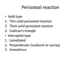
Plain X-ray
• size
•Site of the lesion
• Borders of the lesion/zone of transition
• Type of bone destruction
• Periosteal reaction
• Matrix of the lesion
• Nature and extent of soft tissue involvement
• Multiplicity
24.
Size
• The largerlesion the more likely to be
aggressive or malignant
Borders of thelesion/zone of transition
• Sharp
• Narrow
• wide
29.
Pattern of bone
destruction
•Geographic
: destructive lesion with sharply
defined border. Less aggressive,
slow growing
• Moth-eaten
: area of destruction with
ragged border. More rapid
growth
• Permeative
: illed defined lesion with
multiple worm holes. Spread
through marrow space. Implied
a aggressive malignancy
Tumor matrix
• Osteoblastic
:Fluffy, cotton like or cloud-like densities
• Catilagenous
: comma-shaped, punctate, annular, popcorn-
like
34.
Osteosarcoma
• Most isIntramedullary tumor
• Highly malignant tumor arising within the
bone and spreading rapidly outwards to
periosteum and surface around soft tissue.
• Age 10-25 years , male > female
• Common involves long-bone metaphyses,
especially around the knee and proximal end
of humerus
35.
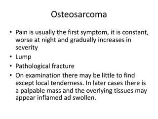
Osteosarcoma
• Pain isusually the first symptom, it is constant,
worse at night and gradually increases in
severity
• Lump
• Pathological fracture
• On examination there may be little to find
except local tenderness. In later cases there is
a palpable mass and the overlying tissues may
appear inflamed ad swollen.
36.
Variants Usual age
atdiagnosis
Common
Primary Sites
Radiographic Appearance Distinctive
Features
Clinical
Course
I. Conventional
Osteoblastic
Chondroblastic
Fibroblastic
Second and
third decade
Around knee
jointand shoulder
Variable,depending on degree of
mineralization of osteoid
Tumor osteoid present; variable
degrees of osteoblastic,
chondroblastic and fibroblastic
differentiation
Early
dissemination
to lungs
Skeleton
II. Telangiectatic Second and
third decade
Similar to
conventional
osteosarcoma
Predominantly lytic lesionwith
littleor no sclerosis
Cystic, cavity-liketumor;
blood-filled spaces in tumor
Similar to
conventional
osteoSarcoma
III. Small cell Second and
third decade
Similar to con-
ventional
osteosarcoma
May be predominantly lytic May be confused with Ewing’s
sarcoma
Similar to
conventional
osteosarcoma;
radioresponsive
IV. Multifocal Synchronous
involvementof
multiplebones
Multiple skeletal sites showing
densely sclerotic lesions
Multiple primary tumors vs,
metastatic primary tumor
Uniformly Fatal
V. Parosteal
(juxtacortical
osteosarcoma)
Third decade
or older
Posterior aspect
of distalfemur
Arises from cortex; encircles
involved bone; pronounced
ossification
Low-grade tumor with
characteristicradiograph and
pathology
Indolent clinical
course with low
Propensity for
metastases
VI. Periosteal
(juxtacortical
chondrosarcoma)
First-seventh
decade
Tibia and femur Tumor located superficially in
cortex
Tumor limited to periphery of
cortex
Intermediate
prognosis
37.
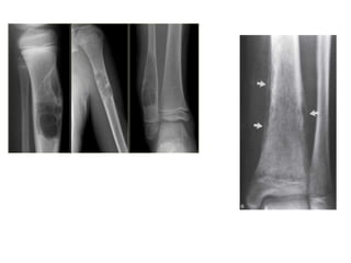
Osteosarcoma
• X-rays
: variable
:hazy osteolytic areas may alternate with
unusually dense osteoblastic area.
: ill-defined margin
: Sunburst and Codman’s triangle are typical
osteosarcoma
Diagnosis
• Most :X-ray**
: exclude post-traumatic swellings, infection,
stress fracture ,and the more aggressive cystic
lesion
• Radioisotrope scan
• CT
• MRI : show the extent of the tumor
• Biopsy :
41.
• Bone scan
•CXR
: routinely for detected lung metastasis
• CT chest : more sensitivity
• LDH , ALP rising
42.
Treatment
• Surgery
Amputation
Limb salvage
• Tumor resectable and no skip lesion
: wide resection
depend on the site of tumor >> bone graft ,
custom made implant
43.
• Radioresistent
• Chemotherapy
80%of patient only amputation was mestastsis
and death in 6-9 mo.
: Multiagent neoadjuvant Chemotherapy 8-12 mo.
Preop CMT response,if tumor necrosis is >
90%,CMT continued for 6-12 mo. If poor response,
a different chemotherapeutic regime is substituted