This document provides guidelines for the treatment of central nervous system cancers. It was last updated on September 11, 2020 and is version 3.2020. It includes treatment guidelines for various types of brain and spinal cord tumors in adults and children. For each tumor type, it provides recommendations on diagnosis, staging, treatment including surgery, radiation therapy, chemotherapy, targeted therapy, immunotherapy and clinical trials. It also includes principles and recommendations regarding imaging, pathology, surgery and radiation therapy for brain and spinal cord tumors.


![UPDATES
Continued
BRAIN-C 1 of 8 (continued)
Anaplastic Gliomas/Glioblastoma High-Grade (Grades III/IV)
• Tumor volumes are best defined using pre- and postoperative MRI
imaging using enhanced post-contrast T1 with/without and FLAIR/T2
sequences to define GTV. To account for sub-diagnostic tumor infiltration,
the GTV is expanded 1–2 cm (CTV) for grade III up to 2-2.5 cm (CTV) for
grade...
• Consider proton therapy for patients with good long-term prognosis
(grade III IDH-mutant tumors [Buckner 2016] and grade III 1p19q co-
deleted tumors [Shih 2015]) to better spare un-involved brain and
preserve cognitive function, is new to the page.
RT Dosing Information
• 4th bullet, second sentence modified: Typical fractionation schedules are
34 Gy/10 fx, 40.05 Gy/15 fx, or 50 Gy/20 fx.
BRAIN-C 4 of 8
• WHO Grade 1 Meningiomas
2nd, sub,sub-bullet modifed (25–30 Gy in 5 fractions)
BRAIN-C 6 of 8
• 1st sub-bullet under RT dosing modified: ..spinal cord and/or nerve root
• 4th sub-bullet modified: When lower BED regimens are utilized upfront
(ie, BED ≤60 Gy2
, which includes up to 20 Gy in 5 fractions but does not
include 30 Gy in 10 fractions), retreatment with similar BED regimens,
such as 20 Gy in 5 fractions or 8 Gy in 1 fraction, can safely be considered
as early as 4 weeks to 6 weeks from initial treatment for pain relief.
• 5th sub-bullet modified: In other cases of retreatment, doses ranging from
15 Gy in 1 fraction with SBRT to 40 Gy in 20 fractions with a conformal
approach have been utilized for tumor control, with careful consideration
of tolerance of the spinal cord and/or nerve roots. In these instances,
it is generally recommended that 6 months or more of time between
treatments is required.
BRAIN-D (1 of 15)
Adjuvant Treatment
Useful in Certain Circumstances; modified as follows:
• Pilocytic astrocytoma, PXA, ganglioglioma if BRAF V600E activiting
mutation
BRAF/MEK inhibitors:
◊◊ dabrafenib/trametinib
◊◊ vemurafenib/cobimetinib
BRAIN-D (2 of 15)
Anaplastic Gliomas
• Carboplatin category 2B removed under Other Recommended Regimens
for Recurrence Therapy (Also for BRAIN-D 3 of 15)
Footnotes
j: modified: Bevacizumab + chemotherapy can be considered if
bevacizumab monotherapy fails and it is desirable to continue the steroid
sparing effects of bevacizumab (Also for BRAIN-D 3 of 15).
BRAIN-D (3 of 15)
Adjuvant Treatment for Glioblastoma
Preferred Regimens; modified as follows:
• RT with concurrent and adjuvant TMZ ± TTF
Useful in Certain Circumstances; the following is new to the page:
• RT with concurrent and adjuvant lomustine and TMZ (for patients with
MGMT promoter methylated tumors, KPS ≥60, and age ≤70 years)
(category 2B), with the following corresponding footnote: Moderate to
significant myelosuppression was observed, but the toxicity profile for
this regimen is not yet fully defined.
Recurrence Therapy for Glioblastoma
Preferred Regimens:
Regorafenib is new to the page
BRAIN-D (5 of 15)
Induction Therapy for Primary CNS Lymphoma
Useful in Certain Circumstances
• Patient is unsuitable for or intolerant to high-dose methotrexate
See Other Recommended Regimens for Relapsed or Refractory
Disease
Relapsed or Refractory Disease
Other Recommended Regimens; modified as follows:
• Retreat with high-dose methotrexate
with or without rituximab and ibrutinib
• Topotecan
• Dexamethasone, high-dose cytarabine, cisplatin
BRAIN-D (7 of 15)
Other Recommended Regimens for Meningioma; modified as follows:
• Interferon alfa (category 2B)
• Somatostatin analogue, if octreotide scan positive
Useful in Certain Circumstances; modified as follows:
• Somatostatin analogue (category 2B) if octreotide scan positive
Updates in Version 1.2020 of the NCCN Guidelines for Central Nervous System Cancers from Version 3.2019 include:
NCCN Guidelines Version 3.2020
Central Nervous System Cancers
Version 3.2020 , 09/11/20 © 2020 National Comprehensive Cancer Network®
(NCCN®
), All rights reserved. NCCN Guidelines®
and this illustration may not be reproduced in any form without the express written permission of NCCN.
NCCN Guidelines Index
Table of Contents
Discussion
Printed by Matteo De notaris on 11/13/2020 6:02:56 AM. For personal use only. Not approved for distribution. Copyright © 2020 National Comprehensive Cancer Network, Inc., All Rights Reserved.](https://image.slidesharecdn.com/cns-201113111756/85/Cns-7-320.jpg)


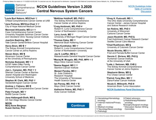
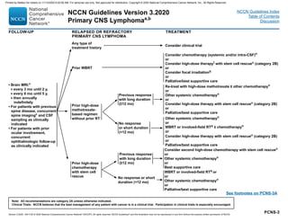
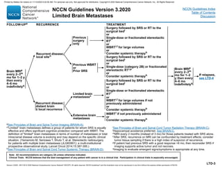
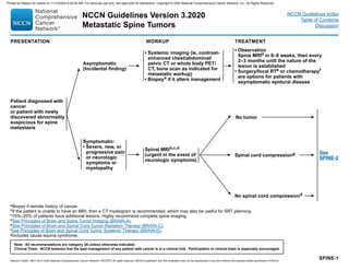
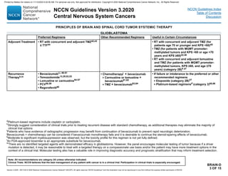
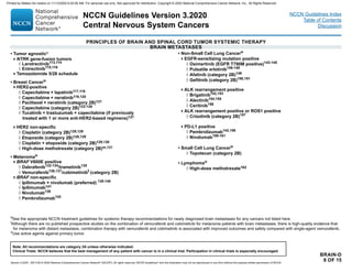
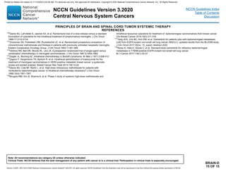
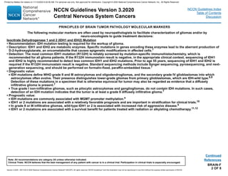
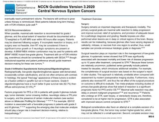
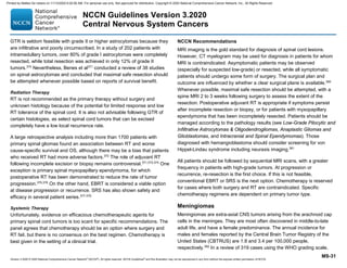
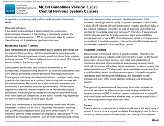
![GLIO-2
PATHOLOGYd ADJUVANT TREATMENT FOLLOW-UPb
See
Recurrence
(GLIO-5)
Anaplastic
oligodendroglioma
(1p19q codeleted)
Consider clinical trial (preferred for eligible patients)
or
Standard RTl and neoadjuvant or adjuvantm PCV (category 1)n
or
Standard RTl with concurrent and adjuvant temozolomiden
or
Standard RTl and adjuvant temozolomiden
Brain MRI 2–8 wks
after RT,p then every
2–4 mo for 3 y, then
every 3–6 months
indefinitely
Anaplastic astrocytoma,
Anaplastic
oligoastrocytoma, NOSj
Consider clinical trial (preferred for eligible patients)
or
Standard RT followed by adjuvant temozolomiden
or
Standard RTl with concurrent and adjuvant temozolomiden
or
Standard RTl + neoadjuvant or adjuvantm PCV
Anaplastic gliomasa
Poor performance
status (KPS 60)
RTl(hypofractionated [preferred] or standard)
or
Temozolomide (category 2B)n,o
or
Palliative/best supportive care
ANAPLASTIC GLIOMAS (SEE GLIO-3/GLIO-4 FOR GLIOBLASTOMA)
aThis pathway includes the classification of mixed AOA, AA, AO, and other rare
anaplastic gliomas.
bSee Principles of Brain and Spine Tumor Imaging (BRAIN-A).
dSee Principles of Brain Tumor Pathology (BRAIN-F).
jThe 2016 WHO Classification of Tumors of the CNS has deleted oligoastrocytoma as
a category, although “anaplastic oligoastrocytoma, NOS” may continue to be used
for 1) patients with mixed histology and no available molecular data (ie, no tissue
available for analysis) for determining whether to classify as oligodendroglioma versus
astrocytoma; or 2) rare instances in which the tumor has regions with histologic
features of oligoastrocytoma with 1p19q-codeletion, and distinct regions with
histologic features of astrocytoma without 1p19q-codeletion.
lSee Principles of Brain and Spinal Cord Tumor Radiation Therapy (BRAIN-C).
mThe panel recommends that PCV be administered after RT (as per EORTC 26951)
since the intensive PCV regimen given prior to RT (RTOG 9402) was not tolerated as
well.
nSee Principles of Brain and Spinal Cord Tumor Systemic Therapy (BRAIN-D).
oConsider temozolomide if tumor is MGMT promoter methylated.
pWithin the first 3 months after completion of RT and concomitant temozolomide,
diagnosis of recurrence can be indistinguishable from pseudoprogression on
neuroimaging.
Note: All recommendations are category 2A unless otherwise indicated.
Clinical Trials: NCCN believes that the best management of any patient with cancer is in a clinical trial. Participation in clinical trials is especially encouraged.
Version 3.2020 , 09/11/20 © 2020 National Comprehensive Cancer Network®
(NCCN®
), All rights reserved. NCCN Guidelines®
and this illustration may not be reproduced in any form without the express written permission of NCCN.
NCCN Guidelines Version 3.2020
Anaplastic Gliomas
a
/Glioblastoma
NCCN Guidelines Index
Table of Contents
Discussion
Printed by Matteo De notaris on 11/13/2020 6:02:56 AM. For personal use only. Not approved for distribution. Copyright © 2020 National Comprehensive Cancer Network, Inc., All Rights Reserved.](https://image.slidesharecdn.com/cns-201113111756/85/Cns-13-320.jpg)


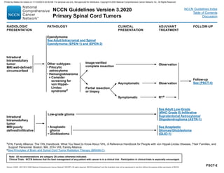
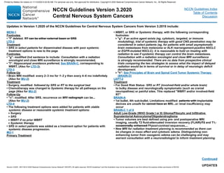
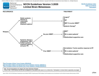
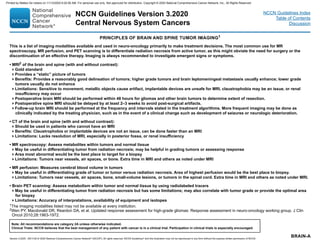
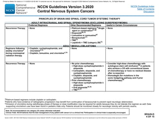
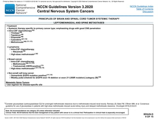
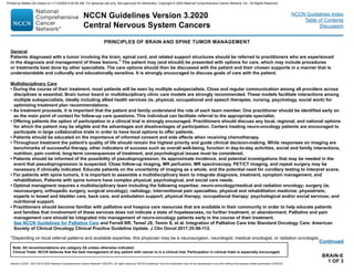
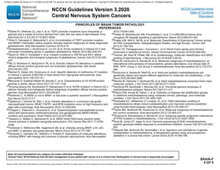
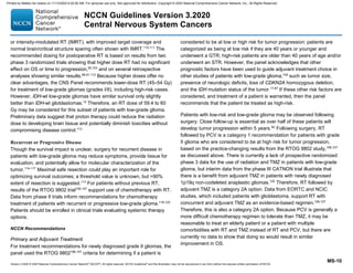
![PSCT-4
aSee Principles of Brain and Spine Tumor Imaging (BRAIN-A).
eSee Principles of Brain and Spinal Cord Tumor Radiation Therapy (BRAIN-C).
gSee Principles of Brain and Spinal Cord Tumor Systemic Therapy (BRAIN-D) for options according to disease histology.
Patients managed by:
Observation
or
Maximum safe
resection for
intradural
intramedullary tumor
or intradural
extramedullary tumor
FOLLOW-UPa
Spine MRI 2–6 wk after treatment,
then every 2–4 mo until 2–3 y,
then every 3–6 mo until 5 y, then
every 6–12 mo indefinitely
Spine MRI every 3–6 mo until
5 y, then at least annually
indefinitely
Low-grade
tumors (I–II)
High-grade
tumors (III–IV)
RECURRENCE
New/worsening
symptoms
or radiographic
progression
TREATMENT FOR RECURRENCE
Re-resection
or
RTe or re-irradiation (include
stereotactic radiotherapy [SRT]), if
surgery not possible
or
Chemotherapyg relative to cell type if
further surgery or RT not possible
Note: All recommendations are category 2A unless otherwise indicated.
Clinical Trials: NCCN believes that the best management of any patient with cancer is in a clinical trial. Participation in clinical trials is especially encouraged.
Version 3.2020 , 09/11/20 © 2020 National Comprehensive Cancer Network®
(NCCN®
), All rights reserved. NCCN Guidelines®
and this illustration may not be reproduced in any form without the express written permission of NCCN.
NCCN Guidelines Version 3.2020
Primary Spinal Cord Tumors
NCCN Guidelines Index
Table of Contents
Discussion
Printed by Matteo De notaris on 11/13/2020 6:02:56 AM. For personal use only. Not approved for distribution. Copyright © 2020 National Comprehensive Cancer Network, Inc., All Rights Reserved.](https://image.slidesharecdn.com/cns-201113111756/85/Cns-32-320.jpg)
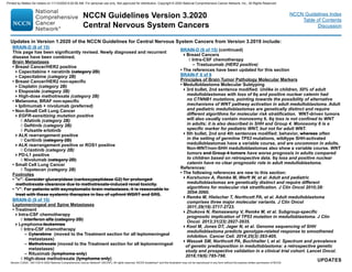
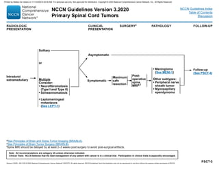
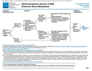
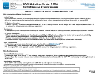
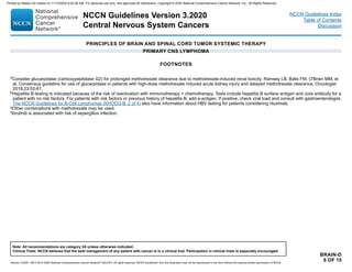
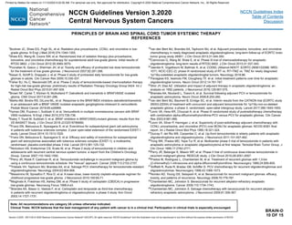
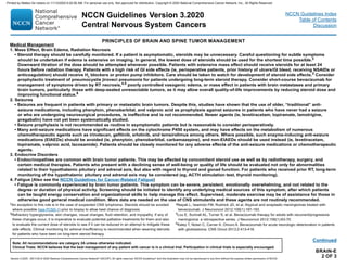
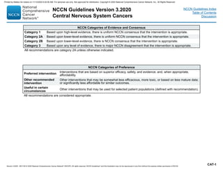
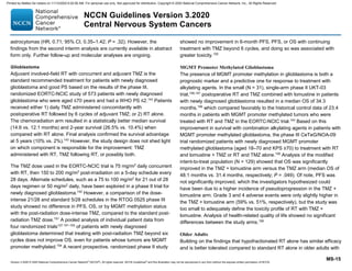
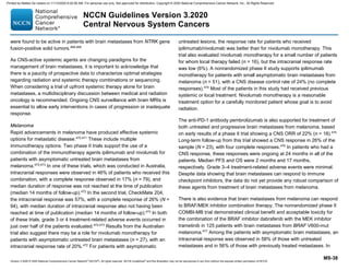
![MENI-1
aMultidisciplinary input for treatment planning if feasible.
bTreatment selection should be based on assessment of a variety of inter-related
factors, including patient features (eg, age, performance score, comorbidities,
treatment preferences), tumor features (eg, size, grade, growth rate, location
[proximity to critical structures], potential for causing neurologic consequences
if untreated, presence and severity of symptoms), and treatment-related factors
(eg, potential for neurologic consequences from surgery/RT, likelihood of
complete resection and/or complete irradiation with SRS, treatability of tumor if it
progresses, available surgical or radiation oncology expertise and resources). The
decision to administer RT after surgery also depends on the extent of resection
achieved. Multidisciplinary input for treatment planning is recommended.
cFor asymptomatic meningiomas, observation is preferred for small tumors,
with a suggested cutoff of ≤3 cm. Active treatment with surgery and/or RT is
recommended in cases with one or more tumor- and/or treatment-related risk
factors, such as proximity to the optic nerve.
dPostoperative brain MRI within 48 hours after surgery.
eSee Principles of Brain and Spine Tumor Imaging (BRAIN-A).
fSee Principles of Brain Tumor Radiation Therapy (BRAIN-C).
gWHO Grade I = Benign meningioma, WHO Grade II = Atypical meningioma, WHO
Grade III = Malignant (anaplastic) meningioma.
PRESENTATIONa TREATMENTb
Radiographic
diagnosis by brain
MRI:
• Dural-based mass
• Homogeneously
contrast-enhancing
• Dural tail
• CSF cleft
Meningioma by
radiographic criteria
or
Possible meningioma:
• Consider resection
• Consider octreotide
scan if diagnostic
doubt exists
Consider RTf
depending on factors in footnote b
In general, postoperative management depends
on grade,g
extent of resection, and symptoms, as
follows:
• Grade I: observation or consider RT (for
symptomatic patients)
• Grade II with complete resection: consider RT
• Grade II with incomplete resection: RT
• Grade III: RT
Follow-up
(See MENI-2)
Observe (preferred for
small asymptomatic
tumors; not generally
recommended for
symptomatic tumors)c
or
Surgeryd,e
(if accessible)f
or
RTf
TREATMENTb ADJUVANT TREATMENT
Note: All recommendations are category 2A unless otherwise indicated.
Clinical Trials: NCCN believes that the best management of any patient with cancer is in a clinical trial. Participation in clinical trials is especially encouraged.
Version 3.2020 , 09/11/20 © 2020 National Comprehensive Cancer Network®
(NCCN®
), All rights reserved. NCCN Guidelines®
and this illustration may not be reproduced in any form without the express written permission of NCCN.
NCCN Guidelines Version 3.2020
Meningiomas
NCCN Guidelines Index
Table of Contents
Discussion
Printed by Matteo De notaris on 11/13/2020 6:02:56 AM. For personal use only. Not approved for distribution. Copyright © 2020 National Comprehensive Cancer Network, Inc., All Rights Reserved.](https://image.slidesharecdn.com/cns-201113111756/85/Cns-33-320.jpg)
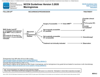


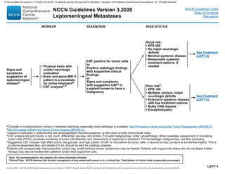


![PRINCIPLES OF RADIATION THERAPY FOR BRAIN AND SPINAL CORD
BRAIN-C
6 OF 8
Metastatic Spine Tumors
• General Treatment Information
Doses to vertebral body metastases will depend on patient’s PS, spine stability, location in relationship to spinal cord, primary histology,
presence of epidural disease, and overall treatment intent (pain relief, long-term local control, or cure).
Stereotactic radiation approaches (SRS/stereotactic body radiotherapy [SBRT]) for spinal cases may be preferred for patients with
oligometastatic disease where tumor ablation is a goal of treatment and in tumors considered radioresistant (eg, renal cell, melanoma,
sarcoma, hepatocellular, and some colorectal and NSCLC cases).
Stereotactic radiation approaches may also be preferred in the setting of tumor recurrence after prior radiation as a strategy to limit
radiation dose to the spinal cord or other critical structures. Careful adherence to consensus guidelines for radiosurgery planning and
delivery is recommended.33-35
• RT Dosing:
Generally, conventional external beam radiation doses of 8 Gy/1 fx, 20 Gy/5 fx, or 30 Gy/10 fx can be used. It is critical to consider tolerance
at the spinal cord and/or nerve root. In selected cases, or recurrences after previous radiation, SBRT is appropriate.
Common recommended doses for spine SRS/SBRT may include:
◊◊ 16–24 Gy x 1 fx;
◊◊ 24 Gy in 2 fx;
◊◊ 24–27 Gy in 3 fx;
◊◊ 30–35 Gy in 5 fx
In patients with uncomplicated spine metastases that are treated primarily for pain relief, 8 Gy in 1 fraction has been shown to provide
equivalent pain control to longer fractionation schedules. Single fraction treatment is more convenient for patients and an important
consideration for patients with poor prognoses. This treatment may be associated with higher rates of retreatment, and a consideration for
patients with a prognosis that exceeds 6 months or greater.
When lower BED regimens are utilized upfront (ie, BED ≤60 Gy, which includes up to 20 Gy in 5 fractions but does not include 30 Gy in
10 fractions), retreatment with similar BED regimens, such as 20 Gy in 5 fractions or 8 Gy in 1 fraction, can safely be considered as early
as 6 weeks from initial treatment for pain relief.
In other cases of retreatment, doses ranging from 15 Gy in 1 fraction with SBRT to 40 Gy in 20 fractions with a conformal approach have
been utilized for tumor control, with careful consideration of tolerance of the spinal cord and/or nerve roots. In these instances, it is
generally recommended that 6 months or more of time between treatments is required.
NCCN Guidelines Version 3.2020
Central Nervous System Cancers
Version 3.2020 , 09/11/20 © 2020 National Comprehensive Cancer Network®
(NCCN®
), All rights reserved. NCCN Guidelines®
and this illustration may not be reproduced in any form without the express written permission of NCCN.
Note: All recommendations are category 2A unless otherwise indicated.
Clinical Trials: NCCN believes that the best management of any patient with cancer is in a clinical trial. Participation in clinical trials is especially encouraged.
NCCN Guidelines Index
Table of Contents
Discussion
Printed by Matteo De notaris on 11/13/2020 6:02:56 AM. For personal use only. Not approved for distribution. Copyright © 2020 National Comprehensive Cancer Network, Inc., All Rights Reserved.](https://image.slidesharecdn.com/cns-201113111756/85/Cns-54-320.jpg)


![BRAIN-D
11 OF 15
35
Vaccaro V, Fabi A, Vidiri A, et al. Activity and safety of bevacizumab plus fotemustine for recurrent
malignant gliomas. Biomed Res Int 2014;2014:351252.
36
Liu Y, Feng F, Ji P, et al. Improvement of health related quality of life in patients with recurrent glioma
treated with bevacizumab plus daily temozolomide as the salvage therapy. Clin Neurol Neurosurg
2018;169:64-70.
37
Fulton D, Urtasun R, Forsyth P. Phase II study of prolonged oral therapy with etoposide (VP16) for
patients with recurrent malignant glioma. J Neurooncol 1996;27:149-155.
38
Leonard A, Wolff JE. Etoposide improves survival in high-grade glioma: a meta-analysis. Anticancer
Res 2013;33:3307-3315.
39
Murray LJ, Bridgewater CH, Levy D. Carboplatin chemotherapy in patients with recurrent high-grade
glioma. Clin Oncol (R Coll Radiol) 2011;23:55-61.
40
Roci E, Cakani B, Brace G, et al. Platinum-based chemotherapy in recurrent high-grade glioma
patients: retrospective study. Med Arch 2014;68:140-143.
41
Soffietti R, Nobile M, Ruda R, et al. Second-line treatment with carboplatin for recurrent or
progressive oligodendroglial tumors after PCV (procarbazine, lomustine, and vincristine)
chemotherapy: a phase II study. Cancer 2004;100:807-813.
42
Stupp R, Mason WP, van den Bent MJ, et al. Radiotherapy plus concomitant and adjuvant
temozolomide for glioblastoma. N Engl J Med 2005;352:987-996.
43
Stupp R, Hegi ME, Mason WP, et al. Effects of radiotherapy with concomitant and adjuvant
temozolomide versus radiotherapy alone on survival in glioblastoma in a randomised phase III study:
5-year analysis of the EORTC-NCIC trial. Lancet Oncol 2009;10:459-466.
44
Stupp R, Taillibert S, Kanner A, et al. Effect of tumor-treating fields plus maintenance temozolomide
vs maintenance temozolomide alone on survival in patients with glioblastoma: a randomized clinical
trial. JAMA 2017;318:2306-2316.
45
Muni R, Minniti G, Lanzetta G, et al. Short-term radiotherapy followed by adjuvant chemotherapy in
poor-prognosis patients with glioblastoma. Tumori 2010;96:60-64.
46
Malmstrom A, Gronberg BH, Marosi C, et al. Temozolomide versus standard 6-week radiotherapy
versus hypofractionated radiotherapy in patients older than 60 years with glioblastoma: the Nordic
randomised, phase 3 trial. Lancet Oncol 2012;13:916-926.
47
Herrlinger U, Tzaridis T, Mack F, et al. Lomustine-temozolomide combination therapy versus standard
temozolomide therapy in patients with newly diagnosed glioblastoma with methylated MGMT
promoter (CeTeG/NOA-09): a randomised, open-label, phase 3 trial. Lancet 2019;393:678-688.
48
Cloughesy T, Prados MD, Mikkelsen T. A phase 2 randomized non-comparative clinical trial of the
effect of bevacizumab alone or in combination with irinotecan on 6-month progression free survival in
recurrent refractory glioblastoma [abstract]. J Clin Oncol 2008;26(Suppl 15):2010b.
49
Friedman HS, Prados MD, Wen PY, et al. Bevacizumab alone and in combination with irinotecan in
recurrent glioblastoma. J Clin Oncol 2009;27:4733-4740.
50
Kreisl TN, Kim L, Moore K, et al. Phase II trial of single-agent bevacizumab followed by bevacizumab
plus irinotecan at tumor progression in recurrent glioblastoma. J Clin Oncol 2009;27:740-745.
51
Vredenburgh JJ, Desjardins A, Herndon JE, 2nd, et al. Phase II trial of bevacizumab and irinotecan in
recurrent malignant glioma. Clin Cancer Res 2007;13:1253-1259.
52
Yung WK, Albright RE, Olson J, et al. A phase II study of temozolomide vs. procarbazine in patients
with glioblastoma multiforme at first relapse. Br J Cancer 2000;83:588-593.
53
Weller M, Tabatabai G, Kastner B, et al. MGMT promoter methylation is a strong prognostic
biomarker for benefit from dose-intensified temozolomide rechallenge in progressive glioblastoma: the
DIRECTOR trial. Clin Cancer Res 2015;21:2057-2064.
54
Wick W, Puduvalli VK, Chamberlain MC, et al. Phase III study of enzastaurin compared with
lomustine in the treatment of recurrent intracranial glioblastoma. J Clin Oncol 2010;28:1168-1174.
55
Brandes AA, Tosoni A, Amista P, et al. How effective is BCNU in recurrent glioblastoma in the modern
era? A phase II trial. Neurology 2004;63:1281-1284.
56
Reithmeier T, Graf E, Piroth T, et al. BCNU for recurrent glioblastoma multiforme: efficacy, toxicity and
prognostic factors. BMC Cancer 2010;10:30.
57
Taal W, Oosterkamp HM, Walenkamp AM, et al. Single-agent bevacizumab or lomustine versus a
combination of bevacizumab plus lomustine in patients with recurrent glioblastoma (BELOB trial): a
randomised controlled phase 2 trial. Lancet Oncol 2014;15:943-953.
58
Carvalho BF, Fernandes AC, Almeida DS, et al. Second-line chemotherapy in recurrent glioblastoma:
a 2-cohort study. Oncol Res Treat 2015;38:348-354.
59
Schmidt F, Fischer J, Herrlinger U, et al. PCV chemotherapy for recurrent glioblastoma. Neurology
2006;66:587-589.
60
Lombardi G, DeSalvo GL, Brandes AA, et al. Regorafenib compared with lomustine in patients with
relapsed glioblastoma (REGOMA): a multicentre, open-label, randomised, controlled, phase 2 trial.
Lancet Oncol 2019;20(1):110-119.
61
Wick W, Gorlia T, Bendszus M, et al. Lomustine and bevacizumab in progressive glioblastoma. N
Engl J Med 2017;377:1954-1963.
62
Soffietti R, Trevisan E, Bertero L, et al. Bevacizumab and fotemustine for recurrent glioblastoma: a
phase II study of AINO (Italian Association of Neuro-Oncology). J Neurooncol 2014;116:533-541.
63
Badruddoja MA, Pazzi M, Sanan A, et al. Phase II study of bi-weekly temozolomide plus bevacizumab
for adult patients with recurrent glioblastoma. Cancer Chemother Pharmacol 2017;80:715-721.
64
Desjardins A, Reardon DA, Coan A, et al. Bevacizumab and daily temozolomide for recurrent
glioblastoma. Cancer 2012;118:1302-1312.
65
Field KM, Simes J, Nowak AK, et al. Randomized phase 2 study of carboplatin and bevacizumab in
recurrent glioblastoma. Neuro Oncol 2015;17:1504-1513.
66
Kaloshi G, Diamandi P, Cakani B, et al. The added value of bevacizumab concomitantly administered
with carboplatin versus carboplatin alone in patients with recurrent glioblastomas. Tumori
2015;101:41-45.
67
Gornet MK, Buckner JC, Marks RS, et al. Chemotherapy for advanced CNS ependymoma. J
Neurooncol 1999;45:61-67.
68
Brandes AA, Cavallo G, Reni M, et al. A multicenter retrospective study of chemotherapy for recurrent
intracranial ependymal tumors in adults by the Gruppo Italiano Cooperativo di Neuro-Oncologia.
Cancer 2005;104:143-148.
69
Chamberlain MC. Recurrent intracranial ependymoma in children: salvage therapy with oral
etoposide. Pediatr Neurol 2001;24:117-121.
PRINCIPLES OF BRAIN AND SPINAL CORD TUMOR SYSTEMIC THERAPY
REFERENCES
NCCN Guidelines Version 3.2020
Central Nervous System Cancers
Version 3.2020 , 09/11/20 © 2020 National Comprehensive Cancer Network®
(NCCN®
), All rights reserved. NCCN Guidelines®
and this illustration may not be reproduced in any form without the express written permission of NCCN.
Note: All recommendations are category 2A unless otherwise indicated.
Clinical Trials: NCCN believes that the best management of any patient with cancer is in a clinical trial. Participation in clinical trials is especially encouraged.
NCCN Guidelines Index
Table of Contents
Discussion
Printed by Matteo De notaris on 11/13/2020 6:02:56 AM. For personal use only. Not approved for distribution. Copyright © 2020 National Comprehensive Cancer Network, Inc., All Rights Reserved.](https://image.slidesharecdn.com/cns-201113111756/85/Cns-67-320.jpg)

![PRINCIPLES OF BRAIN AND SPINAL CORD TUMOR SYSTEMIC THERAPY
REFERENCES
BRAIN-D
14 OF 15
136
McArthur GA, Maio M, Arance A, et al. Vemurafenib in metastatic melanoma patients with brain
metastases: an open-label, single-arm, phase 2, multicentre study. Ann Oncol 2017;28:634-641.
137
Dummer R, Goldinger SM, Turtschi CP, et al. Vemurafenib in patients with BRAF(V600) mutation-
positive melanoma with symptomatic brain metastases: final results of an open-label pilot study. Eur J
Cancer 2014;50:611-621.
138
Tawbi HA, Forsyth PA, Algazi AP, et al. Combined nivolumab and ipilimumab in melanoma metastatic
to the brain. N Engl J Med 2018;379:722-730.
139
Long GV, Atkinson V, Lo S, et al. Combination nivolumab and ipilimumab or nivolumab alone in
melanoma brain metastases: a multicentre randomised phase 2 study. Lancet Oncol 2018;19:672-
681.
140
Long GV, Atkinson V, Lo S, et al. long-term outcomes from the randomized phase II study of
nivolumab or nivolumab + ipilumumab in patients with melanoma brain metastases: Anti-PD-1 Brain
Collaboration (the ABC trial). ESMO 2019 Congress.
141
Margolin K, Ernstoff MS, Hamid O, et al. Ipilimumab in patients with melanoma and brain
metastases: an open-label, phase 2 trial. Lancet Oncol 2012;13:459-465.
142
Goldberg SB, Gettinger SN, Mahajan A, et al. Pembrolizumab for patients with melanoma or non-
small-cell lung cancer and untreated brain metastases: early analysis of a non-randomised, open-
label, phase 2 trial. Lancet Oncol 2016;17:976-983.
143
Soria JC, Ohe Y, Vansteenkiste J, et al. Osimertinib in untreated EGFR-mutated advanced non-
small-cell lung cancer. N Engl J Med 2018;378:113-125.
144
Goss G, Tsai CM, Shepherd FA, et al. CNS response to osimertinib in patients with T790M-positive
advanced NSCLC: pooled data from two phase II trials. Ann Oncol 2018;29:687-693.
145
Mok TS, Wu YL, Ahn MJ, et al. Osimertinib or platinum-pemetrexed in EGFR T790M-positive lung
cancer. N Engl J Med 2017;376:629-640.
146
Grommes C, Oxnard GR, Kris MG, et al. Pulsatile high-dose weekly erlotinib for CNS metastases
from EGFR mutant non-small cell lung cancer. Neuro Oncol 2011;13:1364-1369.
147
Katayama T, Shimizu J, Suda K, et al. Efficacy of erlotinib for brain and leptomeningeal metastases
in patients with lung adenocarcinoma who showed initial good response to gefitinib. J Thorac Oncol
2009;4:1415-1419.
148
Arbour KC, Kris MG, Riely GJ, et al. Twice weekly pulse and daily continuous-dose erlotinib as
initial treatment for patients with epidermal growth factor receptor-mutant lung cancers and brain
metastases. Cancer 2018;124(1):105-109.
149
Hoffknecht P, Tufman A, Wehler T, et al. Efficacy of the irreversible ErbB family blocker afatinib in
epidermal growth factor receptor (EGFR) tyrosine kinase inhibitor (TKI)-pretreated non-small-cell lung
cancer patients with brain metastases or leptomeningeal disease. J Thorac Oncol 2015;10:156-163.
150
Ceresoli GL, Cappuzzo F, Gregorc V, et al. Gefitinib in patients with brain metastases from non-
small-cell lung cancer: a prospective trial. Ann Oncol 2004;15:1042-1047.
151
Wu C, Li YL, Wang ZM, et al. Gefitinib as palliative therapy for lung adenocarcinoma metastatic to
the brain. Lung Cancer 2007;57:359-364.
152
Camidge DR, Kim HR, Ahn MJ, et al. Brigatinib versus crizotinib in ALK-positive non-small-cell lung
cancer. N Engl J Med 2018;379:2027-2039.
153
Kim DW, Tiseo M, Ahn MJ, et al. Brigatinib in patients with crizotinib-refractory anaplastic lymphoma
kinase-positive non-small-cell lung cancer: a randomized, multicenter phase II trial. J Clin Oncol
2017;35:2490-2498.
154
Peters S, Camidge DR, Shaw AT, et al. Alectinib versus crizotinib in untreated ALK-positive non-
small-cell lung cancers. N Engl J Med 2017;377:829-838.
155
Gandhi L, Ou SI, Shaw AT, et al. Efficacy of alectinib in central nervous system metastases in
crizotinib-resistant ALK-positive non-small-cell lung cancer: comparison of RECIST 1.1 and RANO-
HGG criteria. Eur J Cancer 2017;82:27-33.
156
Kim DW, Mehra R, Tan DSW, et al. Activity and safety of ceritinib in patients with ALK-rearranged
non-small-cell lung cancer (ASCEND-1): updated results from the multicentre, open-label, phase 1
trial. Lancet Oncol 2016;17:452-463.
157
Costa DB, Shaw AT, Ou SH, et al. Clinical experience with crizotinib in patients with advanced ALK-
rearranged non-small-cell lung cancer and brain metastases. J Clin Oncol 2015;33:1881-1888.
158
Kluger HM, Chiang V, Mahajan A, et al. Long-term survival of patients with melanoma with active
brain metastases treated with pembrolizumab on a phase II trial. J Clin Oncol 2019;37:52-60.
159
Gauvain C, Vauleon E, Chouaid C, et al. Intracerebral efficacy and tolerance of nivolumab in non-
small-cell lung cancer patients with brain metastases. Lung Cancer 2018;116:62-66.
160
Goldman JW, Crino L, Vokes EE, et al. P2.36: Nivolumab (nivo) in patients (pts) with advanced (adv)
NSCLC and central nervous system (CNS) metastases (mets). J Thorac Oncol 2016;11:S238-S239.
161
Rizvi NA, Mazieres J, Planchard D, et al. Activity and safety of nivolumab, an anti-PD-1 immune
checkpoint inhibitor, for patients with advanced, refractory squamous non-small-cell lung cancer
(CheckMate 063): a phase 2, single-arm trial. Lancet Oncol 2015;16:257-265.
162
Bokstein F, Lossos A, Lossos IS, Siegal T. Central nervous system relapse of systemic non-
Hodgkin's lymphoma: results of treatment based on high-dose methotrexate combination
chemotherapy. Leuk Lymphoma 2002;43:587-593.
163
Chamberlain MC. Leptomeningeal metastasis. Curr Opin Neurol 2009;22:665-674.
164
Gutin PH, Weiss HD, Wiernik PH, Walker MD. Intrathecal N, N', N-triethylenethiophosphoramide
[thio-TEPA (NSC 6396)] in the treatment of malignant meningeal disease: phase I-II study. Cancer
1976;38:1471-1475.
165
Groves MD, Glantz MJ, Chamberlain MC, et al. A multicenter phase II trial of intrathecal topotecan in
patients with meningeal malignancies. Neuro Oncol 2008;10:208-215.
166
Chamberlain MC, Tsao-Wei DD, Groshen S. Phase II trial of intracerebrospinal fluid etoposide in the
treatment of neoplastic meningitis. Cancer 2006;106:2021-2027.
167
Jaeckle KA, Phuphanich S, Bent MJ, et al. Intrathecal treatment of neoplastic meningitis due to
breast cancer with a slow-release formulation of cytarabine. Br J Cancer 2001;84:157-163.
168
Chamberlain MC, Johnston SK, Van Horn A, Glantz MJ. Recurrent lymphomatous meningitis treated
with intra-CSF rituximab and liposomal ara-C. J Neurooncol 2009;91:271-277.
169
Glantz MJ, Jaeckle KA, Chamberlain MC, et al. A randomized controlled trial comparing intrathecal
sustained-release cytarabine (DepoCyt) to intrathecal methotrexate in patients with neoplastic
meningitis from solid tumors. Clin Cancer Res 1999;5:3394-3402.
NCCN Guidelines Version 3.2020
Central Nervous System Cancers
Version 3.2020 , 09/11/20 © 2020 National Comprehensive Cancer Network®
(NCCN®
), All rights reserved. NCCN Guidelines®
and this illustration may not be reproduced in any form without the express written permission of NCCN.
Note: All recommendations are category 2A unless otherwise indicated.
Clinical Trials: NCCN believes that the best management of any patient with cancer is in a clinical trial. Participation in clinical trials is especially encouraged.
NCCN Guidelines Index
Table of Contents
Discussion
Printed by Matteo De notaris on 11/13/2020 6:02:56 AM. For personal use only. Not approved for distribution. Copyright © 2020 National Comprehensive Cancer Network, Inc., All Rights Reserved.](https://image.slidesharecdn.com/cns-201113111756/85/Cns-70-320.jpg)
![BRAIN-E
3 OF 3
Medical Management—continued
5. Psychiatric Disorders (Also see the NCCN Guidelines for Distress Management including NCCN Distress Thermometer [DIS-A])
• Depression and/or anxiety is common in neuro-oncology patients. These symptoms are greater than simple sadness or anxiety associated
with the diagnosis of a tumor. The vegetative symptoms associated with depression or severe anxiety may become very disabling for
the patient and distressing for the family. These symptoms will respond to psychotropic medications as they do in non-tumor patients.
If less severe, strong support from behavioral health allies and other qualified counselors is also extremely beneficial. Physicians, and
other members of their health care teams, should be sensitive to these symptoms and inquire about them in follow-up visits in order to
determine if the patient may be a candidate for psychological or psychiatric treatment. Communication between members of the patient's
health care team regarding the patient's response to treatment is important. See Andersen BL, DeRubeis RJ, Berman BS, et al. Screening,
assessment, and care of anxiety and depressive symptoms in adults with cancer: an American Society of Clinical Oncology guideline
adaptation. J Clin Oncol 2014;32:1605-1619.
• Anti-seizure medications, anxiolytics, some chemotherapy agents, antiemetics, and other agents used directly in cancer therapy may
affect mental status, alertness, and mood. Alterations in thought processes should trigger an investigation for any treatable causes,
including endocrine disorders, infection, side effects of medication, or tumor progression.
6. Venous Thromboembolism (VTE)
• See the NCCN Guidelines for Cancer-Associated Venous Thromboembolic Disease.
Allied Services
• Physical therapy, occupational therapy, and speech therapy may be helpful for many patients with CNS tumors, either benign or malignant.
Surgical intervention is not a prerequisite for referral, and these therapies should not be withheld from patients because of the uncertain
course of certain malignant tumors. Many patients with aggressive, malignant primary brain tumors or CNS metastases can benefit from
inpatient rehabilitation.
• Practitioners are encouraged to serve as a resource and to refer patients to social services, support groups, and cancer patient advocacy
organizations. Institutional or community resources that can assist patients and families in dealing with financial, insurance, and legal
issues are important.
• Practitioners should be familiar with their state laws concerning seizures and driving so that they can advise patients and families
appropriately.
PRINCIPLES OF BRAIN AND SPINE TUMOR MANAGEMENT
NCCN Guidelines Version 3.2020
Central Nervous System Cancers
Version 3.2020 , 09/11/20 © 2020 National Comprehensive Cancer Network®
(NCCN®
), All rights reserved. NCCN Guidelines®
and this illustration may not be reproduced in any form without the express written permission of NCCN.
Note: All recommendations are category 2A unless otherwise indicated.
Clinical Trials: NCCN believes that the best management of any patient with cancer is in a clinical trial. Participation in clinical trials is especially encouraged.
NCCN Guidelines Index
Table of Contents
Discussion
Printed by Matteo De notaris on 11/13/2020 6:02:56 AM. For personal use only. Not approved for distribution. Copyright © 2020 National Comprehensive Cancer Network, Inc., All Rights Reserved.](https://image.slidesharecdn.com/cns-201113111756/85/Cns-74-320.jpg)


![H3F3A Mutation
• Recommendation: H3F3A and HIST1H3B mutation testing is recommended in the appropriate clinical context.
• Description: The most common histone mutation in brain tumors, H3K27M, is caused by a lysine-to-methionine substitution in the H3F3A
gene and inhibits the trimethylation of H3.3 histone. G34 mutations are more common in cortical gliomas in children.32-34
• Detection: Although a K27M histone antibody is available,35 it is not 100% specific and interpretation can be difficult for non-experts.
Therefore, screening by H3F3A and HIST1H3B sequencing is a viable alternative and the preferred approach, especially since it will also
detect mutations in G34.
• Diagnostic value: Histone mutations most commonly occur in pediatric midline gliomas (eg, diffuse intrinsic pontine gliomas [DIPG]),
although midline gliomas in adults can also contain histone mutations.36 Their presence can be considered solid evidence of an infiltrative
glioma, which is often helpful in small biopsies of midline lesions that may not be fully diagnostic with light microscopy or do not fully
resemble infiltrative gliomas.32,33,36
• Prognostic value: K27M gliomas typically do not have MGMT promoter methylation, and the mutation is an adverse prognostic marker in
children and adults. The G34 mutation does not appear to have any prognostic significance once the diagnosis of a glioblastoma has been
established.33,36,37
BRAF Mutation
• Recommendation: BRAF fusion and/or mutation testing is recommended in the appropriate clinical context.
• Description: Activating mutations in BRAF, most commonly the V600E variant seen in other cancers (eg, melanoma), are present in 60%–
80% of supratentorial grade II–III pleomorphic xanthoastrocytomas (PXA), 30% of dysembryoplastic neuroepithelial tumors, 20% of grade I
gangliogliomas, and 5% of grade I pilocytic astrocytomas (PA). Diffusely infiltrative gliomas can also harbor a BRAF mutation, especially in
children. BRAF V600E has even been found in nonneoplastic cortical dysplasia. In contrast, activating BRAF fusions occur predominately in
PA of the posterior fossa, although some supratentorial PA also have this fusion.38-40
• Detection: BRAF V600E is best detected by sequencing, and BRAF fusions can be detected with RNA-Seq or other PCR-based breakpoint
methods that capture the main 16–9, 15–9, and 16–11 breakpoints between BRAF and its main fusion partner, KIAA1549. FISH is too
unreliable to detect BRAF fusions.38
• Diagnostic value: The presence of a BRAF fusion is reliable evidence that the tumor is a pilocytic astrocytoma, provided the histology is
compatible. BRAF V600E is more complicated, as it can occur in a variety of tumors over all four WHO grades and requires integration with
histology.38
• Prognostic value: Tumors with BRAF fusions tend to be indolent, with occasional recurrence but only rare progression to lethality. BRAF
V600E tumors show a much greater range of outcomes and need to be considered in context with other mutations and clinicopathologic
findings (eg, CDKN2A/B deletion). BRAF V600E tumors may respond to BRAF inhibitors such as vemurafenib, but comprehensive clinical
trials are still ongoing.41-43
BRAIN-F
5 OF 8
The following molecular markers are often used by neuropathologists to facilitate characterization of gliomas and/or by
neuro-oncologists to guide treatment decisions:
PRINCIPLES OF BRAIN TUMOR PATHOLOGY MOLECULAR MARKERS
References
Continued
NCCN Guidelines Version 3.2020
Central Nervous System Cancers
Version 3.2020 , 09/11/20 © 2020 National Comprehensive Cancer Network®
(NCCN®
), All rights reserved. NCCN Guidelines®
and this illustration may not be reproduced in any form without the express written permission of NCCN.
Note: All recommendations are category 2A unless otherwise indicated.
Clinical Trials: NCCN believes that the best management of any patient with cancer is in a clinical trial. Participation in clinical trials is especially encouraged.
NCCN Guidelines Index
Table of Contents
Discussion
Printed by Matteo De notaris on 11/13/2020 6:02:56 AM. For personal use only. Not approved for distribution. Copyright © 2020 National Comprehensive Cancer Network, Inc., All Rights Reserved.](https://image.slidesharecdn.com/cns-201113111756/85/Cns-79-320.jpg)


![Version 3.2020 © 2020 National Comprehensive Cancer Network©
(NCCN©
), All rights reserved. NCCN Guidelines®
and this illustration may not be reproduced in any form without the express written permission of NCCN.
NCCN Guidelines Version 3.2020
Central Nervous System Cancers
MS-2
Overview
In the year 2020, an estimated 23,890 people in the United States will be
diagnosed with a malignant primary central nervous system (CNS) tumor,
and these tumors will be responsible for approximately 18,020 deaths.1
The incidence of primary brain tumors has been increasing over recent
decades,2
especially in older adults.3,4
However, this growth in incidence
rates may be explained by increased use of CT and MRI and changes in
WHO classification of CNS tumors.2,5,6
Literature Search Criteria and Guidelines Update
Methodology
Prior to the update of this version of the NCCN Guidelines for Central
Nervous System Cancers, an electronic search of the PubMed database
was performed to obtain key literature in the field of neuro-oncology, using
the following search terms: {[(brain OR spine OR spinal OR supratentorial
OR cranial OR intracranial OR leptomeningeal) AND (cancer OR
carcinoma OR tumor OR metastases OR lesion)] OR glioma OR
astrocytoma OR oligodendroglioma OR glioblastoma OR ependymoma
OR medulloblastoma OR (primary central nervous system lymphoma) OR
meningioma}. The PubMed database was chosen because it remains the
most widely used resource for medical literature and indexes peer-
reviewed biomedical literature.7
The search results were narrowed by selecting studies in humans
published in English. Results were confined to the following article types:
Clinical Trial, Phase II; Clinical Trial, Phase III; Clinical Trial, Phase IV;
Practice Guideline; Guidelines; Randomized Controlled Trial; Meta-
Analysis; Systematic Reviews; and Validation Studies.
The data from key PubMed articles and articles from additional sources
deemed as relevant to these Guidelines and discussed by the panel have
been included in this version of the Discussion section (eg, e-publications
ahead of print, meeting abstracts). Recommendations for which high-level
evidence is lacking are based on the panel’s review of lower-level
evidence and expert opinion.
The complete details of the Development and Update of the NCCN
Guidelines are available on the NCCN website (www.NCCN.org).
Tumor Types
The NCCN Guidelines for CNS Cancers focus on management of the
following adult CNS cancers: pilocytic and infiltrative supratentorial
astrocytomas, oligodendrogliomas, anaplastic gliomas and glioblastoma,
ependymomas, medulloblastoma, brain metastases, leptomeningeal
metastases, non–AIDS-related primary CNS lymphomas (PCNSLs),
metastatic spinal tumors, meningiomas, and primary spinal cord tumors.
These guidelines are updated annually to include new information or
treatment philosophies as they become available. However, because this
field continually evolves, practitioners should use all of the available
information to determine the best clinical options for their patients.
Principles of Management
Primary brain tumors are a heterogeneous group of neoplasms with varied
outcomes and management strategies. Primary malignant brain tumors
range from pilocytic astrocytomas, which are very uncommon,
noninvasive, and surgically curable, to glioblastoma, the most common
malignant brain tumor in adults, which is highly invasive and virtually
incurable. Likewise, patients with metastatic brain disease may have
rapidly progressive systemic disease or no systemic cancer at all. These
patients may have one or dozens of brain metastases, and they may have
a malignancy that is highly responsive or, alternatively, highly resistant to
radiation therapy (RT) or chemotherapy. Because of this marked
heterogeneity, the prognostic features and treatment options for primary
and metastatic brain tumors must be carefully reviewed on an individual
Printed by Matteo De notaris on 11/13/2020 6:02:56 AM. For personal use only. Not approved for distribution. Copyright © 2020 National Comprehensive Cancer Network, Inc., All Rights Reserved.](https://image.slidesharecdn.com/cns-201113111756/85/Cns-85-320.jpg)
![Version 3.2020 © 2020 National Comprehensive Cancer Network©
(NCCN©
), All rights reserved. NCCN Guidelines®
and this illustration may not be reproduced in any form without the express written permission of NCCN.
NCCN Guidelines Version 3.2020
Central Nervous System Cancers
MS-3
basis and sensitively communicated to each patient. In addition, these
CNS tumors are associated with a range of symptoms such as seizures,
fatigue, psychiatric disorders, impaired mobility, neuro-cognitive
dysfunction, difficulty speaking, and short-term memory problems, as well
as complications such as intracerebral edema, endocrinopathies, and
venous thromboembolism that can seriously impact patients’ quality of life.
The involvement of an interdisciplinary team, including neurosurgeons, RT
therapists, oncologists, neurologists, and neuroradiologists, is a key factor
in the appropriate management of these patients. For any subtype of
malignant brain tumors, the NCCN Panel strongly recommends brain
tumor board or multidisciplinary review of each patient’s case once the
pathology is available. Further discussion of multidisciplinary care and
allied services, as well as guidelines on medical management of various
disease complications, can be found in the section, Principles of Brain and
Spine Tumor Management in the algorithm.
Treatment Principles
Several important principles guide surgical management and treatment
with RT and systemic therapy for adults with brain tumors. Regardless of
tumor histology, neurosurgeons generally provide the best outcome for
their patients if they remove as much tumor as safely possible (ideally
achieving a gross total resection [GTR]) and thereby provide sufficient
representative tumor tissue to ensure an accurate diagnosis. Decisions
regarding aggressiveness of surgery for primary brain tumors are complex
and depend on the: 1) age and performance status (PS) of the patient; 2)
proximity to “eloquent” areas of the brain; 3) feasibility of decreasing the
mass effect with aggressive surgery; 4) resectability of the tumor
(including the number and location of lesions); and 5) time since last
surgery in patients with recurrent disease.8
Further discussion can be
found in the Principles of Brain Tumor Surgery in the algorithm. It is
recommended to consult neurosurgeons with extensive experience in the
management of intracranial and spine neoplasms.
The surgical options include stereotactic biopsy, open biopsy, subtotal
resection (STR), or GTR. The pathologic diagnosis is critical and may be
difficult to accurately determine without sufficient tumor tissue. Review of
the tumor tissue by an experienced neuropathologist is highly
recommended. The Principles of Brain Tumor Pathology describe guiding
principles for diagnosis of CNS tumor pathology, given the 2016 addition
of molecular parameters to the WHO classification of CNS tumors.9
Radiation oncologists use several different treatment modalities in patients
with primary brain tumors, including fractionated stereotactic RT and
stereotactic radiosurgery (SRS). Standard fractionated external beam RT
(EBRT) is the most common approach. Hypofractionated radiation is an
appropriate option for select patients (ie, older adults and patients with a
poor PS). RT for patients with primary brain tumors is administered within
a limited field (covering tumor or surgical cavity and a small margin of
adjacent brain tissue), while whole-brain RT (WBRT) and SRS are used
primarily for treatment of brain metastases. The dose of RT administered
varies depending on the pathology as seen in Principles of Radiation
Therapy for Brain and Spinal Cord.
The information contained in the algorithms and principles of management
sections are designed to help clinicians navigate through the complex
management of patients with CNS tumors. Standard systemic therapy
options for each tumor subtype are listed in the Principles of Brain and
Spinal Cord Tumor Systemic Therapy; however, enrollment in a clinical
trial is the preferred treatment for eligible patients.
Gliomas
The NCCN Guidelines for CNS Cancers include recommendations for
management of the following gliomas:9
Printed by Matteo De notaris on 11/13/2020 6:02:56 AM. For personal use only. Not approved for distribution. Copyright © 2020 National Comprehensive Cancer Network, Inc., All Rights Reserved.](https://image.slidesharecdn.com/cns-201113111756/85/Cns-86-320.jpg)


![Version 3.2020 © 2020 National Comprehensive Cancer Network©
(NCCN©
), All rights reserved. NCCN Guidelines®
and this illustration may not be reproduced in any form without the express written permission of NCCN.
NCCN Guidelines Version 3.2020
Central Nervous System Cancers
MS-11
Since the design of RTOG 9802106,107
did not address whether all patients
should be treated with RT followed by PCV immediately after a tissue
diagnosis (an observation arm was not included for patients with high-risk
glioma [defined as older than 40 years of age and/or underwent an STR]92
in the study), observation after tissue diagnosis may be a reasonable
option for some patients with high-risk grade II glioma who are
neurologically asymptomatic or who have stable disease. However, close
monitoring of such patients with brain MRI is important. Results from
EORTC 22845, which showed that treatment with RT at diagnosis versus
at progression did not significantly impact OS, provide rationale for
observation in select cases with low-grade gliomas as an initial approach,
deferring RT.104
Long-term toxicity from radiation needs to be a
consideration, especially for young patients with 1p19q codeletion, for
whom there is slightly higher risk of radiation necrosis.128
Recurrence
At the time of recurrence, surgery is recommended if resectable disease is
present. Because recurrence on neuroimaging may be confounded by
treatment effects, biopsy of unresectable disease should be considered to
confirm recurrence. There is a propensity for low-grade gliomas to
transform to higher-grade gliomas over time. Therefore, documenting the
histopathologic transformation of a low-grade glioma to a high-grade
glioma may also enable patients to have clinical trial opportunities, since
most clinical trials in the recurrent setting are for patients with high-grade
gliomas. Moreover, sampling of tumor tissue to confirm recurrence is
encouraged to obtain tissue for next-generation sequencing, the results of
which may inform treatment selection and/or clinical trial eligibility.
Surgery for recurrent disease may be followed by the following treatment
options for patients previously treated with fractionated EBRT: 1)
chemotherapy; 2) consideration of reirradiation with or without
chemotherapy; and 3) palliative/best supportive care. Reirradiation is a
good choice if the new lesion is outside the target of previous RT or if the
recurrence is small and geometrically favorable. For patients with low-risk
features for whom GTR was achieved, observation with no further
treatment may be considered.
Based on the strength of the RTOG 9802 results,106,107
RT with
chemotherapy is a treatment option for patients with recurrent or
progressive low-grade gliomas who have not had prior RT. Options
include RT + adjuvant PCV, RT + adjuvant TMZ, and RT + concurrent and
adjuvant TMZ. RT alone is generally not the preferred treatment option
except in select cases, such as a patient with a poor PS, or who does not
want to undergo chemotherapy treatment. Chemotherapy alone (eg, TMZ,
PCV, carmustine/lomustine) is also a treatment option for these patients,
though this is a category 2B option based on less panel consensus.
Anaplastic Gliomas and Glioblastomas
High-grade gliomas (defined as WHO grade III and IV gliomas) are the
most common type of brain cancer, accounting for more than half of all
malignant primary tumors of the CNS.86
Whereas the prognosis for
glioblastoma (grade IV glioma) is grim (5-year survival rates between 1%–
19%, depending on age), outcomes for anaplastic gliomas (grade III
gliomas) are typically better, depending on the molecular features of the
tumor.62
Challenges regarding treatment of glioblastoma include the
inability of most systemic therapy agents to penetrate the blood-brain
barrier (BBB) and heterogeneity among genetic drivers.129
High-grade astrocytomas diffusely infiltrate surrounding tissues and
frequently cross the midline to involve the contralateral brain. Patients with
these neoplasms often present with symptoms of increased intracranial
pressure, seizures, or focal neurologic findings related to the size and
location of the tumor and associated vasogenic edema. High-grade
astrocytomas usually do not have associated hemorrhage or calcification
Printed by Matteo De notaris on 11/13/2020 6:02:56 AM. For personal use only. Not approved for distribution. Copyright © 2020 National Comprehensive Cancer Network, Inc., All Rights Reserved.](https://image.slidesharecdn.com/cns-201113111756/85/Cns-94-320.jpg)


![Version 3.2020 © 2020 National Comprehensive Cancer Network©
(NCCN©
), All rights reserved. NCCN Guidelines®
and this illustration may not be reproduced in any form without the express written permission of NCCN.
NCCN Guidelines Version 3.2020
Central Nervous System Cancers
MS-14
PCV was added to RT for the upfront management of 1p19q codeleted
tumors.
The EORTC 26951 trial showed that, among the entire group of 368
histopathologically diagnosed study patients with anaplastic
oligodendroglioma or anaplastic oligoastrocytoma, RT followed by 6 cycles
of PCV significantly improved median PFS and OS (42.3 vs. 30.6 months;
HR, 0.75; 95% CI, 0.60–0.95; P = .018) compared with RT alone.145
Moreover, in an exploratory subgroup analysis of the 80 patients whose
tumors were 1p19q codeleted, the benefit was even more pronounced
(OS not reached in the RT + PCV group vs. 112 months in the RT group;
HR, 0.56; 95% CI, 0.31–1.03).22,144,145
RTOG 9402 randomized 291 patients with histopathologically diagnosed
anaplastic oligodendroglioma or anaplastic oligoastrocytoma to treatment
with an intensive PCV regimen followed by RT or RT alone.147
In contrast
to the EORTC 26951 study, no difference in median OS was observed
between the two arms (4.6 years vs. 4.7 years; HR, 0.79; 95% CI, 0.60–
1.04; P = .10). However, an unplanned subgroup analysis of the 126
patients whose tumors were1p19q codeleted found a doubling in median
OS (14.7 vs. 7.3 years; HR, 0.59; 95% CI, 0.37–0.95; P = .03) when PCV
was added to RT as upfront treatment.
As would be predicted, in both studies toxicity was higher in the treatment
arms that included PCV. In EORTC 26951, 70% of patients in the RT
followed by PCV arm did not complete the planned six cycles of
treatment.144,145
In RTOG 9402, there was also a high rate of study
treatment discontinuation and acute toxicities (mainly hematologic),
including two early deaths attributed to PCV-induced neutropenia.146,147
Given the similar efficacy results of the two studies, and the two deaths
that occurred from the intensive PCV regimen in RTOG 9402, PCV
administered after RT is optimal, as per EORTC 26951.
The phase III CODEL study was designed to assess the efficacy of TMZ
for the treatment of newly diagnosed anaplastic oligodendrogliomas. The
initial treatment arms were RT alone, RT + TMZ, and TMZ alone. Initial
results showed that patients who received TMZ alone had significantly
shorter PFS than patients treated with RT (either RT alone or with TMZ)
(2.9 years vs. not reached, respectively; HR, 3.12; 95% CI, 1.26–7.69; P =
.009).148
When the results of RTOG 9402 and EORTC 26951 were
reported showing significant improvement in median OS with RT + PCV
upfront, the CODEL study was redesigned to compare RT + PCV to RT +
TMZ in patients with anaplastic oligodendroglioma as well as low-grade
oligodendroglioma. This study is ongoing.
Anaplastic Astrocytoma
The RTOG 9813 trial showed that RT with concurrent TMZ resulted in
similar outcomes as RT with concurrent nitrosourea (either CCNU
[lomustine] or BCNU [carmustine]) therapy in patients with newly
diagnosed anaplastic astrocytomas, with perhaps slightly better PFS with
TMZ (HR, 0.70; 95% CI, 0.50–0.98; P = .039).59
However, the toxicity of
nitrosourea was significantly worse than for TMZ, and resulted in higher
rates of discontinuation due to toxicity (79% vs. 40%, respectively; P
.001). The ongoing CATNON phase 3 randomized trial is testing RT alone,
as well as RT with adjuvant TMZ, concurrent TMZ, or both, in patients with
newly diagnosed anaplastic astrocytoma. An initial interim analysis
showed that adjuvant TMZ significantly improved PFS (HR, 0.62; 95% CI,
0.50–0.76) and OS (HR, 0.67; 95% CI, 0.51–0.88).125
Median OS for the
group of patients treated with post-RT TMZ had not been reached, but
median OS at 5 years was 55.9% (95% CI, 47.2–63.8) with and 44.1%
(36.3–51.6) without adjuvant TMZ. A second interim analysis showed that
patients with IDH-mut anaplastic astrocytomas benefit from treatment with
adjuvant TMZ (HR, 0.41; 95% CI, 0.27–0.64), but not those with tumors
that are IDH-wt (HR, 1.05; 95% CI, 0.73–1.52).149
There was also no
definite benefit to concurrent TMZ in patients with IDH-mut anaplastic
Printed by Matteo De notaris on 11/13/2020 6:02:56 AM. For personal use only. Not approved for distribution. Copyright © 2020 National Comprehensive Cancer Network, Inc., All Rights Reserved.](https://image.slidesharecdn.com/cns-201113111756/85/Cns-97-320.jpg)
![Version 3.2020 © 2020 National Comprehensive Cancer Network©
(NCCN©
), All rights reserved. NCCN Guidelines®
and this illustration may not be reproduced in any form without the express written permission of NCCN.
NCCN Guidelines Version 3.2020
Central Nervous System Cancers
MS-16
newly diagnosed glioblastoma, a phase III randomized trial with 562 newly
diagnosed patients 65 years of age or older compared hypofractionated
RT with concurrent and adjuvant TMZ to hypofractionated radiation alone.
Patients in the combination therapy arm had better PFS (5.3 months vs.
3.9 months; HR, 0.50; 95% CI, 0.41–0.60; P .001) and median OS (9.3
months vs. 7.6 months; HR, 0.67; 95% CI, 0.56–0.80; P .001) compared
to patients treated with hypofractionated RT alone.126
The greatest
improvement in median OS was seen in patients with MGMT promoter
methylated tumors (13.5 months RT + TMZ vs. 7.7 months RT alone; HR,
0.53; 95% CI, 0.38–0.73; P .001). The benefit of adding TMZ to RT was
smaller in patients with MGMT promoter unmethylated tumors and did not
quite reach statistical significance (10.0 months vs. 7.9 months,
respectively; HR, 0.75; 95% CI, 0.56–1.01; P = .055; P = .08 for
interaction).
Two phase III studies in elderly newly diagnosed glioblastoma patients
assessed treatment with TMZ alone versus radiation.45,46
The Nordic trial
randomized 291 patients aged 60 years and older with good PS across
three treatment groups: TMZ, hypofractionated RT, or standard RT.45
Patients older than 70 years had better survival with TMZ or
hypofractionated RT compared to standard RT, and patients whose
tumors were MGMT promoter methylated benefitted more from treatment
with TMZ compared to patients with MGMT promoter unmethylated tumors
(median OS 9.7 vs. 6.8 months; HR, 0.56; 95% CI, 0.34–0.93; P = .02).
The NOA-08 study assessed the efficacy of TMZ alone compared to
standard RT in 373 patients aged 65 years and older.46
TMZ was found to
be non-inferior to standard RT; median OS was similar in both groups (8.6
months in the TMZ arm vs. 9.6 months in the standard RT arm; HR, 1.09;
95% CI, 0.84–1.42; P [non-inferiority] = .033). For patients whose tumors
were MGMT promoter methylated, event-free survival was longer with
TMZ treatment compared to standard RT (8.4 months vs. 4.6 months).
Neither the Nordic trial nor the NOA-08 trial included a combination RT
and TMZ control arm, which is the treatment regimen typically offered to
patients who are fit enough to tolerate it, regardless of age. Although
radiation in combination with TMZ is recommended over single-modality
therapy for newly diagnosed patients with glioblastoma who are older than
70 years of age and have good PS, the results of these two phase III
studies support the recommendation that TMZ alone as initial therapy may
be a reasonable option for those elderly patients who have MGMT
promoter methylated tumors and would initially prefer to delay treatment
with radiation.45,46
Alternating Electric Field Therapy
In 2015, the FDA approved alternating electric field therapy for the
treatment of patients with newly diagnosed glioblastoma based on the
results of the open-label phase III EF-14 clinical trial. This portable medical
device generates low-intensity alternating electric fields to stop mitosis/cell
division. In the EF-14 trial, 695 patients with newly diagnosed glioblastoma
and good PS (KPS ≥70) were randomized to TMZ alone on a 5/28-day
schedule or the same TMZ and alternating electric field therapy, following
completion of standard focal brain radiation and daily TMZ.160
The results
of the study showed an improvement in median PFS (6.7 vs. 4.0 months,
respectively; HR, 0.63; 95% CI, 0.52–0.76; P .001) and OS (20.9 vs.
16.0 months, respectively; HR, 0.63; 95% CI, 0.53–0.76; P .001) in
patients who received TMZ plus alternating electric field therapy.161
The
number of adverse events was not statistically different between the two
treatment groups except for a greater frequency of mild to moderate local
skin irritation/itchiness in the patients treated with the alternating electric
fields.162
There was no increased frequency of seizures.163,164
Based on
the results of this study, concurrent treatment with adjuvant TMZ and
alternating electric fields is a category 1 recommendation for newly
diagnosed glioblastoma patients 70 years of age or younger who have a
good PS. This is also considered a reasonable treatment option for
patients older than 70 years of age with good PS and newly diagnosed
Printed by Matteo De notaris on 11/13/2020 6:02:56 AM. For personal use only. Not approved for distribution. Copyright © 2020 National Comprehensive Cancer Network, Inc., All Rights Reserved.](https://image.slidesharecdn.com/cns-201113111756/85/Cns-99-320.jpg)


![Version 3.2020 © 2020 National Comprehensive Cancer Network©
(NCCN©
), All rights reserved. NCCN Guidelines®
and this illustration may not be reproduced in any form without the express written permission of NCCN.
NCCN Guidelines Version 3.2020
Central Nervous System Cancers
MS-29
unconfirmed complete response, partial response, progressive disease,
and relapsed disease.297
Given the dramatic effect of steroids on symptom
relief, they are commonly administered concurrently with workup. A high-
dose methotrexate-containing regimen is the recommended induction
treatment. In the case of methotrexate-induced renal dysfunction, consider
glucarpidase to aid clearance. Non–methotrexate-based regimens may be
used if the patient cannot tolerate methotrexate, usually those with
impaired renal function.
If a patient is found to have malignant uveitis, orbital RT may be
considered because of poor penetration of systemic chemotherapy into
the uveal fluid. However, there are reports of clearance of ocular
lymphoma in patients who were treated with systemic high-dose
methotrexate.298
Therefore, for a patient with PCNSL who has
asymptomatic ocular involvement, a reasonable strategy is to delay RT to
the globe in order to see if high-dose methotrexate is effective. Referral to
a neuro-ophthalmologist or ophthalmologic oncologist for intraocular
injection of chemotherapy (category 2B) is also an option.
WBRT may be used in patients who are not candidates for chemotherapy.
For a patient treated with WBRT, consideration of intra-CSF
chemotherapy plus focal spinal RT are treatment options if the lumbar
puncture or spinal MRI are positive. Intrathecal chemotherapy options
include methotrexate, cytarabine, and rituximab.
Treatment following induction high-dose methotrexate-based therapy
depends on disease response.297
Given the rarity of this disease, there are
few high-quality studies to inform treatment decision-making. For patients
who have a complete or unconfirmed complete response, consolidation
therapy options that may be considered include high-dose chemotherapy
(carmustine/thiotepa or thiotepa/busulfan/cyclophosphamide [TBC]) with
stem cell rescue307,314-320
or low-dose WBRT. However, WBRT in this
setting may increase neurotoxicity,356,363
especially in patients older than
60 years.301,324,325
High-dose cytarabine with or without etoposide is also a
consolidation treatment option for patients who had a complete response
to induction high-dose methotrexate-based therapy (this regimen may also
be considered in patients who do not have a complete response).300-302,331
If there is not a complete or unconfirmed complete disease response
following induction therapy, it is recommended to pursue another systemic
therapy or WBRT in order to rapidly induce a response, diminish
neurologic morbidity, and optimize quality of life. Best supportive care is
another option for patients with residual disease following methotrexate-
based treatment who are not candidates for other reasonable rescue
therapies.
Relapsed or Refractory Disease
Patients should be followed using brain MRI. Imaging of the spine and
CSF sampling may be done as clinically indicated for patients with spine
disease. If there is ocular involvement, ophthalmologic exams may also be
carried out.
For patients who are treated with prior WBRT and ultimately relapse, they
may consider further chemotherapy (systemic and/or intrathecal), focal
reirradiation, or palliative/best supportive care.
For patients who were initially treated with high-dose methotrexate-based
chemotherapy but did not receive WBRT, the decision about whether to
use other systemic therapy or proceed to RT at the time of relapse
depends on the duration of response to initial chemotherapy. If a patient
had experienced a relatively long-term response of one year or more, then
treating either with the same (in most cases, high-dose methotrexate-
based therapy) or another regimen is reasonable. However, for patients
who either have no response or relapsed within a very short time after
systemic therapy, recommendations include WBRT, switching to a
different chemotherapy regimen, or involved-field RT with or without
Printed by Matteo De notaris on 11/13/2020 6:02:56 AM. For personal use only. Not approved for distribution. Copyright © 2020 National Comprehensive Cancer Network, Inc., All Rights Reserved.](https://image.slidesharecdn.com/cns-201113111756/85/Cns-112-320.jpg)


![Version 3.2020 © 2020 National Comprehensive Cancer Network©
(NCCN©
), All rights reserved. NCCN Guidelines®
and this illustration may not be reproduced in any form without the express written permission of NCCN.
NCCN Guidelines Version 3.2020
Central Nervous System Cancers
MS-44
patients with focal leptomeningeal disease, particularly in the setting of
focal disease causing CSF flow disruption.531
Surgery
The role of neurosurgery for leptomeningeal metastases is mainly limited
to intraventricular catheter and subcutaneous reservoir placement for drug
administration.532
This is preferred over lumbar punctures because of
improved drug delivery, safety, superior pharmacokinetics, lower inter-
patient variability, and patient comfort.533
Systemic Therapy
Some systemically administered agents are able to reach the
leptomeninges, while others do not traverse the blood CSF barrier.
Intrathecal (intra-CSF) chemotherapy can be used to address particularly
non-bulky leptomeningeal disease, although it is important to note that it is
an effective treatment for brain parenchymal disease. Some drugs have
good CNS penetration, particularly organ-specific targeted therapies or
systemically administered chemotherapies given in high doses.528
Intrathecal therapy can involve either administration via a lumbar puncture
or intraventricular injections via an Ommaya reservoir. However, both
intra-CSF therapy and high-dose systemic therapy are associated with
significant toxicity or complications and are therefore generally restricted
to patients with good PS.
Agents used for intra-CSF therapy are often histology-specific and,
because they are directly injected into the CSF, have good drug
bioavailability. The panel included intrathecal options deemed appropriate
based on moderate benefit: methotrexate534-536
; cytarabine535,537,538
;
thiotepa536,539
; rituximab for lymphoma540
; topotecan541
; etoposide542
; and
trastuzumab for HER2-positive breast cancer.543
Interferon alfa was
removed as an intra-CSF chemotherapy option in 2020 due to
discontinuation.
Breast cancers521,544
and lymphomas537,545
are also particularly responsive
to high-dose methotrexate. In addition, osimertinib and weekly pulse
erlotinib have been used for metastatic NSCLC with EGFR sensitizing
mutations [exon 19 deletion or exon 21 L858R mutation only for erlotinib
(category 2B)].546-549
NCCN Recommendations
Patient Evaluation
Patients present with signs and symptoms ranging from injury to nerves
that traverse the subarachnoid space, direct tumor invasion of the brain or
spinal cord, alteration of the local blood supply, obstruction of normal CSF
flow pathways leading to increased intracranial pressure, or interference
with normal brain function. Patients should have a physical examination
with a careful neurologic evaluation. MRI of the brain and spine should
also be performed for accurate staging, particularly if the patient is a
candidate for active treatment. A definitive diagnosis is most commonly
made by CSF analysis via lumbar puncture if it is safe for the patient. The
CSF protein is typically increased, and there may be a pleocytosis or
decreased glucose levels and ultimately positive CSF cytology for tumor
cells. Assessment of circulating tumor cells increases the sensitivity of
tumor cell detection in CSF.550-552
This assessment is now CLIA-approved
in some states and should be done when it is available. CSF cytology
testing has approximately 50% sensitivity with the first lumbar puncture,
and up to 90% sensitivity after repeated CSF analyses in affected
patients.530
Clinicians should be aware that lumbar punctures may be
contraindicated in patients with anticoagulation, thrombocytopenia, or
bulky intracranial disease. In these cases, suspicious CSF biochemical
results combined with suggestive clinical and/or radiologic features should
be taken into consideration. Although a positive CSF cytology in patients
with solid tumors is virtually always diagnostic, reactive lymphocytes from
infections (for example, herpes zoster infection) can often be mistaken for
malignant lymphocytes.
Printed by Matteo De notaris on 11/13/2020 6:02:56 AM. For personal use only. Not approved for distribution. Copyright © 2020 National Comprehensive Cancer Network, Inc., All Rights Reserved.](https://image.slidesharecdn.com/cns-201113111756/85/Cns-127-320.jpg)

![Version 3.2020 © 2020 National Comprehensive Cancer Network©
(NCCN©
), All rights reserved. NCCN Guidelines®
and this illustration may not be reproduced in any form without the express written permission of NCCN.
NCCN Guidelines Version 3.2020
Central Nervous System Cancers
MS-48
median survival of 4 months. Efficacy of RT was highly dependent on the
histology: 70% of patients with nonambulatory breast cancer recovered
mobility compared to only 20% of hepatocellular carcinoma patients. In
general, solid tumors are considered either moderately radiosensitive (eg,
breast and prostate cancers) or radioresistant (eg, melanoma;
osteosarcomas; cancers of the thyroid, colon, and kidney).572
On the other
hand, hematologic malignancies such as lymphomas and multiple
myelomas are highly responsive to RT. Hence, RT alone is routinely
utilized as therapy for these cancers, even in the presence of cord
compression.
Where there is no compression, fracture, or instability, EBRT is effective in
achieving local control as primary treatment. A mean 77% local control
rate from seven retrospective studies including 885 patients was found in
a systematic review by Gerszten and colleagues.572
RT is also a mainstay
of palliative treatment for patients with poor PS, significant comorbidities,
and/or limited life expectancy (3–4 months). Klimo’s meta-analysis,
including 543 patients treated by RT, revealed pain control rates of 54% to
83%.566
Unlike surgery, RT has no immediate significant treatment-related
complications and very few local recurrences. However, it increases
surgical complications as it impairs wound healing.
Stereotactic radiation approaches (SRS or stereotactic body RT [SBRT])
allow precise high-dose targeting in one or two fractions while minimizing
exposure of the surrounding cord.573
This is especially important in pre-
irradiated patients. A review including 59 publications with 5655 patients
who received SRS for spinal metastases showed 1-year local control rates
of 80% to 90% for newly diagnosed disease, 80% following surgery, and
65% for previously irradiated disease.574
Single-institution reports suggest
that SRS is safe and offers more durable response than conventional
therapy,572,575,576
and results of the phase II RTOG 0631 trial demonstrated
the feasibility of SRS for these patients.577
The phase III component of this
trial is ongoing and is comparing single-dose stereotactic RT of 16 Gy to
single-dose EBRT of 8 Gy in patients with one to three spinal metastases
(NCT00922974). Consensus guidelines should be followed for stereotactic
radiation planning and delivery.573,578,579
Reasonable dosing schedules for
the postoperative setting have been published by Redmond et al.579
Vertebral Augmentation
Percutaneous vertebroplasty and kyphoplasty involve injection of cement
(polymethyl methacrylate) into the vertebral body. Vertebroplasty is a
direct injection, while kyphoplasty involves inserting a balloon that
provides a cavity for the injection. These vertebral augmentation
procedures immediately reinforce and stabilize the column, thereby
relieving pain and preventing further fractures.580
They are suitable in poor
surgical candidates with painful fractures, but are relatively contraindicated
in the case of spinal cord compression because they do not achieve
decompression. Symptomatic complications occur in up to 8% of patients
(mostly with vertebroplasty), including embolization of the cement and
local metastasis along the needle tract.
Systemic Therapy
Corticosteroids remain a routine initial prescription for patients presenting
with cord compression, with a number of theoretical benefits including anti-
inflammation, reduction in edema, short-term neurologic function
improvement, and enhanced blood flow. However, the preference between
high-dose (96 mg daily) and low-dose (10–16 mg daily) is still unclear.581-
583
Chemotherapy has a limited role in metastatic spinal tumors except for
chemosensitive tumors such as lymphoma, myeloma, and germ cell
tumors. Agents efficacious for the primary tumor are used.
Printed by Matteo De notaris on 11/13/2020 6:02:56 AM. For personal use only. Not approved for distribution. Copyright © 2020 National Comprehensive Cancer Network, Inc., All Rights Reserved.](https://image.slidesharecdn.com/cns-201113111756/85/Cns-131-320.jpg)


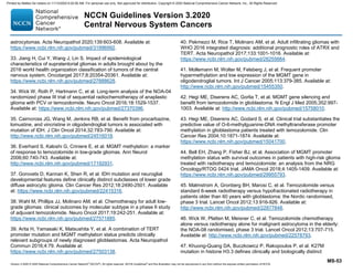


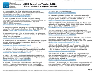

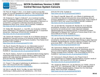


![Version 3.2020 © 2020 National Comprehensive Cancer Network©
(NCCN©
), All rights reserved. NCCN Guidelines®
and this illustration may not be reproduced in any form without the express written permission of NCCN.
NCCN Guidelines Version 3.2020
Central Nervous System Cancers
MS-64
first relapse. Br J Cancer 2000;83:588-593. Available at:
http://www.ncbi.nlm.nih.gov/pubmed/10944597.
193. Prados M, Rodriguez L, Chamberlain M, et al. Treatment of recurrent
gliomas with 1,3-bis(2-chloroethyl)-1-nitrosourea and alpha-
difluoromethylornithine. Neurosurgery 1989;24:806-809. Available at:
https://www.ncbi.nlm.nih.gov/pubmed/2546093.
194. Chamberlain MC, Johnston S. Bevacizumab for recurrent alkylator-
refractory anaplastic oligodendroglioma. Cancer 2009;115:1734-1743.
Available at: http://www.ncbi.nlm.nih.gov/pubmed/19197992.
195. Chamberlain MC, Johnston S. Salvage chemotherapy with
bevacizumab for recurrent alkylator-refractory anaplastic astrocytoma. J
Neurooncol 2009;91:359-367. Available at:
http://www.ncbi.nlm.nih.gov/pubmed/18953491.
196. Norden AD, Young GS, Setayesh K, et al. Bevacizumab for recurrent
malignant gliomas: efficacy, toxicity, and patterns of recurrence. Neurology
2008;70:779-787. Available at:
http://www.ncbi.nlm.nih.gov/pubmed/18316689.
197. Cloughesy T, Prados MD, Mikkelsen T. A phase 2 randomized non-
comparative clinical trial of the effect of bevacizumab alone or in
combination with irinotecan on 6-month progression free survival in
recurrent refractory glioblastoma [abstract]. J Clin Oncol 2008;26(Suppl
15):2010b. Available at:
http://meeting.ascopubs.org/cgi/content/abstract/26/15_suppl/2010b.
198. Kreisl TN, Kim L, Moore K, et al. Phase II trial of single-agent
bevacizumab followed by bevacizumab plus irinotecan at tumor
progression in recurrent glioblastoma. J Clin Oncol 2009;27:740-745.
Available at: http://www.ncbi.nlm.nih.gov/pubmed/19114704.
199. Vredenburgh JJ, Desjardins A, Herndon JE, 2nd, et al. Phase II trial
of bevacizumab and irinotecan in recurrent malignant glioma. Clin Cancer
Res 2007;13:1253-1259. Available at:
http://www.ncbi.nlm.nih.gov/pubmed/17317837.
200. Carvalho BF, Fernandes AC, Almeida DS, et al. Second-line
chemotherapy in recurrent glioblastoma: a 2-cohort study. Oncol Res
Treat 2015;38:348-354. Available at:
https://www.ncbi.nlm.nih.gov/pubmed/26278578.
201. Schmidt F, Fischer J, Herrlinger U, et al. PCV chemotherapy for
recurrent glioblastoma. Neurology 2006;66:587-589. Available at:
https://www.ncbi.nlm.nih.gov/pubmed/16505319.
202. DeVita VT, Lawrence TS, Rosenberg SA. DeVita, Hellman, and
Rosenberg's Cancer: Principles and Practice of Oncology (ed 8).
Philadelphia: Lippincott Williams Wilkins; 2008.
203. Packer RJ, Hoffman HJ, Friedman HS. Tumors of the fourth ventricle.
In: Levin VA, ed. Cancer in the Nervous System. New York: Churchill
Livingstone; 1996:153-156.
204. Pajtler KW, Witt H, Sill M, et al. Molecular classification of ependymal
tumors across all CNS compartments, histopathological grades, and age
groups. Cancer Cell 2015;27:728-743. Available at:
https://www.ncbi.nlm.nih.gov/pubmed/25965575.
205. Hubner JM, Kool M, Pfister SM, Pajtler KW. Epidemiology, molecular
classification and WHO grading of ependymoma. J Neurosurg Sci
2018;62:46-50. Available at:
https://www.ncbi.nlm.nih.gov/pubmed/28895660.
206. Kawabata Y, Takahashi JA, Arakawa Y, Hashimoto N. Long-term
outcome in patients harboring intracranial ependymoma. J Neurosurg
2005;103:31-37. Available at:
http://www.ncbi.nlm.nih.gov/pubmed/16121970.
207. Metellus P, Figarella-Branger D, Guyotat J, et al. Supratentorial
ependymomas: prognostic factors and outcome analysis in a retrospective
series of 46 adult patients. Cancer 2008;113:175-185. Available at:
http://www.ncbi.nlm.nih.gov/pubmed/18470910.
208. Metellus P, Barrie M, Figarella-Branger D, et al. Multicentric French
study on adult intracranial ependymomas: prognostic factors analysis and
Printed by Matteo De notaris on 11/13/2020 6:02:56 AM. For personal use only. Not approved for distribution. Copyright © 2020 National Comprehensive Cancer Network, Inc., All Rights Reserved.](https://image.slidesharecdn.com/cns-201113111756/85/Cns-147-320.jpg)


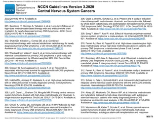


![Version 3.2020 © 2020 National Comprehensive Cancer Network©
(NCCN©
), All rights reserved. NCCN Guidelines®
and this illustration may not be reproduced in any form without the express written permission of NCCN.
NCCN Guidelines Version 3.2020
Central Nervous System Cancers
MS-77
Eur J Nucl Med Mol Imaging 2010;37:310-318. Available at:
http://www.ncbi.nlm.nih.gov/pubmed/19763565.
389. Gehler B, Paulsen F, Oksuz MO, et al. [68Ga]-DOTATOC-PET/CT
for meningioma IMRT treatment planning. Radiat Oncol 2009;4:56.
Available at: http://www.ncbi.nlm.nih.gov/pubmed/19922642.
390. Nakamura M, Roser F, Michel J, et al. The natural history of
incidental meningiomas. Neurosurgery 2003;53:62-70; discussion 70-61.
Available at: http://www.ncbi.nlm.nih.gov/pubmed/12823874.
391. Olivero WC, Lister JR, Elwood PW. The natural history and growth
rate of asymptomatic meningiomas: a review of 60 patients. J Neurosurg
1995;83:222-224. Available at:
http://www.ncbi.nlm.nih.gov/pubmed/7616265.
392. Stafford SL, Perry A, Suman VJ, et al. Primarily resected
meningiomas: outcome and prognostic factors in 581 Mayo Clinic patients,
1978 through 1988. Mayo Clin Proc 1998;73:936-942. Available at:
http://www.ncbi.nlm.nih.gov/pubmed/9787740.
393. Mahmood A, Qureshi NH, Malik GM. Intracranial meningiomas:
analysis of recurrence after surgical treatment. Acta Neurochir (Wien)
1994;126:53-58. Available at:
http://www.ncbi.nlm.nih.gov/pubmed/8042555.
394. Mathiesen T, Lindquist C, Kihlstrom L, Karlsson B. Recurrence of
cranial base meningiomas. Neurosurgery 1996;39:2-7; discussion 8-9.
Available at: http://www.ncbi.nlm.nih.gov/pubmed/8805134.
395. Perry A, Stafford SL, Scheithauer BW, et al. Meningioma grading: an
analysis of histologic parameters. Am J Surg Pathol 1997;21:1455-1465.
Available at: http://www.ncbi.nlm.nih.gov/pubmed/9414189.
396. Simpson D. The recurrence of intracranial meningiomas after surgical
treatment. J Neurol Neurosurg Psychiatry 1957;20:22-39. Available at:
http://www.ncbi.nlm.nih.gov/pubmed/13406590.
397. Condra KS, Buatti JM, Mendenhall WM, et al. Benign meningiomas:
primary treatment selection affects survival. Int J Radiat Oncol Biol Phys
1997;39:427-436. Available at:
http://www.ncbi.nlm.nih.gov/pubmed/9308947.
398. Soyuer S, Chang EL, Selek U, et al. Radiotherapy after surgery for
benign cerebral meningioma. Radiother Oncol 2004;71:85-90. Available
at: http://www.ncbi.nlm.nih.gov/pubmed/15066300.
399. Pirzkall A, Debus J, Haering P, et al. Intensity modulated
radiotherapy (IMRT) for recurrent, residual, or untreated skull-base
meningiomas: preliminary clinical experience. Int J Radiat Oncol Biol Phys
2003;55:362-372. Available at:
https://www.ncbi.nlm.nih.gov/pubmed/12527049.
400. Aghi MK, Carter BS, Cosgrove GR, et al. Long-term recurrence rates
of atypical meningiomas after gross total resection with or without
postoperative adjuvant radiation. Neurosurgery 2009;64:56-60; discussion
60. Available at: http://www.ncbi.nlm.nih.gov/pubmed/19145156.
401. Hug EB, Devries A, Thornton AF, et al. Management of atypical and
malignant meningiomas: role of high-dose, 3D-conformal radiation
therapy. J Neurooncol 2000;48:151-160. Available at:
http://www.ncbi.nlm.nih.gov/pubmed/11083080.
402. Rogers CL, Won M, Vogelbaum MA, et al. High-risk meningioma:
initial outcomes from NRG Oncology/RTOG 0539. Int J Radiat Oncol Biol
Phys 2020;106:790-799. Available at:
https://www.ncbi.nlm.nih.gov/pubmed/31786276.
403. Selch MT, Ahn E, Laskari A, et al. Stereotactic radiotherapy for
treatment of cavernous sinus meningiomas. Int J Radiat Oncol Biol Phys
2004;59:101-111. Available at:
https://www.ncbi.nlm.nih.gov/pubmed/15093905.
404. Pollock BE, Stafford SL, Utter A, et al. Stereotactic radiosurgery
provides equivalent tumor control to Simpson Grade 1 resection for
patients with small- to medium-size meningiomas. Int J Radiat Oncol Biol
Printed by Matteo De notaris on 11/13/2020 6:02:56 AM. For personal use only. Not approved for distribution. Copyright © 2020 National Comprehensive Cancer Network, Inc., All Rights Reserved.](https://image.slidesharecdn.com/cns-201113111756/85/Cns-160-320.jpg)


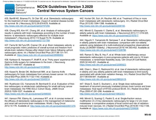


![Version 3.2020 © 2020 National Comprehensive Cancer Network©
(NCCN©
), All rights reserved. NCCN Guidelines®
and this illustration may not be reproduced in any form without the express written permission of NCCN.
NCCN Guidelines Version 3.2020
Central Nervous System Cancers
MS-87
537. Glantz MJ, LaFollette S, Jaeckle KA, et al. Randomized trial of a
slow-release versus a standard formulation of cytarabine for the
intrathecal treatment of lymphomatous meningitis. J Clin Oncol
1999;17:3110-3116. Available at:
http://www.ncbi.nlm.nih.gov/pubmed/10506606.
538. Jaeckle KA, Phuphanich S, Bent MJ, et al. Intrathecal treatment of
neoplastic meningitis due to breast cancer with a slow-release formulation
of cytarabine. Br J Cancer 2001;84:157-163. Available at:
http://www.ncbi.nlm.nih.gov/pubmed/11161370.
539. Gutin PH, Weiss HD, Wiernik PH, Walker MD. Intrathecal N, N', N-
triethylenethiophosphoramide [thio-TEPA (NSC 6396)] in the treatment of
malignant meningeal disease: phase I-II study. Cancer 1976;38:1471-
1475. Available at: https://www.ncbi.nlm.nih.gov/pubmed/825215.
540. Chamberlain MC, Johnston SK, Van Horn A, Glantz MJ. Recurrent
lymphomatous meningitis treated with intra-CSF rituximab and liposomal
ara-C. J Neurooncol 2009;91:271-277. Available at:
http://www.ncbi.nlm.nih.gov/pubmed/18820836.
541. Groves MD, Glantz MJ, Chamberlain MC, et al. A multicenter phase
II trial of intrathecal topotecan in patients with meningeal malignancies.
Neuro Oncol 2008;10:208-215. Available at:
http://www.ncbi.nlm.nih.gov/pubmed/18316473.
542. Chamberlain MC, Tsao-Wei DD, Groshen S. Phase II trial of
intracerebrospinal fluid etoposide in the treatment of neoplastic meningitis.
Cancer 2006;106:2021-2027. Available at:
http://www.ncbi.nlm.nih.gov/pubmed/16583432.
543. Zagouri F, Sergentanis TN, Bartsch R, et al. Intrathecal
administration of trastuzumab for the treatment of meningeal
carcinomatosis in HER2-positive metastatic breast cancer: a systematic
review and pooled analysis. Breast Cancer Res Treat 2013;139:13-22.
Available at: http://www.ncbi.nlm.nih.gov/pubmed/23588955.
544. Mrugala MM, Kim B, Sharma A, et al. Phase II study of systemic
high-dose methotrexate and intrathecal liposomal cytarabine for treatment
of leptomeningeal carcinomatosis from breast cancer. Clin Breast Cancer
2019;19:311-316. Available at:
https://www.ncbi.nlm.nih.gov/pubmed/31175053.
545. Bokstein F, Lossos A, Lossos IS, Siegal T. Central nervous system
relapse of systemic non-Hodgkin's lymphoma: results of treatment based
on high-dose methotrexate combination chemotherapy. Leuk Lymphoma
2002;43:587-593. Available at:
http://www.ncbi.nlm.nih.gov/pubmed/12002763.
546. Grommes C, Oxnard GR, Kris MG, et al. Pulsatile high-dose weekly
erlotinib for CNS metastases from EGFR mutant non-small cell lung
cancer. Neuro Oncol 2011;13:1364-1369. Available at:
http://www.ncbi.nlm.nih.gov/pubmed/21865399.
547. Yang JCH, Kim SW, Kim DW, et al. Osimertinib in patients with
epidermal growth factor receptor mutation-positive non-small-cell lung
cancer and leptomeningeal metastases: the BLOOM study. J Clin Oncol
2020;38:538-547. Available at:
https://www.ncbi.nlm.nih.gov/pubmed/31809241.
548. Nanjo S, Hata A, Okuda C, et al. Standard-dose osimertinib for
refractory leptomeningeal metastases in T790M-positive EGFR-mutant
non-small cell lung cancer. Br J Cancer 2018;118:32-37. Available at:
https://www.ncbi.nlm.nih.gov/pubmed/29190637.
549. Ahn MJ, Chiu CH, Cheng Y, et al. Osimertinib for patients with
leptomeningeal metastases associated with EGFR T790M-positive
advanced NSCLC: the AURA leptomeningeal metastases analysis. J
Thorac Oncol 2020;15:637-648. Available at:
https://www.ncbi.nlm.nih.gov/pubmed/31887431.
550. Lee JS, Melisko ME, Magbanua MJ, et al. Detection of cerebrospinal
fluid tumor cells and its clinical relevance in leptomeningeal metastasis of
breast cancer. Breast Cancer Res Treat 2015;154:339-349. Available at:
https://www.ncbi.nlm.nih.gov/pubmed/26520840.
551. Lin X, Fleisher M, Rosenblum M, et al. Cerebrospinal fluid circulating
tumor cells: a novel tool to diagnose leptomeningeal metastases from
Printed by Matteo De notaris on 11/13/2020 6:02:56 AM. For personal use only. Not approved for distribution. Copyright © 2020 National Comprehensive Cancer Network, Inc., All Rights Reserved.](https://image.slidesharecdn.com/cns-201113111756/85/Cns-170-320.jpg)

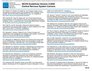


![Human Reproduction [ Reproductive System ] Notes @irfanullah_mehar Irfanullah...](https://cdn.slidesharecdn.com/ss_thumbnails/humanreproductionreproductivesystemnotesirfanullahmeharirfanullahmeharjanantantra-260111172350-56e85778-thumbnail.jpg?width=640&height=640&fit=bounds)


