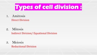
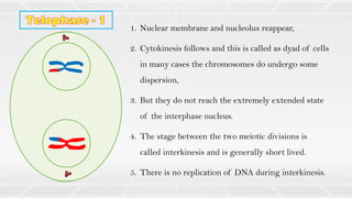
1. The document describes different types of cell division including amitosis, mitosis, and meiosis.
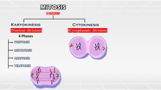
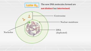
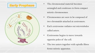
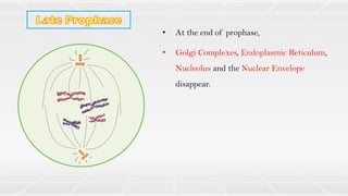
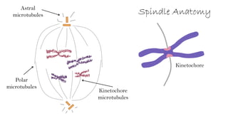
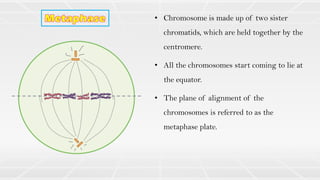
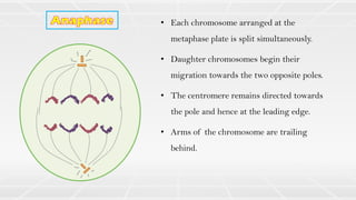
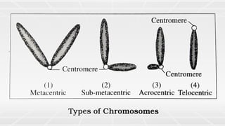
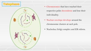
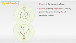
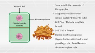
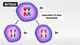
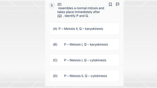
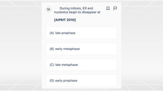
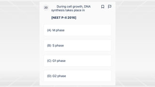
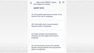
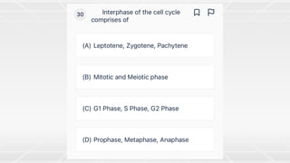
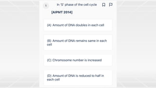
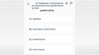
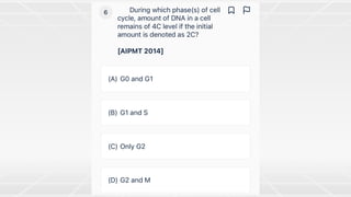
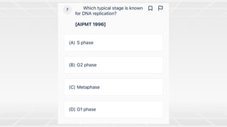
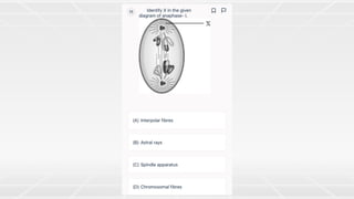
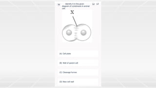
2. Mitosis involves nuclear division (karyokinesis) and cytoplasmic division (cytokinesis) through the phases of prophase, metaphase, anaphase and telophase.
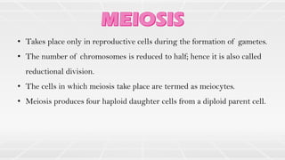
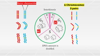
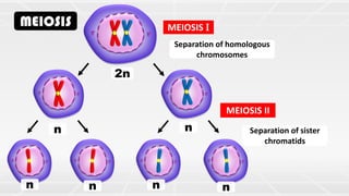
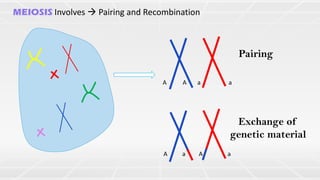
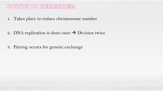
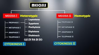
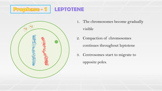
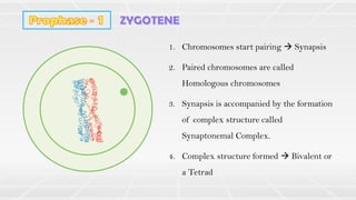
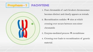
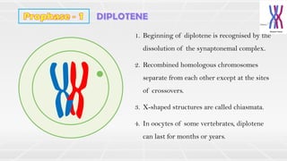
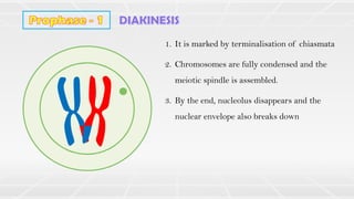
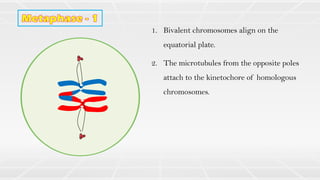
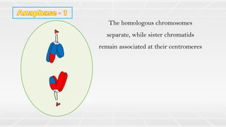
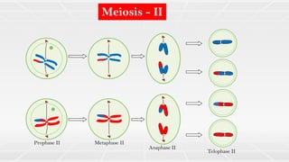
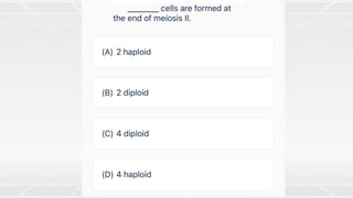
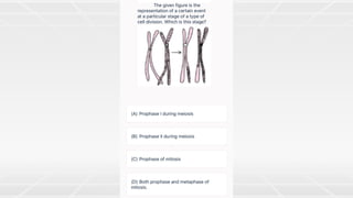
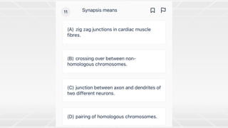
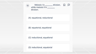
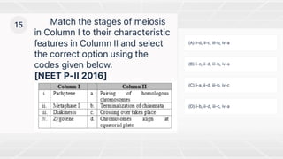
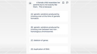
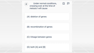
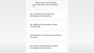
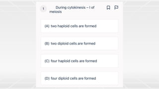
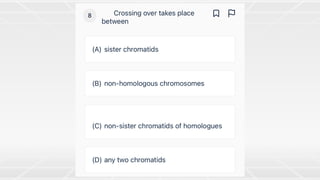
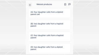
3. Meiosis involves two nuclear divisions and produces four haploid daughter cells from a single diploid parent cell, reducing the chromosome number by half. It includes the processes of pairing, recombination and segregation of homologous chromosomes.


![74676371-Coagulation-and-Flocculation[1].ppt](https://cdn.slidesharecdn.com/ss_thumbnails/74676371-coagulation-and-flocculation1-260116154109-a3cbf55e-thumbnail.jpg?width=640&height=640&fit=bounds)


