Download to read offline
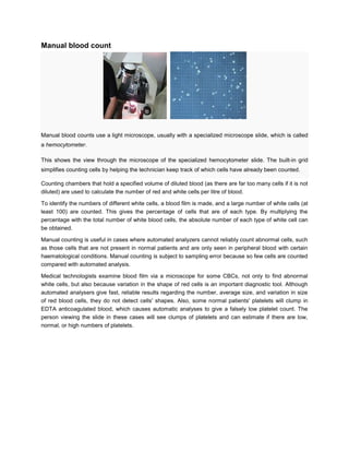

Manual blood counts involve using a light microscope and specialized slides known as hemocytometers to count red blood cells, white blood cells, and platelets. A hemocytometer contains a built-in grid that helps technicians keep track of which cells have been counted. Blood is diluted before counting to allow individual cells to be seen. Manual counts provide cell counts when automated analyzers cannot reliably count abnormal cells or detect variations in cell shape that provide diagnostic information. While more subject to errors than automated methods, manual counts remain useful for certain medical conditions and platelet clumping issues.


