2. patients with ascites, there is a nonhepatic cause of fluid
retention. Successful treatment is dependent on an accu-
rate diagnosis of the cause of ascites; e.g., peritoneal car-
cinomatosis does not respond to diuretic therapy. Patients
with ascites should be questioned about risk factors for
liver disease. Those who lack an apparent cause for cirrho-
sis should also be questioned about lifetime body weight;
nonalcoholic steatohepatitis has been concluded to be
causative in many of these patients.11 Past history of can-
cer, heart failure, renal disease, or tuberculosis is also rel-
evant. Hemophagocytic syndrome can masquerade as
cirrhosis with ascites.12 These patients have fever, jaun-
dice, and hepatosplenomegaly, usually in the setting of
lymphoma or leukemia.12
Physical Examination
The presence of a full, bulging abdomen should lead to
percussion of the flanks. If the amount of flank dullness is
greater than usual (i.e., if the percussed tympany-dullness
interface is higher than normally found on the lateral
aspect of the abdomen with the patient supine), one
should test for “shifting”. The presence of shifting dull-
ness has 83% sensitivity and 56% specificity in detecting
ascites.13 Approximately 1500 mL of fluid must be
present before flank dullness is detected.13 If no flank
dullness is present, the patient has less than a 10% chance
of having ascites.13 The fluid wave and puddle sign are
cumbersome and perform less well when compared to
shifting dullness.13 Ascites due to alcoholic cardiomyop-
athy can mimic that due to alcoholic cirrhosis. Jugular
venous distension is present in the former but not in the
latter. Also measurement of a blood concentration of
brain natriuretic peptide or pro–brain natriuretic peptide
can help distinguish ascites due to heart failure from as-
cites due to cirrhosis.14 The median pro–brain natriuretic
peptide concentration is 6100 pg/mL in the former and
only 166 pg/mL in the latter.14
Giant cysts or pseudocysts can rarely mimic ascites.
Paracentesis may produce fluid with unusual characteris-
tics. Imaging usually provides the correct diagnosis.15
The physical examination for detecting ascites in the
obese patient is problematic. An abdominal ultrasound
may be required to determine with certainty if fluid is
present. Ascites usually is present for only a few weeks
before the patient seeks medical attention. In contrast, a
slowly enlarging abdomen over months to years is most
likely due to obesity not ascites.
The diagnosis of new-onset ascites is suspected on the
basis of the history and physical examination and usually
confirmed by successful abdominal paracentesis and/or
ultrasound. The diagnosis of the etiology of ascites forma-
tion is based on the results of the history, physical exam-
ination, and ascitic fluid analysis. In general, few other
tests are required. However, the liver is commonly imaged
to screen for hepatocellular carcinoma, portal vein throm-
bosis, and hepatic vein thrombosis.
Abdominal Paracentesis
Abdominal paracentesis with appropriate ascitic fluid
analysis is probably the most rapid and cost-effective
method of diagnosing the cause of ascites.16,17 Fluid due
to portal hypertension can be readily differentiated from
fluid due to other causes.10 Also, in view of the high prev-
alence of ascitic fluid infection at the time of admission to
the hospital, an admission surveillance tap may detect
unexpected infection.18
Although older published series reported a relatively
high morbidity, and even mortality, when trocars were
used for paracentesis, more recent studies regarding para-
Table 1. Grading System for Recommendations
Classification Description
Class I Conditions for which there is evidence and/or general
agreement that a given diagnostic evaluation, procedure
or treatment is beneficial, useful, and effective.
Class II Conditions for which there is conflicting evidence and/or a
divergence of opinion about the usefulness/efficacy of a
diagnostic evaluation, procedure or treatment.
Class IIa Weight of evidence/opinion is in favor of
usefulness/efficacy.
Class IIb Usefulness/efficacy is less well established by evidence/
opinion.
Class III Conditions for which there is evidence and/or general
agreement that a diagnostic
evaluation/procedure/treatment is not useful/effective
and in some cases may be harmful.
Level of
Evidence Description
Level A Data derived from multiple randomized clinical trials or meta-
analyses.
Level B Data derived from a single randomized trial, or nonrandomized
studies.
Level C Only consensus opinion of experts, case studies, or standard-
of-care.
Table 2. Differential Diagnosis of Ascites
Cirrhosis
Alcoholic hepatitis
Heart failure
Cancer (peritoneal carcinomatosis, massive liver metastases, etc)
“Mixed” ascites, i. e., cirrhosis plus another cause for ascites
Pancreatitis
Nephrotic syndrome
Tuberculous peritonitis
Acute liver failure
Budd-Chiari syndrome
Sinusoidal obstruction syndrome
Postoperative lymphatic leak
Myxedema
2088 RUNYON HEPATOLOGY, June 2009
3. centesis complications in patients with ascites docu-
mented no deaths or infections caused by the
paracentesis.19 Complications were reported in only
about 1% of patients (abdominal wall hematomas), de-
spite the fact that 71% of the patients had an abnormal
prothrombin time.19 Although more serious complica-
tions (hemoperitoneum or bowel entry by the paracente-
sis needle) occur,20 they are sufficiently unusual
(Ͻ1/1000 paracenteses) that they should not deter per-
formance of this procedure. In a study of 4729 paracen-
teses, investigators reported that eight of nine bleeding
complications occurred in patients with renal failure; per-
haps the qualitative platelet abnormality in this setting
predisposes to more bleeding.21
Although some physicians give blood products (fresh
frozen plasma and/or platelets) routinely before paracen-
tesis in patients with cirrhosis and coagulopathy, this pol-
icy is not data-supported.19,22 Routine tests of coagulation
also do not reflect bleeding risk in patients with cirrhosis;
these patients regularly have normal global coagulation
because of a balanced deficiency of procoagulants and
anticoagulants.23 In a recent survey of the use of blood
products in relation to paracentesis, 50% of approxi-
mately 100 hepatologists attending a conference on co-
agulopathy in liver disease indicated that they either never
used plasma before procedure or used it only if the inter-
national normalized ratio was Ͼ2.5.24 The risks and costs
of prophylactic transfusions may exceed the benefit. Co-
agulopathy should preclude paracentesis only when there
is clinically evident hyperfibrinolysis (three-dimensional
ecchymosis/hematoma) or clinically evident disseminated
intravascular coagulation. A shortened (Ͻ120 minutes)
euglobulin clot lysis time documents hyperfibrinolysis.25
However, this test may not be routinely available. Epsilon
aminocaproic acid can be used to treat hyperfibrinolysis;
paracentesis can be performed after the lysis time has nor-
malized on treatment.26 Bleeding conditions occur in less
than 1/1000 patients who require paracentesis. There is
no data-supported cutoff of coagulation parameters be-
yond which paracentesis should be avoided.19 In a study
of 1100 large-volume paracenteses, there were no hemor-
rhagic complications despite (1) no prophylactic transfu-
sions, (2) platelet counts as low as 19,000 cells/mm3
(19 ϫ 106/L) (54% Ͻ50,000), and (3) international nor-
malized ratios for prothrombin time as high as 8.7 (75%
Ͼ1.5 and 26.5% Ͼ2.0).22
In the past, the avascular midline, midway between the
pubis and the umbilicus, was usually chosen as the site for
paracentesis. Now, because many paracenteses are per-
formed to remove a large volume of fluid and abdominal
obesity increases the midline wall thickness, the left lower
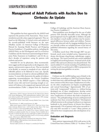
quadrant is the preferred location (Fig. 1). The abdominal
wall in the left lower quadrant, 2 finger breadths (3 cm)
cephalad and 2 finger breadths medial to the anterior
superior iliac spine, has been shown to be thinner and
with a larger pool of fluid than the midline and is usually
a good choice for needle insertion for performance of a
therapeutic paracentesis.27 The right lower quadrant may
be a suboptimal choice in the setting of a dilated cecum
(due to lactulose) or an appendectomy scar. The area of
the inferior epigastric arteries should be avoided; these
vessels are located midway between the pubis and anterior
superior iliac spines and then run cephalad in the rectus
sheath. Visible collaterals should also be avoided. A lapa-
roscopic study found that collaterals can be present in the
midline and thus present a risk for rupture during para-
centesis.28
If the fluid is difficult to localize by examination be-
cause of obesity, ultrasonography can be a useful adjunct
in locating fluid and visualizing the spleen and other
structures to be avoided. There are few contraindications
to paracentesis. The procedure should be performed by a
provider who has been trained in its performance.
Recommendations:
1. Abdominal paracentesis should be performed
and ascitic fluid should be obtained from inpatients
and outpatients with clinically apparent new-onset
ascites. (Class I, Level C)
2. Because bleeding is sufficiently uncommon, the
routine prophylactic use of fresh frozen plasma or
platelets before paracentesis is not recommended.
(Class III, Level C)
Fig. 1. Diagram of the abdomen showing the three usual sites for
abdominal paracentesis. The author prefers the left lower quadrant site.
Reproduced from Thomsen TW, Shaffer RW, White B, Setnik GS. Para-
centesis. N Engl J Med 2006;355:e21, with permission from the Mas-
sachusetts Medical Society. Copyright (2006) Massachusetts Medical
Society. All rights reserved.
HEPATOLOGY, Vol. 49, No. 6, 2009 RUNYON 2089
4. Ascitic Fluid Analysis
An algorithm approach seems preferable to ordering a
large number of tests on most specimens (Table 3). If
uncomplicated ascites due to cirrhosis is suspected, only
screening tests (e.g., cell count and differential, albumin
and total protein concentration) are performed on the
initial specimen. If the results of these tests are unexpect-
edly abnormal, further testing can be performed on an-
other ascitic fluid sample. Also, many laboratories save an
aliquot of fluid for a few days; this fluid can be tested if the
specimen has been handled properly. However, because
most specimens are consistent with uncomplicated cir-
rhotic ascites, no further testing will be needed in the
majority of patients.
If ascitic fluid infection is suspected (fever, abdominal
pain, unexplained encephalopathy, acidosis, azotemia,
hypotension, or hypothermia), bacterial culture of the
fluid in blood culture bottles inoculated at the bedside
should be performed. Use of a urine dipstick to detect
neutrophils in ascitic fluid takes only 90 seconds to 2
minutes.29,30 However, the largest study of a urine dip-
stick (2123 paracenteses) demonstrated a sensitivity of
only 45%.31 Development of an ascites-specific dipstick
in contrast to a urine dipstick is needed. Automated cell
counting has been shown to be accurate in one study; the
result is rapidly available and could replace the manual
cell count if it is further validated.32 Additional testing,
e.g., for total protein, lactate dehydrogenase (LDH), and
glucose to assist in differentiating spontaneous from sec-
ondary bacterial peritonitis, can be performed on the ini-
tial specimen based on clinical judgment.33 An ascitic
fluid carcinoembryonic antigen Ͼ5 ng/mL or ascitic fluid
alkaline phosphatase Ͼ240 U/L has also been shown to be
accurate in detecting gut perforation into ascitic fluid.34
The serum-ascites albumin gradient (SAAG) has been
proved in prospective studies to categorize ascites better
than the total-protein–based exudate/transudate concept
and better than modified pleural fluid exudate/transudate
criteria.11,35 Calculating the SAAG involves measuring
the albumin concentration of serum and ascitic fluid spec-
imens obtained on the same day and subtracting the as-
citic fluid value from the serum value. If the SAAG is
Ն1.1 g/dL (11 g/L), the patient has portal hypertension,
with approximately 97% accuracy.11 Patients who have
portal hypertension plus a second cause for ascites forma-
tion also have a SAAG Ն1.1 g/dL.
Patients undergoing serial outpatient therapeutic para-
centeses probably should be tested only for cell count and
differential36,37 (the author has detected eight episodes of
spontaneous bacterial peritonitis [SBP] in approximately
400 paracenteses in a paracentesis clinic in 2 years [un-
published observations]). Bacterial culture is not neces-
sary in asymptomatic patients undergoing serial large-
volume paracenteses.
The most expensive tests are the cytology and smear
and culture for mycobacteria; these tests should probably
be ordered only when there is a high pretest probability of
occurrence of the disease under consideration. The ascitic
fluid cytology is positive only in the setting of peritoneal
carcinomatosis.38 The sensitivity of cytology in detecting
peritoneal carcinomatosis is 96.7% if three samples are
sent and processed promptly; the first sample is positive in
82.8% and at least one of two samples is positive in
93.3%.38 In this study, 50 mL of fresh warm ascitic fluid
were hand-carried to the laboratory for immediate pro-
cessing. Use of DNA cytometry or magnetic enrichment
may improve the sensitivity of cytology further.39,40 Pa-
tients with peritoneal carcinomatosis usually have a his-
tory of a breast, colon, gastric, or pancreatic primary
carcinoma. The sensitivity of smear for mycobacteria is
approximately 0%; the sensitivity of fluid culture for my-
cobacteria is approximately 50%.41 Only patients at high
risk for tuberculous peritonitis (e.g., recent immigration
from an endemic area or acquired immunodeficiency syn-
drome)42 should have testing for mycobacteria on the first
ascitic fluid specimen. Laparoscopy with biopsy and my-
cobacterial culture of tubercles are the most rapid and
accurate methods of diagnosing tuberculous peritonitis.
Multiple prospective trials have shown that bacterial
growth occurs in only about 50% of instances when as-
citic fluid with a polymorphonuclear leukocyte (PMN)
count Ն250 cells/mm3 (0.25 ϫ 109/L) is cultured by
older methods, i.e., sending a syringe or tube of fluid to
the laboratory, as compared to approximately 80% if the
Table 3. Ascitic Fluid Laboratory Data*
Routine Optional Unusual Unhelpful
Cell count and differential Culture in blood culture bottles AFB smear and culture pH
Albumin Glucose Cytology Lactate
Total protein Lactate dehydrogenase Triglyceride Cholesterol
Amylase Bilirubin Fibronectin
Gram’s stain Glycosaminoglycans
Abbreviation: AFB, acid-fast bacteria. *Adapted from Runyon.17 Reprinted with permission from W.B. Saunders.
2090 RUNYON HEPATOLOGY, June 2009
5. fluid is inoculated into blood culture bottles at the bedside
and prior to administration of antibiotics.43,44
Differential Diagnosis
Although cirrhosis is the cause of ascites formation in
most patients, approximately 15% have a cause other than
liver disease, including cancer, heart failure, tuberculosis,
or nephrotic syndrome (Table 3).10 Approximately 5% of
patients with ascites have two or more causes of ascites
formation, i.e., “mixed” ascites.10 Usually, these patients
have cirrhosis plus one other cause, e.g., peritoneal carci-
nomatosis or peritoneal tuberculosis. Many patients with
enigmatic ascites are eventually found to have two or even
three causes for ascites formation (e.g., heart failure, dia-
betic nephropathy, and cirrhosis due to nonalcoholic ste-
atohepatitis). In this setting, the sum of predisposing
factors leads to sodium and water retention when each
individual factor might not be severe enough to cause
fluid overload.
The cancer antigen 125 (CA125) warrants mention.
Essentially all patients, including men with ascites or
pleural fluid of any cause, have an elevated serum CA125;
when ascites is controlled, the CA125 level decreases dra-
matically.45,46 This test is elevated when mesothelial cells
are under pressure from the presence of fluid; it is very
nonspecific. When this test is found to be abnormal, the
female patient may be unnecessarily referred for gyneco-
logic surgery even if the ovaries were removed decades
earlier; cirrhosis is regularly detected at laparotomy as the
cause for ascites formation (because it is most common
cause) rather than ovarian cancer, and the patient may die
postoperatively. Patients with ascites should not have se-
rum tested for CA125.
Recommendations:
3. The initial laboratory investigation of ascitic
fluid should include an ascitic fluid cell count and
differential, ascitic fluid total protein, and SAAG.
(Class I, Level B)
4. If ascitic fluid infection is suspected, ascitic fluid
should be cultured at the bedside in blood culture
bottles prior to initiation of antibiotics. (Class I, Level
B)
5. Other studies of ascitic fluid can be ordered
based on pretest probability of disease (Table 3).
(Class IIa, Level C)
6. Testing serum for CA125 is not helpful in the
differential diagnosis of ascites. Its use is not recom-
mended in patients with ascites of any type. (Class III,
Level B)
Treatment of Ascites
Appropriate treatment of patients with ascites depends
on the cause of fluid retention. SAAG can be helpful
diagnostically as well as in decision-making regarding
treatment. Patients with low SAAG (Ͻ1.1 g/dL) ascites
usually do not have portal hypertension and, with the
exception of nephrotic syndrome, do not respond to salt
restriction and diuretics.17 In contrast, patients with a
high SAAG (Ն1.1 g/dL) have portal hypertension and
usually are responsive to these measures.17
The remainder of this guideline is applicable only to
patients with cirrhosis as the cause of their ascites. Im-
provement in the outcome of patients with nonportal-
hypertension–related ascites depends on successful
treatment of the underlying disorder.
Alcohol-induced liver injury is one of the most revers-
ible causes of liver disease that leads to high SAAG as-
cites.17 One of the most important steps in treating ascites
in this setting is to treat the underlying liver disease by
ceasing alcohol consumption. In a period of months, ab-
stinence can result in dramatic improvement in the revers-
ible component of alcoholic liver disease. One recent
study demonstrates that patients who have Child-Pugh
class C cirrhosis due to alcohol and who stop drinking
have an approximately 75% 3-year survival, but all those
who continue to drink die in 3 years.47 Ascites may resolve
or become more responsive to medical therapy with ab-
stinence and time. Decompensated hepatitis B cirrhosis
can also have a dramatic response to antiviral treatment.48
Liver diseases other than those that are related to alcohol,
hepatitis B, and autoimmune hepatitis are less reversible;
by the time ascites is present, these patients may be best
served by referral for liver transplantation evaluation
rather than protracted medical therapy.
The mainstays of treatment of patients with cirrhosis
and ascites include (1) education regarding dietary so-
dium restriction (2000 mg/day [88 mmol/day]) and (2)
oral diuretics.16,17 More stringent dietary sodium restric-
tion can speed mobilization of ascites, but is not recom-
mended because it is less palatable and may further
worsen the malnutrition that is usually present in these
patients. Fluid loss and weight change are directly related
to sodium balance in patients with portal hypertension–
related ascites. It is sodium restriction, not fluid restric-
tion, which results in weight loss, as fluid follows sodium
passively.49 Measurement of urinary sodium excretion is a
helpful parameter to follow when rapidity of weight loss is
less than desired.16,17 Random urinary sodium concentra-
tions are of value when they are 0 mmol/L or Ͼ100
mmol/L but are much less helpful when they are interme-
diate because of lack of uniformity of sodium excretion
HEPATOLOGY, Vol. 49, No. 6, 2009 RUNYON 2091
6. during the day and lack of knowledge of total urine vol-
ume, which may vary from 300 mL to greater than 3000
mL. Twenty-four-hour collections of urine for determi-
nation of sodium excretion are much more informative
than random specimens; however, full-day collections are
cumbersome. Providing patients with verbal and written
instructions, a container, and a lab order slip to turn in
with the completed specimen helps insure compliance.
Completeness of collection of the 24-hour specimen can
be assessed by measurement of urinary creatinine. Men
with cirrhosis should excrete more than 15 mg creatinine
per kilogram of body weight per day, and women with
cirrhosis should excrete more than 10 mg/kg/day. Less
creatinine is indicative of an incomplete collection. Total
nonurinary sodium excretion is less than 10 mmol/day in
afebrile patients with cirrhosis without diarrhea.50 One of
the goals of treatment is to increase urinary excretion of
sodium so that it exceeds 78 mmol/day (88 mmol intake/
day Ϫ 10 mmol nonurinary excretion per day). Only the
10%-15% of patients who have spontaneous natriuresis
Ͼ78 mmol/day can be considered for dietary sodium re-
striction alone (i.e., without diuretics). However, when
given a choice, most patients would prefer to take some
diuretics and have a more liberal sodium intake than take
no pills and have a more severe sodium restriction.
A random “spot” urine sodium concentration that is
greater than the potassium concentration correlates with a
24-hour sodium excretion greater than 78 mmol/day with
approximately 90% accuracy.51 This urine sodium/potas-
sium ratio may replace the cumbersome 24-hour collec-
tion.
Fluid restriction is not necessary in treating most pa-
tients with cirrhosis and ascites. The chronic hyponatre-
mia usually seen in patients with cirrhosis and ascites is
seldom morbid unless it is rapidly corrected in the oper-
ating room at the time of liver transplantation.52 A study
of 997 patients with cirrhosis and ascites demonstrates
that the serum sodium is Յ120 mmol/L in only 1.2% of
patients and Յ125 mmol/L in only 5.7%.53 Attempts to
rapidly correct hyponatremia in this setting with hyper-
tonic saline can lead to more complications than the hy-
ponatremia itself.54 Preliminary data suggest that
aquaretic drugs have the promise of correcting hyponatre-
mia. The intravenous aquaretic agent conivaptan has been
studied in patients with cirrhosis and is approved for use
for treatment of “euvolemic and hypervolemic hyponatre-
mia in hospitalized patients”.55 Caution is advised by the
manufacturer in the use of this drug in patients with cir-
rhosis. An oral preparation tolvaptan increases serum so-
dium in patients who have pretreatment values Ͻ130
mmol/L.56 However, whether these agents will be effec-
tive without side effects in the subset of patients with
cirrhosis who are more in need of correction of hypona-
tremia (serum sodium Յ120 mmol/L) remains un-
proven. Cost-effectiveness also warrants investigation.
Unfortunately, many drugs that have theoretical promise
in treating ascites, e.g., angiotensin-converting enzyme
inhibitors, have been shown to aggravate hypotension and
have not been clinically useful. Severe hyponatremia does
warrant fluid restriction in the patient with cirrhosis and
ascites; however, there is no data-supported specific
threshold for initiating fluid restriction. A serum sodium
Ͻ120-125 mmol/L is a reasonable threshold. Patients
with cirrhosis do not usually have symptoms from hypo-
natremia until the sodium is Ͻ110 mmol/L or unless the
decline in sodium is very rapid.
Although it is traditional to recommend bed rest
(based on extrapolation from heart failure), this is imprac-
tical and there are no controlled trials to support this
practice. Upright posture may aggravate the plasma renin
elevation found in patients with cirrhosis with ascites.
Theoretically, this may increase sodium avidity. This the-
oretical concern would have to translate into clinically
relevant outcomes before bed rest could be advocated.
The usual diuretic regimen consists of single morning
doses of oral spironolactone and furosemide, beginning
with 100 mg of the former and 40 mg of the latter.16,17
Previously, single-agent spironolactone was advocated,
but hyperkalemia and the long half-life of this drug have
resulted in its use as a single agent only in patients with
minimal fluid overload.57 Single-agent furosemide has
been shown in a randomized controlled trial to be less
efficacious than spironolactone.58 The good oral bioavail-
ability of furosemide in the patient with cirrhosis, to-
gether with the acute reductions in glomerular filtration
rate associated with intravenous furosemide, favor use of
the oral route of administration.59,60 A randomized trial
purports to demonstrate that spironolactone should be
used as a single agent, with furosemide added only for
refractory patients.61 Diuresis was slower in the single-
agent spironolactone group with a lesser need for dose
adjustments; thus, this approach may be useful for outpa-
tients.61 However, another randomized trial indicates that
initial combination treatment shortens the time to mobi-
lization of moderate ascites.62 Most patients require com-
bination treatment eventually. The largest study ever
performed (involving 3860 patients with cirrhosis and
ascites) used combination therapy from the beginning.63
Starting with both drugs appears to be the preferred ap-
proach in achieving rapid natriuresis and maintaining
normokalemia. An alternative approach would be to start
with single-agent spironolactone, in particular in the out-
patient setting.
2092 RUNYON HEPATOLOGY, June 2009
7. The doses of both oral diuretics can be increased simul-
taneously every 3-5 days (maintaining the 100 mg:40 mg
ratio) if weight loss and natriuresis are inadequate. In
general, this ratio maintains normokalemia. Usual maxi-
mum doses are 400 mg/day of spironolactone and 160
mg/day of furosemide.16,17 Furosemide can be tempo-
rarily withheld in patients presenting with hypokalemia;
this is very common in the setting of alcoholic hepatitis.
Patients with parenchymal renal disease (e.g., diabetic ne-
phropathy or immunoglobulin A nephropathy or those
having undergone liver transplantation) may tolerate less
spironolactone than usual because of hyperkalemia. Sin-
gle morning dosing maximizes compliance. Amiloride
(10-40 mg/day) can be substituted for spironolactone in
patients with tender gynecomastia. However, amiloride is
more expensive and has been shown to be less effective
than an active metabolite of spironolactone in a random-
ized controlled trial.64 Triamterene, metolazone, and hy-
drochlorothiazide have also been used to treat ascites.65-67
Hydrochlorothiazide can also cause rapid development of
hyponatremia when added to the combination of spi-
ronolactone and furosemide.67 Eplenerone is a new aldo-
sterone antagonist that has been used in heart failure.68 It
has not been studied in the setting of cirrhosis and ascites.
Newer loop diuretics must be proven to be superior to
current drugs before their expense can be justified. Al-
though an intravenous dose of 80 mg furosemide can
cause an acute reduction in renal perfusion and subse-
quent azotemia in patients with cirrhosis and ascites, this
same dose has been shown in one study to separate diuret-
ic-resistant (Ͻ50 mmol urine sodium in 8 hours) from
diuretic-sensitive patients (Ͼ50 mmol).69 Another study
has confirmed this observation.70 This intravenous furo-
semide “test” may help speed detection of diuretic-resis-
tant patients so that they can more rapidly be given
second-line treatment options.69 However, intravenous
furosemide can cause azotemia (see below), and its re-
peated use should probably be minimized until its safety
and efficacy are evaluated in randomized trials.
In the largest, multicenter, randomized controlled trial
performed in patients with ascites, dietary sodium restric-
tion and a dual diuretic regimen with spironolactone and
furosemide has been shown to be effective in more than
90% of patients in achieving a reduction in the volume of
ascites to acceptable levels.63
An unblinded randomized controlled trial in patients
with new-onset ascites demonstrates that weekly 25 g in-
fusions of albumin for 1 year followed by infusions every
2 weeks improved survival compared to diuretics alone.71
However, further studies including cost-effectiveness
analysis in the United States are required before this ex-
tremely expensive treatment can be advocated.
Outpatient treatment can be attempted initially. How-
ever, some patients with cirrhosis and ascites also have
gastrointestinal hemorrhage, hepatic encephalopathy,
bacterial infection, and/or hepatocellular carcinoma, and
may require hospitalization for definitive diagnosis and
management of their liver disease as well as management
of their fluid overload. Frequently, intensive education is
required to ensure patient understanding that the diet and
diuretics are actually effective and worth the effort.
There is no limit to the daily weight loss of patients
who have massive edema. Once the edema has resolved,
0.5 kg is probably a reasonable daily maximum.72 Uncon-
trolled or recurrent encephalopathy, serum sodium Ͻ120
mmol/L despite fluid restriction, or serum creatinine
Ͼ2.0 mg/dL (180 mol/L) should lead to cessation of
diuretics, reassessment of the situation, and consideration
of second-line options.
In the past, patients with ascites frequently occupied
hospital beds for prolonged periods of time because of
confusion regarding diagnosis and treatment and because
of iatrogenic problems. Although an abdomen without
clinically detectable fluid is a reasonable ultimate goal, it
should not be a prerequisite for discharge from the hospi-
tal. Patients who are stable, with ascites as their major
problem, can be discharged to the clinic after it has been
determined that they are responding to their medical reg-
imen. However, in order for patients to be discharged
early from the hospital, they should be seen in the outpa-
tient setting promptly, ideally within approximately 1
week of discharge.
Management of Tense Ascites
An initial large-volume paracentesis rapidly relieves
tense ascites. A prospective study has demonstrated that a
single 5-L paracentesis can be performed safely without
post-paracentesis colloid infusion in the patient with di-
uretic-resistant tense ascites.73 Larger volumes of fluid
have been safely removed with the administration of in-
travenous albumin (8 g/L of fluid removed).74 However,
large-volume paracentesis does nothing to correct the un-
derlying problem that led to ascites formation, i.e., so-
dium retention. Large-volume paracentesis predictably
removes the fluid more rapidly (minutes) than does care-
ful diuresis (days to weeks).75 A single large-volume para-
centesis followed by diet and diuretic therapy is
appropriate treatment for patients with tense ascites.73,75
In the diuretic-sensitive patient, to serially remove fluid
by paracentesis when it could be removed with diuretics
seems inappropriate.
In order to prevent reaccumulation of fluid, sodium
intake should be reduced and urinary sodium excretion
should be increased with diuretics. Determining the op-
HEPATOLOGY, Vol. 49, No. 6, 2009 RUNYON 2093
8. timal diuretic doses for each patient, by titrating the doses
upward every 3-5 days until natriuresis and weight loss are
achieved, can take some time. The intravenous furo-
semide “test” may shorten this time. However, this should
be tested in the context of a randomized trial.69 Although
a controlled trial has demonstrated that large-volume
paracentesis is predictably faster than diuretic therapy for
patients with cirrhosis and tense ascites, it should not be
viewed as first-line therapy for all patients with ascites.75
In the outpatient clinic, body weight, orthostatic
symptoms, and serum electrolytes, urea, and creatinine
are monitored. If weight loss is inadequate, a random spot
urine sodium/potassium ratio or 24-hour urine sodium
can be measured. Patients who are excreting urine sodi-
um/potassium Ͼ1 or 24-hour urine sodium Ͼ78 mmol/
day and are not losing weight are consuming more
sodium in their diet than 88 mmol/day and should be
counseled further about dietary sodium restriction. These
patients should not be labeled as diuretic-resistant and
should not proceed to second-line therapy until it is doc-
umented that they are compliant with the diet.
Patients who do not lose weight and excrete Ͻ78
mmol sodium/day should receive an attempt at a higher
dose of diuretics. Frequency of follow-up is determined
by response to treatment and stability of the patient.
Some patients warrant evaluation every 2-4 weeks until it
is clear that they are responding to treatment and not
developing problems. Thereafter, evaluation every few
months may be appropriate. Intensive outpatient treat-
ment, in particular with regard to diet education, may
help prevent subsequent hospitalizations.
Development of ascites as a complication of cirrhosis is
associated with a poor prognosis.9 Liver transplantation
should be considered in the treatment options for these
patients.
Recommendations:
7. Patients with ascites who are thought to have an
alcohol component to their liver injury should abstain
from alcohol consumption. (Class I, Level B)
8. First-line treatment of patients with cirrhosis
and ascites consists of sodium restriction (88 mmol/
day [2000 mg/day]) and diuretics (oral spironolactone
with or without oral furosemide). (Class IIa, Level A)
9. Fluid restriction is not necessary unless serum
sodium is less than 120-125 mmol/L. (Class III, Level
C)
10. An initial therapeutic abdominal paracentesis
should be performed in patients with tense ascites.
Sodium restriction and oral diuretics should then be
initiated. (Class IIa, Level C)
11. Diuretic-sensitive patients should preferably be
treated with sodium restriction and oral diuretics
rather than with serial paracenteses. (Class IIa, Level
C)
12. Liver transplantation should be considered in
patients with cirrhosis and ascites. (Class I, Level B)
Refractory Ascites
Refractory ascites is defined as fluid overload that (1) is
unresponsive to sodium-restricted diet and high-dose di-
uretic treatment (400 mg/day spironolactone and 160
mg/day furosemide) or (2) recurs rapidly after therapeutic
paracentesis.76 Prostaglandin inhibitors such as nonste-
roidal anti-inflammatory drugs can reduce urinary so-
dium excretion in patients with cirrhosis and can induce
azotemia.77 These drugs can convert patients from diuret-
ic-sensitive to refractory and should be avoided in this
setting. Failure of diuretic therapy may be manifested by
(1) minimal to no weight loss together with inadequate
(Ͻ78 mmol/day) urinary sodium excretion despite di-
uretics or (2) development of clinically significant com-
plications of diuretics, e.g., encephalopathy, serum
creatinine Ͼ2.0 mg/dL, serum sodium Ͻ120 mmol/L, or
serum potassium Ͼ6.0 mmol/L. Randomized trials have
shown that fewer than 10% of patients with cirrhosis and
ascites are refractory to standard medical therapy.58,63 Op-
tions for patients refractory to routine medical therapy
include (1) serial therapeutic paracenteses, (2) liver trans-
plantation, (3) transjugular intrahepatic portasystemic
stent-shunt (TIPS), (4) peritoneovenous shunt, and (5)
experimental medical therapy.
Serial therapeutic paracenteses are effective in control-
ling ascites. This has been known since the time of the
ancient Greeks. Controlled trials demonstrating the safety
of this approach have now been published.75 Even in pa-
tients with no urine sodium excretion, paracenteses per-
formed approximately every 2 weeks control ascites.16,17
Frequency of paracentesis provides insight into the pa-
tient’s degree of compliance with the diet. The sodium
concentration of ascitic fluid is approximately equivalent
to that of plasma in these patients: 130 mmol/L. A 6-L
paracentesis removes 780 mmol of sodium (130 mmol/
L ϫ 6 L ϭ 780 mmol). A 10-L paracentesis removes 1300
mmol. Patients consuming 88 mmol of sodium per day,
excreting approximately 10 mmol/day in nonurinary
losses, and excreting no urinary sodium retain a net of 78
mmol/day. Therefore, a 6-L paracentesis removes 10 days
(780 mmol or 78 mmol/day) of retained sodium and a
10-L paracentesis removes approximately 17 days of re-
tained sodium (1300 mmol or 78 mmol/day ϭ 16.7 days)
in patients with no urinary sodium excretion. Patients
with some urinary sodium excretion should require para-
2094 RUNYON HEPATOLOGY, June 2009
9. centeses even less frequently. Patients requiring paracen-
teses of approximately 10 L more frequently than every 2
weeks are clearly not complying with the diet.
In recent years, new paracentesis equipment (e.g., mul-
tihole, large-bore needle and a pump) has become avail-
able that may improve the ease and speed of paracentesis.
Although one might predict that therapeutic paracentesis
would have a higher complication rate than diagnostic
paracentesis, this has not been borne out by prospective
studies.19,22
One controversial issue regarding therapeutic paracen-
tesis is that of colloid replacement. In one study, 105
patients with tense ascites were randomized to receive
albumin (10 g/L of fluid removed) versus no albumin,
after therapeutic paracentesis.78 Being refractory to di-
uretic treatment was not a prerequisite for entry into this
study; in fact, 31.4% of patients had not received diuret-
ics.78 The group that received no albumin developed sta-
tistically significantly more (although asymptomatic)
changes in electrolytes, plasma renin, and serum creati-
nine than the albumin group, but no more clinical mor-
bidity or mortality.78 Although another study has
documented that the subset of patients who develop a rise
in plasma renin after total paracentesis have decreased life
expectancy, there has been no study large enough to dem-
onstrate decreased survival in patients who are given no
plasma expander compared to patients given albumin af-
ter paracentesis.79 Furthermore, the activation in vaso-
constrictor systems that can follow large-volume
paracentesis may not be related to a decreased intravascu-
lar volume.80 Also, albumin infusions markedly increase
albumin degradation, and albumin is very expen-
sive.74,81-83 In a study performed more than 40 years ago,
58% of infused albumin was accounted for by increased
degradation, and a 15% increase in serum albumin led to
a 39% increase in degradation.81 Additionally, in vitro
studies have shown that increasing albumin concentra-
tion in cell culture media has been shown to decrease
albumin synthesis.83
A systematic review of 79 randomized trials of albumin
use in multiple settings, including 10 trials in patients
with ascites, did not make definitive statements about its
use except in the setting of SBP (see section below).84 The
American Thoracic Society’s consensus statement in-
cluded the 7000-patient Saline versus Albumin Fluid
Evaluation (SAFE) trial, which demonstrated no differ-
ence in 28-day mortality in the critical care setting.85 In
view of the extremely high cost of albumin, future studies
also should include cost analyses. Nevertheless, albumin is
being used after therapeutic paracentesis. While more
studies are awaited, it is reasonable although not manda-
tory to give it for paracenteses greater than 5 L.78
Studies have infused between 5 and 10 g of albumin
per liter of fluid removed.78-80 No study has compared
doses. If albumin is infused, providing 6-8 g/L of fluid
removed seems appropriate. In Europe, only a 20% intra-
venous solution is available. In the United States, 5% and
25% intravenous solutions are available. Both are iso-
tonic. Infusion of the 5% solution increases the sodium
load five-fold.
Nonalbumin plasma expanders such as dextran 70, hy-
droxyethylstarch, and even saline have been advocated,
also without demonstration of a survival advantage.79,86
Hydroxyethylstarch can fill Kupffer cells and cause portal
hypertension even in patients without underlying liver
disease.87 Terlipressin appears to be as effective as albu-
min in suppressing plasma renin elevation in a random-
ized trial; this drug is not currently available in the United
States.88
Part of the controversy regarding post-paracentesis
plasma expanders relates to study design. More studies are
needed, in particular studies that target survival as the
specific study endpoint in patients with truly diuretic-
resistant ascites. Chronic therapeutic paracenteses should
be reserved for the 10% of patients who truly fail diuretic
treatment. Some patients may benefit from albumin in-
fusion after large-volume paracentesis. What are needed
are risk factors that permit preparacentesis identification
of the subset of patients who are at higher risk of postpara-
centesis circulatory dysfunction. Serial paracenteses also
deplete proteins, may aggravate malnutrition, and predis-
pose to infection.89
Liver transplantation should be considered in the treat-
ment options of patients with ascites. Once patients be-
come refractory to routine medical therapy, 21% die
within 6 months.90 Referral should not be delayed in
patients with refractory ascites.
TIPS is a side-to-side portacaval shunt that is usually
placed by an interventional radiologist using local anes-
thesia.91-96 In some centers, especially in Europe, the pro-
cedure may be performed by hepatologists. General
anesthesia is used in some centers. One randomized trial
comparing TIPS to large-volume paracentesis demon-
strated higher mortality in the TIPS group, but this study
was very small and took place very early in our experience
with this relatively new technique.93 Four large-scale,
multicenter randomized controlled trials comparing
TIPS to sequential large-volume paracenteses have been
completed and published.91,92,94,95 (Table 4). All of these
report better control of ascites in the TIPS group. One
reports no survival advantage by univariate analysis but a
statistically significant survival advantage for the TIPS
group by multivariate analysis.91 Another reports preven-
tion of hepatorenal syndrome but with higher costs in the
HEPATOLOGY, Vol. 49, No. 6, 2009 RUNYON 2095
10. TIPS group: there were similar rates of encephalopathy
overall but more severe hepatic encephalopathy in the
TIPS group.92 Another study shows no survival advantage
with TIPS, but a trend (P ϭ 0.058) toward more moder-
ate or severe encephalopathy in the TIPS group and no
effect on quality of life.94 The most recently published
study reports a survival advantage in the TIPS group with
similar hospitalization rates but more severe encephalop-
athy with TIPS.95 Multiple meta-analyses have been pub-
lished regarding these trials.96-101 They all report better
control of ascites and more encephalopathy in the TIPS
group. Unfortunately recurrent tense ascites is frequently
a manifestation of noncompliance on the part of the pa-
tient rather than refractory ascites. The most recent meta-
analysis, which used individual patient data, reports
significantly (P ϭ 0.035) improved transplant-free sur-
vival with TIPS and similar cumulative probability of
developing a first episode of encephalopathy.101
Only one trial required a specific cutoff of cardiac ejec-
tion fraction (Ͼ50%) for eligibility for enrollment.94 Due
to their hyperdynamic circulation, the ejection fraction of
the patient with cirrhosis is usually greater than 70%-
75%.102 An ejection fraction of greater than 60% may be
more appropriate as an inclusion criterion for entry into a
TIPS study, because patients with an ejection fraction
between 50% and 60% and those with diastolic dysfunc-
tion may have a higher risk of post-TIPS heart failure and
reduced survival.103,104
Patients with parenchymal renal disease, especially
those on dialysis, may not respond as well to TIPS as those
with functional renal insufficiency.105
Meanwhile, a polytetrafluoroethylene-covered stent
has been developed that has more than twice the interval
of patency of the uncoated stent at 1 year in a randomized
trial; this shunt is associated with a greater 2-year survival
than that seen with uncoated stents in a retrospective
multicenter study.106,107 Also, a scoring system, Model for
End-Stage Liver Disease (MELD), has been developed
and validated to predict 3-month mortality after TIPS.108
All of the trials mentioned above were initiated before the
coated shunt was available and before this scoring system
was popularized. Furthermore, some investigators and
some trials have withheld diuretics after TIPS. This fur-
ther limits its efficacy. TIPS usually converts diuretic-
resistant patients into diuretic-sensitive patients. Giving
diuretics after TIPS and titrating the doses to achieve
natriuresis is appropriate.
As the experience with TIPS continues, the level of
sophistication of patient screening improves (e.g., ejec-
tion fraction and MELD), and the technology of the stent
itself improves, the results of future trials may be better
than past trials. More randomized trials are planned.
Meanwhile, TIPS should be second-line therapy. There is
a more detailed discussion of TIPS in the practice guide-
line on this topic.109
Peritoneovenous shunt, e.g., LeVeen or Denver, was
popularized in the 1970s as a physiologic treatment of
ascites. Shunt placement has been shown in controlled
trials to decrease the duration of hospitalization, decrease
the number of hospitalizations, and decrease the dose of
diuretics.63,110 However, poor long-term patency, exces-
sive complications, and no survival advantage compared
to medical therapy in controlled trials have led to near
abandonment of this procedure.70,110 A randomized con-
trolled trial of uncoated TIPS versus peritoneovenous
shunt reports better long-term efficacy in the TIPS
group.111 There are no trials of coated TIPS versus peri-
toneovenous shunt. Shunt-related fibrous adhesions and
even “cocoon” formation after peritoneous shunt can
make subsequent liver transplantation difficult. Peritone-
ovenous shunting should probably now be reserved for
diuretic-resistant patients who are not candidates for
transplant or TIPS, and who are not candidates for serial
therapeutic paracenteses because of multiple abdominal
Table 4. Large-Scale Randomized Controlled Trials of TIPS Versus Serial Large-Volume Paracenteses
Ref.
Number Inclusion Criteria
Method of Randomization
and Analysis N Control of Ascites Survival Encephalopathy
91 Tense ascites and
failure of 4
weeks of therapy
No details 60 61% vs. 18% (P ϭ 0.006) 69% vs. 52% (P ϭ 0.11) 58% vs. 48%*
92 Ascites refractory to
medical therapy
Sealed opaque envelope; 70 51% vs. 17% (P ϭ 0.003) 41% vs. 35%* All 77% vs. 66% (P ϭ 0.29)
Intention to treat Severe 60% vs. 34% (P ϭ
0.03)
94 Refractory ascites No details; Intention to
treat
109 58% vs. 16% (P Ͻ 0.001) 40% vs. 37%* Moderate-severe 38% vs. 12%
(P ϭ 0.058)
95 Refractory or
recidivant
No details 66 79% vs. 42% (P ϭ 0.0012) 77% vs. 52% (P ϭ 0.021) Severe (P ϭ 0.039)
*P value not significant.
2096 RUNYON HEPATOLOGY, June 2009
11. surgical scars or distance from a physician willing to per-
form and capable of performing paracenteses. Peritone-
ovenous shunting could also be considered before
transplant in patients who are not candidates for TIPS,
with the realization that surgery in the right upper quad-
rant can make subsequent transplant more difficult. Re-
cent experience in shunt insertion by the surgeon may also
be a factor in optimizing results in the rare patient who is
selected to undergo this procedure.
Interventional radiologists have reported the possibil-
ity of performing a peritoneovenous shunt without the
participation of a surgeon.112 Radiologists are also placing
plastic subcutaneous access ports for paracentesis.113 Ra-
diologists and surgeons have collaborated to develop a
device that drains ascitic fluid into the urinary bladder.114
None of these new techniques has been studied in ran-
domized trials. We await the results of such studies before
placing these innovations into our algorithm.
There are several experimental treatment options for
patients with refractory ascites. In addition to the un-
blinded randomized controlled trial (mentioned above) of
regular albumin infusion in patients with new-onset as-
cites, there is a retrospective study demonstrating efficacy
of weekly albumin infusions of 50 g in reducing body
weight in patients with refractory ascites who were not
candidates for TIPS.71,115 Regular infusions of albumin
for treatment of new-onset or refractory ascites should be
considered experimental until more studies demonstrate
efficacy and cost-effectiveness. A pilot randomized trial of
0.075 mg of oral clonidine twice per day versus placebo in
patients with cirrhosis, ascites, and a plasma norepineph-
rine level of Ͼ300 pg/mL demonstrated more rapid mo-
bilization of ascites with fewer complications.116 Another
pilot randomized trial of paracentesis plus albumin versus
clonidine plus spironolactone in patients with cirrhosis,
refractory ascites, and a plasma norepinephrine level of
Ͼ300 pg/mL demonstrated fewer hospitalizations in the
latter group.117 A pilot study of subcutaneous octreotide
in two patients with refractory ascites demonstrated an
improvement in renal function and hemodynamics and a
reduction in renin and aldosterone.118 Clearly, more data
are needed before these experimental options can be
placed in the treatment algorithm.
Recommendations
13. Serial therapeutic paracenteses are a treatment
option for patients with refractory ascites. (Class I,
Level C)
14. Postparacentesis albumin infusion may not be
necessary for a single paracentesis of less than 4-5 L.
(Class I, Level C)
15. For large-volume paracenteses, an albumin in-
fusion of 6-8 g/L of fluid removed can be considered.
(Class IIa, Level C)
16. Referral for liver transplantation should be
expedited in patients with refractory ascites. (Class
IIa, Level C)
17. TIPS may be considered in appropriately se-
lected patients who meet criteria similar to those of
published randomized trials. (Class I, Level A)
18. Peritoneovenous shunt, performed by a surgeon
experienced with this technique, should be considered
for patients with refractory ascites who are not can-
didates for paracenteses, transplant, or TIPS. (Class
IIb, Level A)
Hepatorenal Syndrome
Diagnosis
The major criteria for the diagnosis of hepatorenal syn-
drome in the setting of cirrhosis were updated in 2007
and include (1) cirrhosis with ascites; (2) serum creatinine
Ͼ1.5 mg/dL; (3) no improvement of serum creatinine
(decrease to a level of 1.5 mg/dL or less) after at least 2
days with diuretic withdrawal and volume expansion with
albumin119 (The recommended dose of albumin is 1 g/kg
body weight/day up to a maximum of 100 g/day); (4)
absence of shock; (5) no current or recent treatment with
nephrotoxic drugs; and (6) absence of parenchymal kid-
ney disease as indicated by proteinuria Ͼ500 mg/day,
microhematuria (Ͼ50 red blood cells per high power
field), and/or abnormal renal ultrasonography.119 Many
of the older studies did not involve measurement of car-
diac filling pressures to exclude the possibility of intravas-
cular volume depletion. A more recent study used
albumin to achieve a central venous pressure of Ͼ3 cm of
water.120 Two types of hepatorenal syndrome have been
described. Type I is characterized by rapidly progressive
reduction in renal function as defined by a doubling of the
initial serum creatinine to a level Ͼ2.5 mg/dL or a 50%
reduction of the initial 24-hour creatinine clearance to a
level Ͻ20 mL/minute in less than 2 weeks. Type II does
not have a rapidly progressive course and is a common
cause of death in patients who do not die of other com-
plications of cirrhosis.119
Treatment
Hemodialysis is frequently used to control azotemia
and maintain electrolyte balance before liver transplanta-
tion. Many patients require it for a variable interval after
transplant. Hypotension during dialysis is a common
problem. However, without transplantation, survival is
dismal; one older series reported no survivors out of 25
patients.121 A more recent study reports that eight of 30
HEPATOLOGY, Vol. 49, No. 6, 2009 RUNYON 2097
12. patients with hepatorenal syndrome survived 30 days
with use of hemodialysis or continuous venovenous he-
modialysis in the intensive care unit setting.122 Continu-
ous venovenous hemofiltration/hemodialysis causes less
hypotension but requires the continuous involvement of a
dialysis nurse.123 In a study that screened 3860 patients
with cirrhosis and ascites and included an arm for patients
with hepatorenal syndrome, peritoneovenous shunting
was not shown to improve survival in hepatorenal syn-
drome; however, there were only 33 patients with hepa-
torenal syndrome, and a type II error could not be
excluded.63 Furthermore, this study was performed before
the types of hepatorenal syndrome were delineated.
Many pharmaceutical treatments, predominantly va-
soconstrictors, including some that are not available in the
United States, have been studied. Usually, short case se-
ries with or without historical controls are reported. Re-
cently, treatments have been much more successful for
type I hepatorenal syndrome. Dopamine is the traditional
drug that has been used clinically. The drug combination,
along with albumin infusion, that has been reported from
Europe and the United States is octreotide and mido-
drine.124,125 In the initial study, five patients received
10-20 g intravenous albumin per day for 20 days, plus
octreotide with a target dose of 200 g subcutaneously
three times per day, and midodrine titrated up to a max-
imum of 12.5 mg orally three times per day to achieve an
increase in mean blood pressure of 15 mm Hg.124 Results
were superior to those of eight patients treated with do-
pamine and albumin.124 This regimen can be adminis-
tered outside of an intensive care unit and can even be
given at home.124 A retrospective study from the United
States involving 60 patients treated with octreotide/mido-
drine/albumin and 21 concurrent nonrandomized albu-
min-treated controls reported reduced mortality in the
treatment group (43% versus 71%, P Ͻ 0.05).125 An
uncontrolled pilot study of this drug combination fol-
lowed by TIPS reported improved renal function and
natriuresis.126 Two studies, including one with random-
ization and crossover design, demonstrate that octreotide
alone is not beneficial for hepatorenal syndrome; mido-
drine appears to be required in addition.127,128 Another
pilot study, this one using norepinephrine plus albumin,
reports 83% (10 of 12 patients) success in reversing type I
hepatorenal syndrome; this treatment requires that the
patient be in an intensive care unit.129 An uncontrolled
trial using terlipressin (not available in the United States)
also reports success with type I hepatorenal syndrome.130
A U.S. multicenter, randomized, controlled trial of terli-
pressin versus placebo in 112 patients with type I hepato-
renal syndrome nearly achieved significance (P ϭ 0.059)
in its primary endpoint (survival at 14 days with serum
creatinine Ͻ1.5 mg/dL on two occasions); unfortunately,
there was no survival advantage.131 A European multi-
center, randomized, controlled trial of terlipressin and
albumin versus albumin alone in 46 patients with type I
or type II hepatorenal syndrome demonstrated an im-
provement in renal function but no survival advantage at
3 months.132 A meta-analysis of studies of terlipressin
demonstrated a 52% efficacy in reversing hepatorenal
syndrome.133 Whether terlipressin will become available
in the United States remains to be seen. TIPS alone has
also been reported to be effective in treatment of type I
hepatorenal syndrome in an uncontrolled study of seven
patients.134
Two studies have now been published involving pa-
tients with type II hepatorenal syndrome. One uncon-
trolled study involved terlipressin treatment of 11 patients
followed by TIPS in nine; renal function improved sig-
nificantly compared to pretreatment levels.135 Another
pilot uncontrolled study of TIPS in 18 patients awaiting
liver transplantation reported “total remission of ascites”
in eight patients and “partial response. . .without the need
of paracentesis” in 10 patients.136
Enthusiasm is high for these new treatments.137 What
are needed are more well-designed, randomized con-
trolled trials before we know where to place these options
in the treatment algorithm. Until further data are avail-
able, albumin, octreotide, and midodrine should be con-
sidered in the treatment of type I hepatorenal syndrome.
It has been known for Ͼ30 years that liver transplan-
tation is an effective treatment for hepatorenal syndrome;
this will probably never be studied in a randomized tri-
al.138
Recommendations:
19. Albumin infusion plus administration of vaso-
active drugs such as octreotide and midodrine should
be considered in the treatment of type I hepatorenal
syndrome. (Class IIa, Level B)
20. Patients with cirrhosis, ascites, and type I he-
patorenal syndrome should have an expedited referral
for liver transplantation. (Class I, Level B)
Spontaneous Bacterial Peritonitis
Diagnosis
Ascitic fluid infection is sufficiently common (12% in
a recent series) at the time of admission of a patient with
cirrhosis and ascites to justify a diagnostic paracentesis.18
The diagnosis of SBP is made when there is a positive
ascitic fluid bacterial culture and an elevated ascitic fluid
absolute PMN count (i.e., Ն250 cells/mm3 [0.25 ϫ 109/
L]) without an evident intra-abdominal, surgically treat-
able source of infection.139 An abdominal paracentesis
2098 RUNYON HEPATOLOGY, June 2009
13. must be performed and ascitic fluid must be analyzed
before a confident diagnosis of ascitic fluid infection can
be made. A “clinical diagnosis” of infected ascitic fluid
without a paracentesis is not adequate. Empiric treatment
of suspected infection without a sample for testing does
not permit narrowing the spectrum of coverage compared
to the situation when an organism is cultured that is sus-
ceptible to a narrow-spectrum antibiotic. Even a single
dose of an effective broad-spectrum drug causes the cul-
ture to produce no growth in 86% of cases; only resistant
flora are detected.33 Dipstick testing of ascitic fluid and
automated cell counts may improve early detection of this
infection.29-32
Empiric Treatment
Patients with ascitic fluid PMN counts Ն250 cells/
mm3 (0.25 ϫ 109/L) in a clinical setting compatible with
ascitic fluid infection should receive empiric antibiotic
therapy (Table 5).17,139 An elevated ascitic fluid PMN
count probably represents evidence of failure of the first
line of defense, the peritoneal macrophages, to kill invad-
ing bacteria. Most of the bacterial cultures of these fluid
samples will grow bacteria if (1) the fluid is cultured in
blood culture bottles, (2) there has been no prior antibi-
otic treatment, and (3) there is no other explanation for an
elevated PMN count, e.g., hemorrhagic ascites, peritoneal
carcinomatosis, pancreatitis, or peritoneal tuberculo-
sis.17,43,140 The patients who meet the above criteria but
have negative cultures have been labeled with a diagnosis
of culture-negative neutrocytic ascites.140 The initial
threshold PMN count for making this diagnosis was 500
cells/mm3 (0.5 ϫ 109/L).140 However, subsequent studies
have revised this threshold to 250 cells/mm3 (0.25 ϫ
109/L).141 Patients with culture-negative neutrocytic as-
cites have similar signs, symptoms, and mortality as pa-
tients with SBP and warrant empiric antibiotic
treatment.140 A prospective study in which two paracen-
teses were performed in rapid sequence (approximately 8
hours apart) before initiation of antibiotic therapy has
demonstrated that only 8% of patients with culture-pos-
itive ascitic fluid with an elevated PMN count become
culture-negative spontaneously.142 The majority of pa-
tients with culture-positive neutrocytic ascites demon-
strate rising bacterial counts and rising PMN counts when
serial samples are obtained in rapid sequence before initi-
ation of antibiotic therapy.142 The majority of patients
with culture-negative neutrocytic ascites continue with
this pattern of ascitic fluid analysis when serial samples are
obtained in rapid sequence before initiation of antibiotic
therapy; 34.5% become culture-positive.143
The ascitic fluid PMN count is more rapidly available
than the culture and appears to be accurate in determin-
ing who really needs empiric antibiotic treatment.17,139
Delaying treatment until the ascitic fluid culture grows
bacteria may result in the death of the patient from over-
whelming infection. In some patients, infection is de-
tected at the bacterascites stage before there is a neutrophil
response, i.e., Ͻ250 cells/mm3 (0.25 ϫ 109/L); this has
been labeled monomicrobial nonneutrocytic bacteras-
cites.143 Most patients—62% in one study—resolve the
colonization without antibiotics and without a neutrophil
response.143 Patients with bacterascites who do not re-
solve the colonization and who progress to SBP have signs
or symptoms of infection at the time of the paracentesis
that documents bacterascites.142,143 Therefore, patients
with cirrhosis and ascites who have convincing signs or
symptoms of infection (fever, abdominal pain, or unex-
Table 5. Treatment of Spontaneous Bacterial Peritonitis (SBP)
Ref
No. Study Design
Method of
Randomization and
Analysis N Results P Mortality P
145 Cefotaxime vs. ampicillin/ Random number table 73 Cure of infection Ͻ0.02 Infection-related mortality 19% vs 31% NS
Tobramycin for severe
infections
85% vs 56% Hospitalization mort 33% vs 43% NS
148 Cefotaxime 5 days vs. 10 days Sealed opaque envelope 100 Cure 93% vs.
91%
NS Infection-related mortality 0% vs 4% NS
For SBP Intention to treat Recurrence 12%
vs 13%
Hospitalization NS
Mortality 33% vs. 43% NS
150 Oral ofloxacin vs Sealed envelope 123 Resolution NS Hospitalization NS
Cefotaxime for SBP 84 vs 85% mortality 19% vs 19% NS
152 Cefotaxime with or without
albumin for SBP
Sealed envelope 126 Resolution 98%
vs 94%
NS Hospitalization mortality 10% vs 29% Ͻ.01
Renal failure
0.002
10% vs 33%
Abbreviations: AFTP, ascitic fluid total protein; NS, not significant; HRS, hepatorenal syndrome.
HEPATOLOGY, Vol. 49, No. 6, 2009 RUNYON 2099
14. plained encephalopathy) should receive empiric treat-
ment until the culture results are known regardless of the
PMN count in ascitic fluid.
The patient with alcoholic hepatitis represents a special
case. These patients may have fever, leukocytosis, and
abdominal pain that can masquerade as SBP. In addition,
they can develop SBP. These patients do not develop
false-positive elevated ascitic fluid PMN counts because
of peripheral leukocytosis144; an elevated PMN count
must be presumed to represent SBP. Empiric antibiotic
treatment (for presumed ascitic fluid infection) of patients
with alcoholic hepatitis who have fever and/or peripheral
leukocytosis can be discontinued after 48 hours if ascitic
fluid, blood, and urine cultures demonstrate no bacterial
growth.
Relatively broad-spectrum therapy is warranted in pa-
tients with suspected ascitic fluid infection until the re-
sults of susceptibility testing are available. Cefotaxime, a
third-generation cephalosporin, has been shown to be su-
perior to ampicillin plus tobramycin in a controlled
trial.145 Cefotaxime or a similar third-generation cepha-
losporin appears to be the treatment of choice for sus-
pected SBP; it covers 95% of the flora including the three
most common isolates: Escherichia coli, Klebsiella pneu-
moniae, and pneumococci145 (Table 5). Widespread use
of quinolones to prevent SBP in high-risk subgroups of
patients (see below) has led to a change in flora with more
gram-positives and quinolone-resistant bacteria in recent
years.146 Dosing of cefotaxime 2 g intravenously every 8
hours has been shown to result in excellent ascitic fluid
levels (20-fold killing power after one dose).147 After sen-
sitivities are known, the spectrum of coverage can usually
be narrowed. A randomized controlled trial involving 100
patients has demonstrated that 5 days of treatment is as
efficacious as 10 days in the treatment of carefully charac-
terized patients with SBP.148 An uncontrolled study dem-
onstrated that 5 days of ceftriaxone 1 g intravenously
twice per day was effective in treating culture-negative
neutrocytic ascites.149 Ceftriaxone is highly protein
bound; this is a potential limitation in its ability to pene-
trate low protein ascitic fluid.
Oral Treatment. Oral ofloxacin (400 mg twice per
day for an average of 8 days) has been reported in a ran-
domized controlled trial to be as effective as parenteral
cefotaxime in the treatment of SBP in patients without
vomiting, shock, grade II (or higher) hepatic encephalop-
athy, or serum creatinine Ͼ3 mg/dL.150 Only 61% of
patients with SBP met study inclusion criteria. All treat-
ment was given in hospitalized patients.150 Intravenous
ciprofloxacin followed by oral administration of this drug
was found to be more cost-effective compared to intrave-
nous ceftazidime in a randomized trial in patients who
had not received quinolone prophylaxis.151 Patients who
have received quinolone prophylaxis may become in-
fected with flora resistant to quinolones and should be
treated with alternative agents.
Intravenous Albumin Infusion in Addition to Ce-
fotaxime. One controlled trial randomized patients with
SBP to receive cefotaxime alone versus cefotaxime plus
1.5 g albumin per kilogram body weight within 6 hours of
enrollment and 1.0 g/kg on day 3.152 A decrease in mor-
tality from 29% to 10% was reported.152 Improving con-
trol of a complication of advanced cirrhosis is commonly
reported; however, dramatically improving survival is sel-
dom shown. A more recent study has shown that albumin
should be given when the serum creatinine is Ͼ1 mg/dL,
blood urea nitrogen Ͼ30 mg/dL, or total bilirubin Ͼ4
mg/dL, but is not necessary in patients who do not meet
these criteria.153 Albumin has been shown to be superior
to hydroxyethylstarch in treatment of SBP.154
Distinction from Secondary Bacterial Peritonitis
Secondary bacterial peritonitis, i.e., ascitic fluid infec-
tion caused by a surgically treatable intra-abdominal
source, can masquerade as SBP. Secondary peritonitis can
be divided into two subsets: those with free perforation of
a viscus (e.g., duodenal ulcer) and those with loculated
abscesses in the absence of perforation (e.g., perinephric
abscess). Signs and symptoms do not help separate pa-
tients who need surgical intervention (both subsets of
secondary peritonitis) from those who have SBP and need
only antibiotic treatment.33 In contrast, the initial ascitic
fluid analysis and the response to treatment can assist with
this important distinction.33 The characteristic analysis in
the setting of free perforation is PMN count Ն250 cells/
mm3 (usually many thousands), multiple organisms (fre-
quently including fungi and enterococcus) on Gram stain
and culture, and at least two of the following criteria: total
protein Ͼ1 g/dL, lactate dehydrogenase greater than the
upper limit of normal for serum, and glucose Ͻ50 mg/
dL.33 It is useful to order an ascitic fluid Gram stain,
culture, total protein, LDH, and glucose in patients with
cirrhosis and ascites and an ascitic fluid PMN count
Ն250 cells/mm3. These criteria have been shown to have
100% sensitivity but only 45% specificity in detecting
perforation in a prospective study.33 An ascitic fluid car-
cinoembryonic antigen Ͼ5 ng/mL or ascitic fluid alkaline
phosphatase Ͼ240 U/L has also been shown to be accu-
rate in detecting gut perforation into ascitic fluid with a
sensitivity of 92% and specificity of 88%; these criteria
would not be predicted to be useful in nonperforation
secondary peritonitis.34 Patients who fulfill either set of
criteria for gut perforation should undergo emergent
2100 RUNYON HEPATOLOGY, June 2009
15. plain and upright films, water-soluble contrast studies of
the gut, and/or computed tomographic scanning.33,34
The total protein, LDH, and glucose criteria are only
50% sensitive in detecting nonperforation secondary
peritonitis; the follow-up PMN count after 48 hours of
treatment assists in detecting these patients.33 The 48-
hour PMN count is essentially always below the pretreat-
ment value in SBP when an appropriate antibiotic is used;
in contrast, the PMN count rises despite treatment in
nonperforation secondary peritonitis.21
Patients documented to have free perforation or non-
perforation secondary peritonitis should receive anaerobic
coverage in addition to a third-generation cephalosporin
and should undergo laparotomy.33 The mortality of sec-
ondary peritonitis treated with antibiotics and surgery is
similar to that of SBP treated with antibiotics.33
Follow-Up Paracentesis
A follow-up ascitic fluid analysis is not needed in many
patients with infected ascites.155 The majority of patients
have SBP in the typical setting (i.e., advanced cirrhosis)
with typical symptoms, typical ascitic fluid analysis (total
protein Յ1 g/dL, LDH less than the upper limit of nor-
mal for serum, and glucose Ն50 mg/dL), a single organ-
ism, and a dramatic clinical response.17,155 Repeat
paracentesis can be performed to document sterility of
culture and dramatic decrease in PMN count in patients
with SBP; however, it is not necessary. In contrast, if the
setting, symptoms, analysis, organism(s), or response are
atypical, repeat paracentesis can be helpful in raising the
suspicion of secondary peritonitis and prompting further
evaluation and surgical intervention when appropriate.33
Recommendations:
21. Patients with ascites admitted to the hospital
should undergo abdominal paracentesis. Paracentesis
should be repeated in patients (whether in the hospital
or not) who develop signs or symptoms or laboratory
abnormalities suggestive of infection (e.g., abdominal
pain or tenderness, fever, encephalopathy, renal fail-
ure, acidosis, or peripheral leukocytosis). (Class I,
Level B)
22. Patients with ascitic fluid PMN counts >250
cells/mm3 (0.25 ؋ 109/L) should receive empiric an-
tibiotic therapy, e.g., an intravenous third-generation
cephalosporin, preferably cefotaxime 2 g every 8
hours. (Class I, Level A)
23. Oral ofloxacin (400 mg twice per day) can be
considered a substitute for intravenous cefotaxime in
inpatients without prior exposure to quinolones, vom-
iting, shock, grade II (or higher) hepatic encephalop-
athy, or serum creatinine >3 mg/dL. (Class IIa, Level
B)
24. Patients with ascitic fluid PMN counts <250
cells/mm3 (0.25 ؋ 109/L) and signs or symptoms of
infection (temperature >100°F or abdominal pain or
tenderness) should also receive empiric antibiotic ther-
apy, e.g., intravenous cefotaxime 2 g every 8 hours,
while awaiting results of cultures. (Class I, Level B)
25. When the ascitic fluid of a patient with cirrho-
sis is found to have a PMN count >250 cells/mm3
(0.25 ؋ 109/L) and there is high suspicion of second-
ary peritonitis, it should also be tested for total pro-
tein, LDH, glucose, Gram stain, carcinoembryonic
antigen, and alkaline phosphatase to assist with the
distinction of SBP from secondary peritonitis. (Class
IIa, Level B)
26. Patients with ascitic fluid PMN counts >250
cells/mm3 (0.25 ؋ 109/L) and clinical suspicion of
SBP, who also have a serum creatinine >1 mg/dL,
blood urea nitrogen >30 mg/dL, or total bilirubin >4
mg/dL should receive 1.5 g albumin/kg body weight
within 6 hours of detection and 1.0 g/kg on day 3.
(Class IIa, Level B)
Prevention of SBP
The identification of risk factors for development of
SBP (including ascitic fluid protein concentration Ͻ1.0
g/dL, variceal hemorrhage, and prior episode of SBP) has
led to randomized controlled trials of prophylactic anti-
biotics156-162 (Table 6). Norfloxacin 400 mg/day orally
has been reported to successfully prevent SBP in (1) pa-
tients with low-protein ascites and (2) patients with prior
SBP.157-158 Norfloxacin 400 mg orally twice per day for 7
days helps prevent infection in patients with variceal hem-
orrhage.159 An antibiotic can be given intravenously while
the patient is actively bleeding; ofloxacin (400 mg/day)
has been validated for this purpose.160 Ceftriaxone given
intravenously 1 g/day for 7 days has been shown to be
superior to oral norfloxacin in a randomized trial.161 Ad-
ministering five doses of double-strength trimethoprim/
sulfamethoxazole or a single oral dose of 750 mg
ciprofloxacin per week has also been reported to be effec-
tive in preventing SBP in patients with cirrhosis and as-
cites.162,163 However, intermittent dosing may select
resistant flora more rapidly.164 Daily dosing of this drug
combination may be better than intermittent dosing. Se-
lective intestinal decontamination with norfloxacin or tri-
methoprim/sulfamethoxazole has not been shown to
prolong survival in humans in individual trials. However,
these studies were not designed to detect a survival advan-
tage. A meta-analysis of five trials in patients with cirrhosis
HEPATOLOGY, Vol. 49, No. 6, 2009 RUNYON 2101
16. and gastrointestinal bleeding has shown a survival advan-
tage of 9.1% in the treated group.165
A group in France reported a reduction in hospitaliza-
tion mortality for patients with variceal hemorrhage from
43% 20 years ago to 15% recently; much of the reduced
mortality was attributed to use of antibiotics to prevent
infections.166
Selective intestinal decontamination does select resis-
tant gut flora, which can subsequently cause spontaneous
infection; fortunately, infection-causing bacteria that are
resistant to quinolones are usually sensitive to cefo-
taxime.167 A report from a center in which selective intes-
tinal decontamination has been routine in high-risk
patients for many years documents a change in the flora of
bacterial infections with a predominance of gram-positive
organisms, compared to a predominance of gram-nega-
tive organisms in the past.146 This is cause for concern and
emphasizes the importance of limiting selective intestinal
decontamination to patients at high risk. Selective intes-
tinal decontamination with norfloxacin or trimethoprim/
sulfamethoxazole in patients with prior SBP or low-
protein ascitic fluid does appear to be cost-effective.168,169
One trial in which patients with low-protein (Յ1
g/dL) ascitic fluid or bilirubin Ͼ2.5 mg/dL were random-
ized either to continuous norfloxacin or to inpatient-only
norfloxacin demonstrated that continuous norfloxacin re-
duced SBP compared to inpatient-only prophylaxis.170
However, patients receiving continuous norfloxacin had a
higher risk of resistant flora when they did develop infec-
tion.170 A more recent randomized trial of daily norfloxa-
cin versus placebo in patients with ascitic fluid protein
Ͻ1.5 g/dL and at least one of the following: serum creat-
inine Ն1.2 mg/dL, blood urea nitrogen Ն25 mg/dL, se-
rum sodium Յ130 mEq/L or Child-Pugh Ն9 points with
bilirubin Ն3 mg/dL demonstrated prevention of SBP as
well as hepatorenal syndrome and a survival advantage in
the norfloxacin group.171 Based on the available literature,
it is reasonable to give norfloxacin (or trimethoprim/sul-
famethoxazole) continuously to patients who meet these
criteria.162,170,171
In a report of liver transplant infections, one risk factor
for posttransplant fungal infection was “prolonged ther-
apy with ciprofloxacin”.172 There are no published ran-
domized trials of selective intestinal decontamination
versus placebo in preventing infections in patients await-
ing liver transplantation. Use of long-term selective intes-
tinal decontamination in this setting in the absence of
prior SBP is not data-supported.
Parenteral antibiotics to prevent sclerotherapy-related
infections do not appear to be warranted, based on a con-
trolled trial.173 It is the active bleeding that appears to be
the risk factor for infection, not sclerotherapy.174 Variceal
banding has largely replaced sclerotherapy; antibiotics
would be even less likely to be of benefit in the setting of
banding.
Recommendations:
27. Intravenous ceftriaxone for 7 days or twice-
daily norfloxacin for 7 days should be given to prevent
bacterial infections in patients with cirrhosis and gas-
trointestinal hemorrhage. (Class I, Level A)
Table 6. Prevention of Spontaneous Bacterial Peritonitis (SBP)
Ref
No. Study Design
Method of
Randomization and
Analysis N Results P Mortality P
157 Norfloxacin vs. no drug No details 63 SBP 0% vs. 23% Ͻ0.05 Infection-related mortality (0% vs. 13%) NS
In inpatients with AFTP Hospitalization mortality (6% vs. 16%) NS
158 Norfloxacin vs. placebo in
patients with prior SBP
No details 80 SBP recurrence
12% vs. 35%
0.014 18% vs. 25% NS
159 Norfloxacin vs. no drug in
patients with cirrhosis and
gut hemorrhage
No details 119 Infection 10% vs.
37%
0.001 7% vs. 12% NS
161 Norfloxacin vs ceftriaxone Computer-generated 111 Infection 0.003 9% vs 11% NS
In patients with cirrhosis and
gut hemorrhage
envelopes 33% vs 11%
162 Trimethoprim/sulfamethoxazole
vs. no drug in patients with
cirrhosis and ascites
No details 67 SBP or bacteremia
(3% vs. 27%)
0.025 7% vs. 20% .15
165 Meta-analysis of antibiotic
prevention of infection in
patients with cirrhosis and
gut hemorrhage
Meta-analysis 534 32% reduction in
infection
Ͻ0.001 9% increase in survival .004
171 Norfloxacin vs placebo in
patients who met criteria
Computer-generated
envelopes
68 SBP 7% vs 61% 0.001 3-mo 94% vs 62% 0.003
HRS 28% vs 7% 0.02 12-mo 60% vs 48% 0.05
Abbreviations: AFTP, ascitic fluid total protein; NS, not significant; HRS, hepatorenal syndrome.
2102 RUNYON HEPATOLOGY, June 2009
17. 28. Patients who have survived an episode of SBP
should receive long-term prophylaxis with daily nor-
floxacin (or trimethoprim/sulfamethoxazole) because
this is the most data-supported indication for long-
term outpatient prophylaxis. (Class I, Level A)
29. In patients with cirrhosis and ascites but no
gastrointestinal bleeding, long-term use of norfloxacin
(or trimethoprim/sulfamethasoxazole) can be justified
if the ascitic fluid protein <1.5 g/dL and at least one
of the following is present: serum creatinine >1.2
mg/dL, blood urea nitrogen >25 mg/dL, serum so-
dium <130 mEq/L or Child-Pugh >9 points with
bilirubin >3 mg/dL. (Class I, Level B)
30. Intermittent dosing of antibiotics to prevent
bacterial infections may be inferior to daily dosing
(due to the development of bacterial resistance) and
thus daily dosing should preferentially be used. (Class
IIb, Level C)
Acknowledgment: This guideline was commissioned
and approved by the AASLD and represents the position
of the Association. This guideline was produced in collab-
oration with the AASLD Practice Guidelines Committee.
Members of the AASLD Practice Guidelines Committee
included Margaret C. Shuhart, M.D., M.S., Chair; Gary
L. Davis, M.D. (Board Liaison); Kiran Bambha, M.D.;
Andres Cardenas, M.D., M.M.Sc.; Timothy J. Davern,
M.D.; Christopher P. Day, M.D., Ph.D.; Steven-Huy B.
Han, M.D.; Charles D. Howell, M.D.; Lawrence U. Liu,
M.D.; Paul Martin, M.D.; Nancy Reau, M.D.; Bruce A.
Runyon, M.D.; Jayant A. Talwalkar, M.D., M.P.H.;
John B. Wong, M.D.; and Colina Yim, RN, MN.
References
1. Eddy DM. A Manual for Assessing Health Practices and Designing Prac-
tice Guidelines: The Explicit Approach. Philadelphia, PA: American Col-
lege of Physicians; 1996.
2. American Gastroenterological Association. Policy statement on the use of
medical practice guidelines by managed care organizations and insurance
carriers. Gastroenterology 1995;108:925-926.
3. American Heart Association. Methodology Manual. http://www-
.heart.org/presenter.jhtml?identifierϭ3039683. Accessed January 2009.
4. Shiffman RN, Shekelle P, Overhage JM, Slutsky J, Grimshaw J, Desh-
pande AM. Standardized reporting of clinical practice guidelines: a pro-
posal from the Conference on Guideline Standardization. Ann Intern
Med 2003;139:493-498.
5. Minino AM, Heron MP, Smith BK. Deaths: preliminary data for 2004.
Natl Vital Stat Rep 2006;54:1-7.
6. Gines P, Quintero E, Arroyo V, Teres J, Bruguera M, Rimola A, et al.
Compensated cirrhosis: natural history and prognostic factors. HEPATOL-
OGY 1987;7:12-18.
7. Lucena MI, Andrade RJ, Tognoni G, Hidalgo R, de la Cuesta FS, Fraile
JM, et al. Multicenter hospital study on prescribing patterns for prophy-
laxis and treatment of complications of cirrhosis. Eur J Clin Pharmacol
2002;58:435-440.
8. Arroyo V, Colmenero J. Ascites and hepatorenal syndrome in cirrhosis:
pathophysiological basis of therapy and current management. J Hepatol
2003;38:S69-S89.
9. Planas R, Montoliu S, Balleste B, Rivera M, Miguel M, Masnou H, et al.
Natural history of patients hospitalized for management of cirrhotic as-
cites. Clin Gastroenterol Hepatol 2006;4:1385-1394.
10. Runyon BA, Montano AA, Akriviadis EA, Antillon MR, Irving MA,
McHutchison JG. The serum-ascites albumin gradient is superior to the
exudate-transudate concept in the differential diagnosis of ascites. Ann
Intern Med 1992;117:215-220.
11. Poonwala A, Nair SP, Thuluvath PJ. Prevalence of obesity and diabetes in
patients with cryptogenic cirrhosis: a case-control study. HEPATOLOGY
2000;32:689-692.
12. de Kerguenec C, Hillaire S, Molinie V, Gardin C, Degott C, Erlinger S,
et al. Hepatic manifestations of hemophagocytic syndrome: a study of 30
cases. Am J Gastroenterol 2001;96:852-857.
13. Cattau EL Jr, Benjamin SB, Knuff TE, Castell DO. The accuracy of the
physical exam in the diagnosis of suspected ascites. JAMA 1982;247:
1164-1166.
14. Sheer TA, Joo E, Runyon BA. Usefulness of serum pro-brain-type natri-
uretic peptide in distinguishing ascites due to cirrhosis from ascites due to
heart failure. J Clin Gastroenterol. In press.
15. Oray-Schrom P, St Martin D, Bartelloni P, Amoateng-Adjepong Y. Gi-
ant nonpancreatic pseudocyst causing acute anuria. Am J Gastroenterol
2002;34:160-163.
16. Runyon BA. Care of patients with ascites. N Engl J Med 1994;330:337-
342.
17. Runyon BA. Ascites and spontaneous bacterial peritonitis. In: Feldman
M, Friedman LS, Brandt LJ, eds. Sleisenger and Fordtran’s Gastrointes-
tinal and Liver Disease. 8th ed. Philadelphia, PA: Saunders; 2006:1935-
1964.
18. Borzio M, Salerno F, Piantoni L, Cazzaniga M, Angeli P, Bissoli F, et al.
Bacterial infection in patients with advanced cirrhosis: a multicentre pro-
spective study. Dig Liver Dis 2001;33:41-48.
19. Runyon BA. Paracentesis of ascitic fluid: a safe procedure. Arch Intern
Med 1986;146:2259-2261.
20. Webster ST, Brown KL, Lucey MR, Nostrant TT. Hemorrhagic compli-
cations of large volume abdominal paracentesis. Am J Gastroenterol
1996;92:366-368.
21. Pache I, Bilodeau M. Severe haemorrhage following abdominal paracen-
tesis for ascites in patients with liver failure. Aliment Pharmacol Ther
2005;21:525-529.
22. Grabau CM, Crago SF, Hoff LK, Simon JA, Melton CA, Ott BJ, et al.
Performance standards for therapeutic abdominal paracentesis. HEPA-
TOLOGY 2004;40:484-488.
23. Mannucci PM. Abnormal hemostasis tests and bleeding in chronic liver
disease: are they related? No. J Thromb Haemost 2006;4:721-723.
24. Caldwell SH, Hoffman M, Lisman T, Macik BG, Northup PG, Reddy
KR, et al. Coagulation disorders and hemostasis in liver disease: patho-
physiology and critical assessment of current management. HEPATOLOGY
2006;44:1039-1046.
25. Hu KQ, Yu AS, Tiyyagura L, Redeker AG, Reynolds TB. Hyperfibrino-
lytic activity in hospitalized cirrhotic patients in a referral liver unit. Am J
Gastroenterol 2001;96:1581-1586.
26. Gunawan B, Runyon B. The efficacy and safety of epsilon-aminocaproic
acid treatment in patients with cirrhosis and hyperfibrinolysis. Aliment
Pharmacol Ther 2006;23:115-120.
27. Sakai H, Sheer TA, Mendler MH, Runyon BA. Choosing the location for
non-image guided abdominal paracentesis. Liver Int 2005;25:984-986.
28. Oelsner DH, Caldwell SH, Coles M, Driscoll CJ. Subumbilical midline
vascularity of the abdominal wall in portal hypertension observed at lapa-
roscopy. Gastrointest Endosc 1998;47:388-390.
29. Castellote J, Lopez C, Gornals J, Tremosa G, Farina ER, Baliellas C, et al.
Rapid diagnosis of spontaneous bacterial peritonitis by use of reagent
strips. HEPATOLOGY 2003;37:893-896.
30. Runyon BA. Strips and tubes: refining the diagnosis of spontaneous bac-
terial peritonitis. HEPATOLOGY 2003;37:745-747.
31. Nousbaum JP, Cadranel JF, Nahon P, Khac EN, Moreau R, Thevenot T,
et al. Diagnostic accuracy of the multistix 8 SG reagent strip in diagnosis
of spontaneous bacterial peritonitis. HEPATOLOGY 2007;45:1275-1281.
HEPATOLOGY, Vol. 49, No. 6, 2009 RUNYON 2103
18. 32. Angeloni S, Nicolini G, Merli M, Nicalao F, Pinto G, Aronne T, et al.
Validation of automated blood cell counter for the determination of
polymorphonuclear cell count in the ascitic fluid of cirrhotic patients with
or without spontaneous bacterial peritonitis. Am J Gastroenterol 2003;
98:1844-1848.
33. Akriviadis EA, Runyon BA. The value of an algorithm in differentiating
spontaneous from secondary bacterial peritonitis. Gastroenterology
1990;98:127-133.
34. Wu SS, Lin OS, Chen Y-Y, Hwang KL, Soon MS, Keeffe EB. Ascitic
fluid carcinoembryonic antigen and alkaline phosphatase levels for the
differentiation of primary from secondary bacterial peritonitis with intes-
tinal perforation. J Hepatol 2001;34:215-221.
35. Hoefs JC. Serum protein concentration and portal pressure determine the
ascitic fluid protein concentration in patients with chronic liver disease.
J Lab Clin Med 1983;102:260-273.
36. Jeffries MA, Stern MA, Gunaratnum NT, Fontana RJ. Unsuspected
infection is infrequent in asymptomatic outpatients with refractory as-
cites undergoing therapeutic paracentesis. Am J Gastroenterol 1999;94:
2972-2976.
37. Evans LT, Kim R, Poterucha JJ, Kamath PS. Spontaneous bacterial peri-
tonitis in asymptomatic outpatients with cirrhotic ascites. HEPATOLOGY
2003;37:897-901.
38. Runyon BA, Hoefs JC, Morgan TR. Ascitic fluid analysis in malignancy-
related ascites. HEPATOLOGY 1988;8:1104-1109.
39. Decker D, Stratmann H, Springer W, Schwering H, Varnai N, Bollman
R. Benign and malignant cells in effusions: diagnostic value of image
DNA cytometry in comparison to cytological analysis. Pathol Res Pract
1998;194:791-795.
40. Kielhorn E, Schofield K, Rimm DL. Use of magnetic enrichment for
detection of carcinoma cells in fluid specimens. Cancer 2002;94:205-
211.
41. Hillebrand DJ, Runyon BA, Yasmineh WG, Rynders G. Ascitic fluid
adenosine deaminase insensitivity in detecting tuberculous peritonitis in
the United States. HEPATOLOGY 1996;24:1408-1412.
42. Cappell MS, Shetty V. A multicenter, case-controlled study of the clinical
presentation and etiology of ascites and of the safety and efficacy of
diagnostic abdominal paracentesis in HIV seropositive patients. Am J
Gastroenterol 1994;89:2172-2177.
43. Runyon BA, Canawati HN, Akriviadis EA. Optimization of ascitic fluid
culture technique. Gastroenterology 1988;95:1351-1355.
44. Runyon BA, Antillon MR, Akriviadis EA, McHutchison JG. Bedside
inoculation of blood culture bottles is superior to delayed inoculation in
the detection of spontaneous bacterial peritonitis. J Clin Microbiol 1990;
28:2811-2812.
45. Runyon BA. Malignancy-related ascites and ascitic fluid humoral tests of
malignancy. J Clin Gastroenterol 1994;18:94-98.
46. Zuckerman E, Lanir A, Sabo E, Rosenvald-Zuckerman T, Matter I, Ye-
shuran D, et al. Cancer antigen 125: a sensitive marker of ascites in
patients with cirrhosis. Am J Gastroenterol 1999;94:1613-1618.
47. Veldt BJ, Laine F, Guillogomarc’h A, Lauvin L, Boudjema K, Messner
M, et al. Indication of liver transplantation in severe alcoholic liver cir-
rhosis: quantitative evaluation and optimal timing. J Hepatol 2002;36:
93-98.
48. Yao F, Bass NM. Lamivudine treatment in patients with severely decom-
pensated cirrhosis due to replicating hepatitis B infection. J Hepatol
2000;33:301-307.
49. Eisenmenger WJ, Ahrens EH, Blondheim SH, Kunkel HG. The effect of
rigid sodium restriction in patients with cirrhosis of the liver and ascites.
J Lab Clin Med 1949;34:1029-1038.
50. Eisenmenger WJ, Blondheim SH, Bongiovanni AM, Kunkel HG. Elec-
trolyte studies on patients with cirrhosis of the liver. J Clin Invest 1950;
29:1491-1499.
51. Stiehm AJ, Mendler MH, Runyon BA. Detection of diuretic-resistance or
diuretic-sensitivity by the spot urine Na/K ratio in 729 specimens from
cirrhotics with ascites: approximately 90% accuracy as compared to 24-hr
urine Na excretion [Abstract]. HEPATOLOGY 2002;36:222A.
52. Abbasoglu O, Goldstein RM, Vodapally MS, Jennings LW, Levy MF,
Husberg BS, et al. Liver transplantation in hyponatremic patients with
emphasis on central pontine myelinolysis. Clin Transplant 1998;12:263-
269.
53. Angeli P, Wong F, Watson H, Gines P, Castelpoggi CHF, Ferraz ML, et
al. Hyponatremia in cirrhosis: results of a patient population survery.
HEPATOLOGY 2006;44:1535-1542.
54. Sterns RH. Severe hyponatremia: treatment and outcome. Ann Intern
Med 187;107:656-664.
55. Wong F, Blei AT, Blendis LM, Thulavath PJ. A vasopressin receptor
antagonist (VPA-985) improves serum sodium concentration in patients
with hyponatremia: a multicenter, randomized, placebo-controlled trial.
HEPATOLOGY 2003;37:182-191.
56. Schrier RW, Gross P, Gheorghiade M, Berl T, Verbalis JG, Czerwiec FS,
et al. Tolvaptan, a selective oral vasopressin V2-receptor antagonist , for
hyponatremia. N Engl J Med 2006;355:2099-2112.
57. Sungaila I, Bartle WR, Walker SE, DeAngelis C, Uetrecht J, Pappas C, et
al. Spironolactone pharmacokinetics and pharmacodynamics in patients
with cirrhotic ascites. Gastroenterology 1992;102:1680-1685.
58. Perez-Ayuso RM, Arroyo V, Planas R, Gaya J, Bory F, Rimola A, et al.
Randomized comparative study of efficacy of furosemide vs. spironolac-
tone in nonazotemic cirrhosis with ascites. Gastroenterology 1983;84:
961-968.
59. Sawhney VK, Gregory PB, Swezey SE, Blaschke TF. Furosemide dispo-
sition in cirrhotic patients. Gastroenterology 1981;81:1012-1016.
60. Daskalopoulos G, Laffi G, Morgan T, Pinzani G, Harley H, Reynolds T,
et al. Immediate effects of furosemide on renal hemodynamics in chronic
liver disease with ascites. Gastroenterology 1987;92:1859-1863.
61. Santos J, Planas R, Pardo A, Durandez R, Cabre E, Morillas RM, et al.
Spironolactone alone or in combination with furosemide in treatment of
moderate ascites in nonazotemic cirrhosis. A randomized comparative
study of efficacy and safety. J Hepatol 2003;39:187-192.
62. Mazza E, Angeli P, Fasolato S, Galioto A, Zola E, Guarda S, et al. Se-
quential versus “ab initio” combined diuretic treatment of moderate as-
cites in cirrhotic patients: final results of a randomized controlled
multicenter clinical study [Abstract]. Gastroenterology 2007;132:A796.
63. Stanley MM, Ochi S, Lee KK, Nemchausky BA, Greenlee HB, Allen JI,
et al. Peritoneovenous shunting as compared with medical treatment in
patients with alcoholic cirrhosis and massive ascites. N Engl J Med 1989;
321:1632-1638.
64. Angeli P, Pria MD, De Bei E, Albino G, Caregaro L, Merkel C, et al.
Randomized clinical study of the efficacy of amiloride and potassium
canrenoate in nonazotemic cirrhotic patients with ascites. HEPATOLOGY
1994;19:72-79.
65. Ginsberg DJ, Saad A, Gabuzda GJ. Metabolic studies with the diuretic
triamterene in patients with cirrhosis and ascites. N Engl J Med 1964;
271:1229-1235.
66. Hillenbrand P, Sherlock S. Use of metolazone in the treatment of ascites
due to liver disease. Br Med J 1971;4:266-270.
67. McHutchison JG, Pinto PC, Reynolds TB. Hydrochlorothiazide as a
third diuretic in cirrhosis with refractory ascites [Abstract]. HEPATOLOGY
1989;10:719.
68. Pitt B, Remme W, Zannad F, Neaton J, Martinez F, Roniker B, et al.
Eplenerone, a selective aldosterone blocker, in patients with left ventric-
ular dysfunction after myocardial infarction. N Engl J Med 2003;348:
1309-1321.
69. Spahr L, Villeneuve JP, Tran HK, Pomier-Layrargues G. Furosemide-
induced natriuresis as a test to identify cirrhotic patients with refractory
ascites. HEPATOLOGY 2001;33:28-31.
70. Toniutto P, Pirisi M, Fabris C, Apollonio L, Sereti K, Baretolli EG. The
significance of the furosemide test for predicting ascites control by diuret-
ics in cirrhosis: a comparison with volume expansion and octreotide in-
fusion. Dig Dis Sci 2006;51:1992-1997.
71. Romanelli RG, La Villa G, Barletta G, Vizzutti F, Lanini F, Arena U, et
al. Long-term albumin infusion improves survival in patients with cirrho-
sis and ascites: an unblinded randomized trial. World J Gastroenterol
2006;12:1403-1407.
2104 RUNYON HEPATOLOGY, June 2009