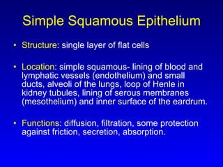
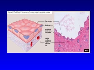
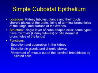
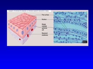
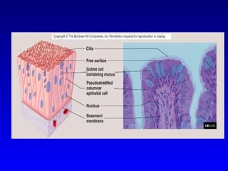
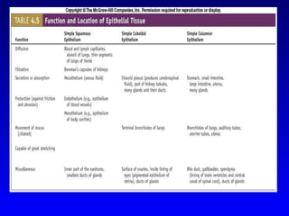
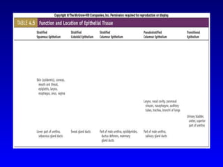
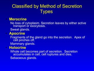
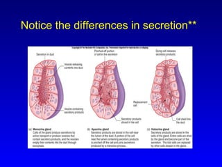
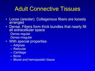
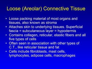
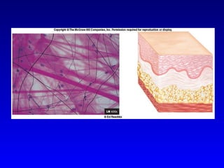
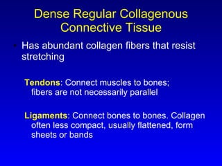
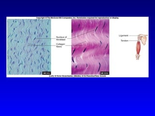
Histology
Objectives: 1. Introduce the four primary tissue types 2. Review the major characteristics of Epithelial tissue 3. Learn the major characteristics of Connective tissue 4. Extracellular Matrix 5. Introduction Muscle and Nervous Tissues 6. Inflammation and Tissue repair
Announcements: The Natural Science Network is hosting a student/faculty get together on Thursday (Aug 28).  This will be in the third floor antrum between 4:00pm – 5:00pm.  Check your schedule online to make sure  everything for the semester is in order. Remove and return one slide at a time in Lab. It is imperative that each slide return to it’s proper position from the box and place in which you retrieved it.
More Announcements:   4. The University is very informative regarding warnings pertaining to weather. Class will be held unless the University specifically alerts you not to attend or travel.
Connective Tissue Very abundant and diverse Found among every organ Contains cells separated by lots of extracellular matrix
Functions of Connective Tissue 1. Enclosing and Separating 2. Connecting tissues 3. Supporting and Moving other tissues 4. Storing 5. Cushioning and Insulating 6. Transporting 7. Protecting
1. Enclosing and Separating Sheets form capsules around other organs dense collagenous connective tissue envelope.  Kidney
2. Connecting tissues Strong cables – Tendons attach muscles to bone,  ligaments attach  bone to bone.
3. Supporting and Moving other tissues Bones and Joints
4. Storing Bones store minerals (calcium and phosphate)
5. Cushioning and Insulating Adipose tissue cushions and protects
6. Transporting Blood transporting – gases, nutrients, enzymes, hormones and cells of the immune system
7. Protection Cells of the immune system and blood along with bones protecting underlying structures from injury
Cell Surface Modifications Microvilli  (brush border):  Increase surface area absorption or secretion Cilia:   Move materials across cell surface
Quick Review of Epithelium Number of layers of cells Simple - one layer of cells. Each extends from basement membrane to the free surface Stratified - more than one layer. Shape of cells of the apical layer used to name the tissue. Includes transitional epithelium where the apical cell layers change shape depending upon distention of the organ which the tissue lines Pseudostratified - tissue appears to be stratified, but all cells contact basement membrane so it is in fact simple Shape of cells Squamous - flat, scale-like Cuboidal -  about equal in height and width Columnar - taller than wide
Functional Characteristics Simple : allows diffusion of gases, filtration of blood, secretion, absorption Stratified : protection, particularly against abrasion Squamous : allows diffusion or acts as filter Cuboidal and columnar : secretion or absorption. May include  goblet cells  that produce and secrete mucus .
Cell Surfaces Free surfaces of epithelium  Smooth : reduce friction Microvilli : increase surface area for absorption or secretion Stereocilia :  elongated microvilli for sensation and absorption Cilia : move materials across the surface  Folds : in transitional epithelium where organ must be able to change shape. Urinary system
Simple Squamous Epithelium Structure : single layer of flat cells Location : simple squamous- lining of blood and lymphatic vessels (endothelium) and small ducts, alveoli of the lungs, loop of Henle in kidney tubules, lining of serous membranes (mesothelium) and inner surface of the eardrum. Functions : diffusion, filtration, some protection against friction, secretion, absorption.
 
Simple Cuboidal Epithelium Locations:  Kidney tubules, glands and their ducts, choroid plexus of the brain, lining of terminal bronchioles of the lungs, and surface of the ovaries.  Structure :  single layer of cube-shaped cells; some types have microvilli (kidney tubules) or cilia (terminal bronchioles of the lungs) Functions: Secretion and absorption in the kidney Secretion in glands and choroid plexus Movement of  mucus out of the terminal bronchioles by ciliated cells
 
Simple Columnar Epithelium Location . Glands and some ducts, bronchioles of lungs, auditory tubes, uterus, uterine tubes, stomach, intestines, gallbladder, bile ducts and ventricles of the brain. Structure:  single layer of tall, narrow cells. Some have cilia (bronchioles of lungs, auditory tubes, uterine tubes, and uterus) or microvilli (intestine). Functions:   Movement of particles out of the bronchioles by ciliated cells Aids in the movement of oocytes through the uterine tubes by ciliated cells Secretion by glands of the stomach and the intestine Absorption by cells of the intestine.
 
Stratified Squamous Epithelium Locations :  Moist- mouth, throat, larynx, esophagus, anus, vagina, inferior urethra, and cornea Keratinized- skin Structure:  multiple layers of cells that are cuboidal in the basal layer and progressively flatten toward the surface. In moist, surface cells retain a nucleus and cytoplasm. In keratinized, surface cells are dead. Functions:  protection against abrasion, caustic chemicals, water loss, and infection.
 
Stratified Cuboidal Epithelium Locations:  sweat gland ducts, ovarian follicular cells, and salivary gland ducts Structure:  multiple layers of somewhat cube-shaped cells. Functions:  secretion, absorption and protection against infections.
 
Stratified Columnar Epithelium Locations:  mammary gland duct, larynx, portion of male urethra. Structure : multiple layers of cells with tall thin cells resting on layers of more cuboidal cells.  Cells ciliated in the larynx. Function : protection and secretion
 
Pseudostratified Columnar Epithelium Locations:  lining of nasal cavity, nasal sinuses, auditory tubes, pharynx, trachea, and bronchi of lungs. Structure:  all cells reach basement membrane. Appears stratified because nuclei are at various levels.  Almost always ciliated and associated with goblet (mucus-producing) cells. Functions:   Synthesize and secrete mucus onto the free surface Move mucus (or fluid) that contains foreign particles over the free surface and from passages
 
Transitional Epithelium Location:  lining of urinary bladder, ureters and superior urethra. Structure:  stratified; cells change shape depending upon amount of distention of the organ. Functions:  accommodates fluctuations in the volume of fluid in an organ or tube; protection against the caustic effects of urine.


Cell Connections Found on lateral and basal surfaces of cells Functions Form permeability layer Bind cells together Provide mechanism for intercellular communication Types Desmosomes Tight junctions Gap junctions
Let’s take a quick look. *
Cell Connections Desmosomes :  disk-shaped regions of cell membrane; often found in areas that are subjected to stress. Contain especially adhesive glycoproteins. Intermediate protein filaments extend into cytoplasm of cells.  Striated squamous epithelium of the skin. Hemidesmosomes : half of a desmosome; attach epithelial cells to basement membrane .
Tight Junctions :   hold cells together,  form permeability barrier. zonula adherens : between adjacent cells, weak glue, hold cells together. Simple epithelium. zonula occludens : permeability barrier, e.g., stomach and urinary bladder, chemicals cannot pass between cells.
Gap Junctions : protein channels that aid in intercellular communication. Allows ions and small molecules to pass through.  Coordinate function of cardiac and smooth muscle.  May help coordinate movement of cilia in ciliated types of epithelium.
Glands Epithelium  with supporting network of C.T. Two types of glands formed by infolding of epithelium:  Endocrine : no open contact with exterior; no ducts; produce hormones Exocrine : open contact maintained with exterior; ducts Exocrine glands classified either by structure or by the method of secretion Classified by structure Unicellular:  goblet cells -- mucous Multicellular: simple and compound – stomach, duodenum Page 121
Multicellular Exocrine Glands Classified on the basis of types of ducts or mode of secretion Types of ducts Simple : ducts with few branches Compound : ducts with many branches If ducts end in tubules or sac-like structures:  acini . Pancreas If ducts end in simple sacs:  alveoli .   Lungs
 
Classified by Method of Secretion Types  Merocrine No loss of cytoplasm. Secretion leaves by either active transport or exocytosis. Sweat glands.  Apocrine Fragments of the gland go into the secretion.  Apex of cell pinches off. Mammary glands.  Holocrine Whole cell becomes part of secretion.  Secretion accumulates in cell, cell ruptures and dies. Sebaceous glands.
Notice the differences in secretion**
Connective Tissue Abundant; found in every organ Consists of cells separated by extracellular matrix Many diverse types Performs variety of important functions
Functions of Connective Tissue Enclose organs as a capsule and separate organs into layers Connect tissues to one another.  Tendons and ligaments. Support and movement. Bones. Storage. Fat. Cushion and insulate. Fat. Transport. Blood. Protect.  Cells of the immune system .
Cells of Connective Tissue Specialized cells produce the extracellular matrix Descriptive word stems Blasts :   create the matrix, example osteoblast Cytes :   maintain the matrix, example chondrocyte Clasts :   break the matrix down for remodeling, example osteoclasts
Cells of Connective Tissue Adipose  or  fat cells  ( adipocytes ). Common in some tissues (dermis of skin); rare in some (cartilage) Mast cells . Common beneath membranes; along small blood vessels. Can release heparin, histamine, and proteolytic enzymes in response to injury. White blood cells (leukocytes) .  Respond to injury or infection Macrophages . Phagocytize or provide protection Fixed : stay in position in connective tissue Wandering : move by amoeboid movement through the connective tissue Undifferentiated mesenchyme   ( stem cells ). Have potential to differentiate into adult cell types
Extracellular Matrix Connective tissue consisting of 3 major components (1) protein fibers (2) ground substance consisting of nonfibrous protein and other molecules (3) Fluid
Very Important Functional Characteristics: Ability of bones and cartilage to bear weight (type II collagen) Tendons and ligaments to withstand tension (type I collagen) Skins dermis to withstand punctures, abrasions and other abuses (type I collagen)
Extracellular Matrix Protein fibers of the matrix Collagen .  Most common  protein in body;  strong, flexible,  Inelastic Fibril vs. Fiber
Reticular . Fill spaces between tissues and organs. Fine collagenous, form branching networks (type III collagen) Very fine – not considered chemically distinct Very short and branch Not as strong yet networks fill spaces between tissues
Elastic .  Returns to its original shape after distension or compression. Contains molecules of protein elastin that resemble coiled springs; molecules are cross-linked
Other Matrix Molecules Most common molecules are called the and include:  ground substance   Hyaluronic acid :  polysaccharide. Good lubricant. Vitreous humor of eye. Proteoglycans : protein and polysaccharides. 80 -100 polysaccharides known as glycosaminoglycans.  Protein part attaches to hyaluronic acid. Trap large amounts of water. Adhesive molecules : hold proteoglycan aggregates together.  Chondronectin  in cartilage,  osteonectin  in bone,  fibronectin  in fibrous connective tissue.
GAGs are unbranched polymers of repeated disaccharide derivatives, including amino sugars, sulfated acetylamino sugars and uronic acids.
 
 
Embryonic Connective Tissue Mesenchyme : source of all adult connective tissue.  Forms primarily from mesoderm Delicate collagen fibers embedded in semifluid matrix  Mucus :  found only in the umbilical cord. Wharton’s jelly.
 
 
Objectives: 09/03/08 1.  Chapter 4:  Review the structure and function of the major connective tissues 2. Introduce the 3 major muscle types and neuralgia cells 3. Tissue repair/aging  4.  Chapter 2:  The Chemical Basis of Life 5. Basic chemistry 6. Electrons and bonding 7. Radioactive Imagery 8. Intermolecular forces
Adult Connective Tissues Loose (areolar). Collagenous fibers are loosely arranged Dense. Fibers form thick bundles that nearly fill all extracellular space Dense regular Dense irregular With special properties Adipose Reticular Cartilage Bone Blood and hemopoietic tissue
Loose (Areolar) Connective Tissue Loose packing material of most organs and tissues, also known as stroma Attaches skin to underlying tissues. Superficial fascia = subcutaneous layer = hypodermis Contains collagen, reticular, elastic fibers and all five types of cells Often seen in association with other types of C.T., like reticular tissue and fat Cells include fibroblasts, mast cells, lymphocytes, adipose cells, macrophages
 
Dense Regular Collagenous Connective Tissue Has abundant collagen fibers that resist stretching Tendons : Connect muscles to bones; fibers are not necessarily parallel Ligaments : Connect bones to bones. Collagen often less compact, usually flattened, form sheets or bands
 
Dense Regular Elastic C.T. Ligaments in vocal folds;  nuchal ligament Collagen fibers give strength (for when you shout), but elastic fibers are more prevalent
 
Dense Irregular Collagenous Connective Tissue Protein fibers arranged in a randomly oriented network Forms innermost layer of the dermis of the skin, scars, capsules of kidney and spleen
 
Dense Irregular Elastic Connective Tissue Bundles and sheets of collagenous and elastic fibers oriented in multiple directions In walls of elastic arteries Strong, yet elastic
 
Connective Tissue with Special Properties: Adipose Predominant cells are  adipocytes Yellow  ( white ).  Most abundant type, has a wide distribution. White at birth and yellows with age.   Carotenes come from plants and can be metabolized into vitamin A.  Scant ring of cytoplasm surrounding single large lipid droplet. Nuclei flattened and eccentric. Brown . Found only in specific areas of body: axillae, neck and near kidneys  Cells are polygonal in shape, have a considerable volume of cytoplasm and contain multiple lipid droplets of varying size. Nuclei are round and almost centrally located.
 
Connective Tissue with Special Properties: Reticular Tissue Forms superstructure of lymphatic and hemopoietic tissues  Network of fine reticular fibers and reticular cells.  Spaces between cells contain white cells and dendritic cells
 
Connective Tissue with Special Properties:  Cartilage Composed of chondrocytes located in matrix-surrounded spaces called lacunae.  Type of cartilage determined by components of the matrix.  Firm consistency.  Ground substance: Proteoglycans and hyaluronic acid complexed together trap large amounts of water. Tissue can spring back after being compressed. Avascular and no nerve supply. Heals slowly. Perichondrium . Dense irregular connective tissue that surrounds cartilage. Fibroblasts of perichondrium can differentiate into chondroblasts.  Types of cartilage Hyaline Fibrocartilage Elastic
Hyaline Cartilage Structure: large amount of collagen fibers evenly distributed in proteoglycan matrix. Smooth surface in articulations Locations: Found in areas for strong support and some flexibility: rib cage, trachea, and bronchi In embryo forms most of skeleton Involved in growth that increases bone length
 
Fibrocartilage Structure:  thick collagen fibers distributed in  proteoglycan matrix; slightly compressible and very tough Locations : found in areas of body where a great deal of pressure is applied to joints Knee, jaw, between vertebrae
 
Elastic Cartilage Structure : elastic and collagen fibers embedded in proteoglycans. Rigid but elastic properties Locations:  external ears and epiglottis
 
Connective Tissue with Special Properties:   Bone Hard connective tissue composed of living cells ( osteocytes ) and mineralized matrix Matrix: gives strength and rigidity; allows bone to support and protect other tissues and organs Organic: collagen fibers Inorganic: hydroxyapatite (Ca plus PO 4 ) Osteocytes located in lacunae Types Cancellous or spongy bone Compact bone
Bone, cont Cancellous   or  spongy  bone : trabeculae of bone with spaces between. Looks like a sponge. Found inside bones. Compact  bone : arranged in concentric circle layers around a central canal that contains a blood vessel. Found on periphery of bones.
 
Connective Tissue with Special Properties: Blood Matrix:  plasma Liquid and lacks fibers. Matrix formed by other tissues, unlike other types of connective tissue. Moves through vessels, but both fluid and cells can move in/out of the vessels. Formed elements : red cells, white cells, and platelets Hemopoietic tissue Forms blood cells Two types of bone marrow Yellow Red
 
Hemopoietic Tissue Forms blood cells Found in bone marrow  Types of bone marrow Red : hemopoietic tissue surrounded by a framework of reticular fibers. Produces red and white cells Yellow : yellow adipose tissue As children grow, yellow marrow replaces much of red marrow
 
Muscle Tissue Characteristics Contracts or shortens with force Moves entire body and pumps blood Types Skeletal : most attached to skeleton, but some attached to other types of connective tissue. Striated and voluntary. Cardiac : muscle of the heart. Striated and involuntary. Smooth : muscle associated with tubular structures and with the skin. Nonstriated and involuntary .
 
Skeletal Muscle
Cardiac Muscle
Smooth Muscle
Nervous Tissue: Neurons Neurons  or  nerve cells  have the ability to produce action potentials Parts: Cell body : contains nucleus Axon : cell process; conducts impulses away from cell body; usually only one per neuron Dendrite : cell process; receive impulses from other neurons; can be many per neuron Types: Multipolar ,  bipolar , and  unipolar
Neurons
Nervous Tissue: Neuroglia Support cells of the brain,  spinal cord  and nerves Nourish, protect,  and insulate  neurons
 
Membranes Mucous Line cavities that open to the outside of body Secrete mucus Contains epithelium with goblet cells,  basement membrane, lamina propria (sometimes with smooth muscle) Found in respiratory, digestive, urinary and reproductive systems. Serous .  simple squamous epithelium called  mesothelium , basement membrane, thin layer of loose C.T. Line cavities not open to exterior Pericardial, pleural, peritoneal Synovial Line freely movable joints Produce fluid rich in hyaluronic acid
 
Inflammation *** Responds  to tissue damage or with an immune response Manifestations Redness, heat, swelling, pain, disturbance of function Mediators Include histamine, kinins, prostaglandins, leukotrienes Stimulate pain receptor and increase blood vessel permeability as well movement of WBCs to affected area.
 
Tissue Repair Substitution  of dead/damaged cells by viable/functional cells Types of cells Labile :  capable of mitosis through life. skin, mucous membranes, hemopoietic tissue, lymphatic tissue Stable : no mitosis after growth ends, but can divide after injury. Liver, pancreas, endocrine cells Permanent : if killed, replaced by a different type of cell.  Limited regenerative ability. nervous, skeletal and cardiac muscle
Skin Repair Primary union : Edges of wound are close together Wound fills with blood Clot forms: fibrin threads start to contract; pull edges together Scab Inflammatory response; pus forms as white cells die Granulation tissue. Replaces clot, delicate C.T. composed of fibroblasts, collagen fibers, capillaries Scar. Formed from granulation tissue. Tissue turns from red to white as capillaries are forced out. Secondary union :  Edges of wound  are not closed; greater chance of infection Clot may not close gap Inflammatory response greater Wound contraction occurs leading to greater scarring
 
Tissue and Aging Cells divide more slowly  Collagen fibers become more irregular in structure, though they may increase in number Tendons and ligaments become less flexible and more fragile Elastic fibers fragment, bind to calcium ions, and become less elastic Arterial walls and elastic ligaments become less elastic Changes in collagen and elastin result in  Atherosclerosis and reduced blood supply to tissues Wrinkling of the skin Increased tendency for bones to break Rate of blood cell synthesis declines in the elderly Injuries don’t heal as readily

Anatomy :O

  • 1.
  • 2.
    Objectives: 1. Introducethe four primary tissue types 2. Review the major characteristics of Epithelial tissue 3. Learn the major characteristics of Connective tissue 4. Extracellular Matrix 5. Introduction Muscle and Nervous Tissues 6. Inflammation and Tissue repair
  • 3.
    Announcements: The NaturalScience Network is hosting a student/faculty get together on Thursday (Aug 28).  This will be in the third floor antrum between 4:00pm – 5:00pm.  Check your schedule online to make sure everything for the semester is in order. Remove and return one slide at a time in Lab. It is imperative that each slide return to it’s proper position from the box and place in which you retrieved it.
  • 4.
    More Announcements: 4. The University is very informative regarding warnings pertaining to weather. Class will be held unless the University specifically alerts you not to attend or travel.
  • 5.
    Connective Tissue Veryabundant and diverse Found among every organ Contains cells separated by lots of extracellular matrix
  • 6.
    Functions of ConnectiveTissue 1. Enclosing and Separating 2. Connecting tissues 3. Supporting and Moving other tissues 4. Storing 5. Cushioning and Insulating 6. Transporting 7. Protecting
  • 7.
    1. Enclosing andSeparating Sheets form capsules around other organs dense collagenous connective tissue envelope. Kidney
  • 8.
    2. Connecting tissuesStrong cables – Tendons attach muscles to bone, ligaments attach bone to bone.
  • 9.
    3. Supporting andMoving other tissues Bones and Joints
  • 10.
    4. Storing Bonesstore minerals (calcium and phosphate)
  • 11.
    5. Cushioning andInsulating Adipose tissue cushions and protects
  • 12.
    6. Transporting Bloodtransporting – gases, nutrients, enzymes, hormones and cells of the immune system
  • 13.
    7. Protection Cellsof the immune system and blood along with bones protecting underlying structures from injury
  • 14.
    Cell Surface ModificationsMicrovilli (brush border): Increase surface area absorption or secretion Cilia: Move materials across cell surface
  • 15.
    Quick Review ofEpithelium Number of layers of cells Simple - one layer of cells. Each extends from basement membrane to the free surface Stratified - more than one layer. Shape of cells of the apical layer used to name the tissue. Includes transitional epithelium where the apical cell layers change shape depending upon distention of the organ which the tissue lines Pseudostratified - tissue appears to be stratified, but all cells contact basement membrane so it is in fact simple Shape of cells Squamous - flat, scale-like Cuboidal - about equal in height and width Columnar - taller than wide
  • 16.
    Functional Characteristics Simple: allows diffusion of gases, filtration of blood, secretion, absorption Stratified : protection, particularly against abrasion Squamous : allows diffusion or acts as filter Cuboidal and columnar : secretion or absorption. May include goblet cells that produce and secrete mucus .
  • 17.
    Cell Surfaces Freesurfaces of epithelium Smooth : reduce friction Microvilli : increase surface area for absorption or secretion Stereocilia : elongated microvilli for sensation and absorption Cilia : move materials across the surface Folds : in transitional epithelium where organ must be able to change shape. Urinary system
  • 18.
    Simple Squamous EpitheliumStructure : single layer of flat cells Location : simple squamous- lining of blood and lymphatic vessels (endothelium) and small ducts, alveoli of the lungs, loop of Henle in kidney tubules, lining of serous membranes (mesothelium) and inner surface of the eardrum. Functions : diffusion, filtration, some protection against friction, secretion, absorption.
  • 19.
  • 20.
    Simple Cuboidal EpitheliumLocations: Kidney tubules, glands and their ducts, choroid plexus of the brain, lining of terminal bronchioles of the lungs, and surface of the ovaries. Structure : single layer of cube-shaped cells; some types have microvilli (kidney tubules) or cilia (terminal bronchioles of the lungs) Functions: Secretion and absorption in the kidney Secretion in glands and choroid plexus Movement of mucus out of the terminal bronchioles by ciliated cells
  • 21.
  • 22.
    Simple Columnar EpitheliumLocation . Glands and some ducts, bronchioles of lungs, auditory tubes, uterus, uterine tubes, stomach, intestines, gallbladder, bile ducts and ventricles of the brain. Structure: single layer of tall, narrow cells. Some have cilia (bronchioles of lungs, auditory tubes, uterine tubes, and uterus) or microvilli (intestine). Functions: Movement of particles out of the bronchioles by ciliated cells Aids in the movement of oocytes through the uterine tubes by ciliated cells Secretion by glands of the stomach and the intestine Absorption by cells of the intestine.
  • 23.
  • 24.
    Stratified Squamous EpitheliumLocations : Moist- mouth, throat, larynx, esophagus, anus, vagina, inferior urethra, and cornea Keratinized- skin Structure: multiple layers of cells that are cuboidal in the basal layer and progressively flatten toward the surface. In moist, surface cells retain a nucleus and cytoplasm. In keratinized, surface cells are dead. Functions: protection against abrasion, caustic chemicals, water loss, and infection.
  • 25.
  • 26.
    Stratified Cuboidal EpitheliumLocations: sweat gland ducts, ovarian follicular cells, and salivary gland ducts Structure: multiple layers of somewhat cube-shaped cells. Functions: secretion, absorption and protection against infections.
  • 27.
  • 28.
    Stratified Columnar EpitheliumLocations: mammary gland duct, larynx, portion of male urethra. Structure : multiple layers of cells with tall thin cells resting on layers of more cuboidal cells. Cells ciliated in the larynx. Function : protection and secretion
  • 29.
  • 30.
    Pseudostratified Columnar EpitheliumLocations: lining of nasal cavity, nasal sinuses, auditory tubes, pharynx, trachea, and bronchi of lungs. Structure: all cells reach basement membrane. Appears stratified because nuclei are at various levels. Almost always ciliated and associated with goblet (mucus-producing) cells. Functions: Synthesize and secrete mucus onto the free surface Move mucus (or fluid) that contains foreign particles over the free surface and from passages
  • 31.
  • 32.
    Transitional Epithelium Location: lining of urinary bladder, ureters and superior urethra. Structure: stratified; cells change shape depending upon amount of distention of the organ. Functions: accommodates fluctuations in the volume of fluid in an organ or tube; protection against the caustic effects of urine.
  • 33.
  • 34.
  • 35.
  • 36.
    Cell Connections Foundon lateral and basal surfaces of cells Functions Form permeability layer Bind cells together Provide mechanism for intercellular communication Types Desmosomes Tight junctions Gap junctions
  • 37.
    Let’s take aquick look. *
  • 38.
    Cell Connections Desmosomes: disk-shaped regions of cell membrane; often found in areas that are subjected to stress. Contain especially adhesive glycoproteins. Intermediate protein filaments extend into cytoplasm of cells. Striated squamous epithelium of the skin. Hemidesmosomes : half of a desmosome; attach epithelial cells to basement membrane .
  • 39.
    Tight Junctions : hold cells together, form permeability barrier. zonula adherens : between adjacent cells, weak glue, hold cells together. Simple epithelium. zonula occludens : permeability barrier, e.g., stomach and urinary bladder, chemicals cannot pass between cells.
  • 40.
    Gap Junctions :protein channels that aid in intercellular communication. Allows ions and small molecules to pass through. Coordinate function of cardiac and smooth muscle. May help coordinate movement of cilia in ciliated types of epithelium.
  • 41.
    Glands Epithelium with supporting network of C.T. Two types of glands formed by infolding of epithelium: Endocrine : no open contact with exterior; no ducts; produce hormones Exocrine : open contact maintained with exterior; ducts Exocrine glands classified either by structure or by the method of secretion Classified by structure Unicellular: goblet cells -- mucous Multicellular: simple and compound – stomach, duodenum Page 121
  • 42.
    Multicellular Exocrine GlandsClassified on the basis of types of ducts or mode of secretion Types of ducts Simple : ducts with few branches Compound : ducts with many branches If ducts end in tubules or sac-like structures: acini . Pancreas If ducts end in simple sacs: alveoli . Lungs
  • 43.
  • 44.
    Classified by Methodof Secretion Types Merocrine No loss of cytoplasm. Secretion leaves by either active transport or exocytosis. Sweat glands. Apocrine Fragments of the gland go into the secretion. Apex of cell pinches off. Mammary glands. Holocrine Whole cell becomes part of secretion. Secretion accumulates in cell, cell ruptures and dies. Sebaceous glands.
  • 45.
    Notice the differencesin secretion**
  • 46.
    Connective Tissue Abundant;found in every organ Consists of cells separated by extracellular matrix Many diverse types Performs variety of important functions
  • 47.
    Functions of ConnectiveTissue Enclose organs as a capsule and separate organs into layers Connect tissues to one another. Tendons and ligaments. Support and movement. Bones. Storage. Fat. Cushion and insulate. Fat. Transport. Blood. Protect. Cells of the immune system .
  • 48.
    Cells of ConnectiveTissue Specialized cells produce the extracellular matrix Descriptive word stems Blasts : create the matrix, example osteoblast Cytes : maintain the matrix, example chondrocyte Clasts : break the matrix down for remodeling, example osteoclasts
  • 49.
    Cells of ConnectiveTissue Adipose or fat cells ( adipocytes ). Common in some tissues (dermis of skin); rare in some (cartilage) Mast cells . Common beneath membranes; along small blood vessels. Can release heparin, histamine, and proteolytic enzymes in response to injury. White blood cells (leukocytes) . Respond to injury or infection Macrophages . Phagocytize or provide protection Fixed : stay in position in connective tissue Wandering : move by amoeboid movement through the connective tissue Undifferentiated mesenchyme ( stem cells ). Have potential to differentiate into adult cell types
  • 50.
    Extracellular Matrix Connectivetissue consisting of 3 major components (1) protein fibers (2) ground substance consisting of nonfibrous protein and other molecules (3) Fluid
  • 51.
    Very Important FunctionalCharacteristics: Ability of bones and cartilage to bear weight (type II collagen) Tendons and ligaments to withstand tension (type I collagen) Skins dermis to withstand punctures, abrasions and other abuses (type I collagen)
  • 52.
    Extracellular Matrix Proteinfibers of the matrix Collagen . Most common protein in body; strong, flexible, Inelastic Fibril vs. Fiber
  • 53.
    Reticular . Fillspaces between tissues and organs. Fine collagenous, form branching networks (type III collagen) Very fine – not considered chemically distinct Very short and branch Not as strong yet networks fill spaces between tissues
  • 54.
    Elastic . Returns to its original shape after distension or compression. Contains molecules of protein elastin that resemble coiled springs; molecules are cross-linked
  • 55.
    Other Matrix MoleculesMost common molecules are called the and include: ground substance Hyaluronic acid : polysaccharide. Good lubricant. Vitreous humor of eye. Proteoglycans : protein and polysaccharides. 80 -100 polysaccharides known as glycosaminoglycans. Protein part attaches to hyaluronic acid. Trap large amounts of water. Adhesive molecules : hold proteoglycan aggregates together. Chondronectin in cartilage, osteonectin in bone, fibronectin in fibrous connective tissue.
  • 56.
    GAGs are unbranchedpolymers of repeated disaccharide derivatives, including amino sugars, sulfated acetylamino sugars and uronic acids.
  • 57.
  • 58.
  • 59.
    Embryonic Connective TissueMesenchyme : source of all adult connective tissue. Forms primarily from mesoderm Delicate collagen fibers embedded in semifluid matrix Mucus : found only in the umbilical cord. Wharton’s jelly.
  • 60.
  • 61.
  • 62.
    Objectives: 09/03/08 1. Chapter 4: Review the structure and function of the major connective tissues 2. Introduce the 3 major muscle types and neuralgia cells 3. Tissue repair/aging 4. Chapter 2: The Chemical Basis of Life 5. Basic chemistry 6. Electrons and bonding 7. Radioactive Imagery 8. Intermolecular forces
  • 63.
    Adult Connective TissuesLoose (areolar). Collagenous fibers are loosely arranged Dense. Fibers form thick bundles that nearly fill all extracellular space Dense regular Dense irregular With special properties Adipose Reticular Cartilage Bone Blood and hemopoietic tissue
  • 64.
    Loose (Areolar) ConnectiveTissue Loose packing material of most organs and tissues, also known as stroma Attaches skin to underlying tissues. Superficial fascia = subcutaneous layer = hypodermis Contains collagen, reticular, elastic fibers and all five types of cells Often seen in association with other types of C.T., like reticular tissue and fat Cells include fibroblasts, mast cells, lymphocytes, adipose cells, macrophages
  • 65.
  • 66.
    Dense Regular CollagenousConnective Tissue Has abundant collagen fibers that resist stretching Tendons : Connect muscles to bones; fibers are not necessarily parallel Ligaments : Connect bones to bones. Collagen often less compact, usually flattened, form sheets or bands
  • 67.
  • 68.
    Dense Regular ElasticC.T. Ligaments in vocal folds; nuchal ligament Collagen fibers give strength (for when you shout), but elastic fibers are more prevalent
  • 69.
  • 70.
    Dense Irregular CollagenousConnective Tissue Protein fibers arranged in a randomly oriented network Forms innermost layer of the dermis of the skin, scars, capsules of kidney and spleen
  • 71.
  • 72.
    Dense Irregular ElasticConnective Tissue Bundles and sheets of collagenous and elastic fibers oriented in multiple directions In walls of elastic arteries Strong, yet elastic
  • 73.
  • 74.
    Connective Tissue withSpecial Properties: Adipose Predominant cells are adipocytes Yellow ( white ). Most abundant type, has a wide distribution. White at birth and yellows with age. Carotenes come from plants and can be metabolized into vitamin A. Scant ring of cytoplasm surrounding single large lipid droplet. Nuclei flattened and eccentric. Brown . Found only in specific areas of body: axillae, neck and near kidneys Cells are polygonal in shape, have a considerable volume of cytoplasm and contain multiple lipid droplets of varying size. Nuclei are round and almost centrally located.
  • 75.
  • 76.
    Connective Tissue withSpecial Properties: Reticular Tissue Forms superstructure of lymphatic and hemopoietic tissues Network of fine reticular fibers and reticular cells. Spaces between cells contain white cells and dendritic cells
  • 77.
  • 78.
    Connective Tissue withSpecial Properties: Cartilage Composed of chondrocytes located in matrix-surrounded spaces called lacunae. Type of cartilage determined by components of the matrix. Firm consistency. Ground substance: Proteoglycans and hyaluronic acid complexed together trap large amounts of water. Tissue can spring back after being compressed. Avascular and no nerve supply. Heals slowly. Perichondrium . Dense irregular connective tissue that surrounds cartilage. Fibroblasts of perichondrium can differentiate into chondroblasts. Types of cartilage Hyaline Fibrocartilage Elastic
  • 79.
    Hyaline Cartilage Structure:large amount of collagen fibers evenly distributed in proteoglycan matrix. Smooth surface in articulations Locations: Found in areas for strong support and some flexibility: rib cage, trachea, and bronchi In embryo forms most of skeleton Involved in growth that increases bone length
  • 80.
  • 81.
    Fibrocartilage Structure: thick collagen fibers distributed in proteoglycan matrix; slightly compressible and very tough Locations : found in areas of body where a great deal of pressure is applied to joints Knee, jaw, between vertebrae
  • 82.
  • 83.
    Elastic Cartilage Structure: elastic and collagen fibers embedded in proteoglycans. Rigid but elastic properties Locations: external ears and epiglottis
  • 84.
  • 85.
    Connective Tissue withSpecial Properties: Bone Hard connective tissue composed of living cells ( osteocytes ) and mineralized matrix Matrix: gives strength and rigidity; allows bone to support and protect other tissues and organs Organic: collagen fibers Inorganic: hydroxyapatite (Ca plus PO 4 ) Osteocytes located in lacunae Types Cancellous or spongy bone Compact bone
  • 86.
    Bone, cont Cancellous or spongy bone : trabeculae of bone with spaces between. Looks like a sponge. Found inside bones. Compact bone : arranged in concentric circle layers around a central canal that contains a blood vessel. Found on periphery of bones.
  • 87.
  • 88.
    Connective Tissue withSpecial Properties: Blood Matrix: plasma Liquid and lacks fibers. Matrix formed by other tissues, unlike other types of connective tissue. Moves through vessels, but both fluid and cells can move in/out of the vessels. Formed elements : red cells, white cells, and platelets Hemopoietic tissue Forms blood cells Two types of bone marrow Yellow Red
  • 89.
  • 90.
    Hemopoietic Tissue Formsblood cells Found in bone marrow Types of bone marrow Red : hemopoietic tissue surrounded by a framework of reticular fibers. Produces red and white cells Yellow : yellow adipose tissue As children grow, yellow marrow replaces much of red marrow
  • 91.
  • 92.
    Muscle Tissue CharacteristicsContracts or shortens with force Moves entire body and pumps blood Types Skeletal : most attached to skeleton, but some attached to other types of connective tissue. Striated and voluntary. Cardiac : muscle of the heart. Striated and involuntary. Smooth : muscle associated with tubular structures and with the skin. Nonstriated and involuntary .
  • 93.
  • 94.
  • 95.
  • 96.
  • 97.
    Nervous Tissue: NeuronsNeurons or nerve cells have the ability to produce action potentials Parts: Cell body : contains nucleus Axon : cell process; conducts impulses away from cell body; usually only one per neuron Dendrite : cell process; receive impulses from other neurons; can be many per neuron Types: Multipolar , bipolar , and unipolar
  • 98.
  • 99.
    Nervous Tissue: NeurogliaSupport cells of the brain, spinal cord and nerves Nourish, protect, and insulate neurons
  • 100.
  • 101.
    Membranes Mucous Linecavities that open to the outside of body Secrete mucus Contains epithelium with goblet cells, basement membrane, lamina propria (sometimes with smooth muscle) Found in respiratory, digestive, urinary and reproductive systems. Serous . simple squamous epithelium called mesothelium , basement membrane, thin layer of loose C.T. Line cavities not open to exterior Pericardial, pleural, peritoneal Synovial Line freely movable joints Produce fluid rich in hyaluronic acid
  • 102.
  • 103.
    Inflammation *** Responds to tissue damage or with an immune response Manifestations Redness, heat, swelling, pain, disturbance of function Mediators Include histamine, kinins, prostaglandins, leukotrienes Stimulate pain receptor and increase blood vessel permeability as well movement of WBCs to affected area.
  • 104.
  • 105.
    Tissue Repair Substitution of dead/damaged cells by viable/functional cells Types of cells Labile : capable of mitosis through life. skin, mucous membranes, hemopoietic tissue, lymphatic tissue Stable : no mitosis after growth ends, but can divide after injury. Liver, pancreas, endocrine cells Permanent : if killed, replaced by a different type of cell. Limited regenerative ability. nervous, skeletal and cardiac muscle
  • 106.
    Skin Repair Primaryunion : Edges of wound are close together Wound fills with blood Clot forms: fibrin threads start to contract; pull edges together Scab Inflammatory response; pus forms as white cells die Granulation tissue. Replaces clot, delicate C.T. composed of fibroblasts, collagen fibers, capillaries Scar. Formed from granulation tissue. Tissue turns from red to white as capillaries are forced out. Secondary union : Edges of wound are not closed; greater chance of infection Clot may not close gap Inflammatory response greater Wound contraction occurs leading to greater scarring
  • 107.
  • 108.
    Tissue and AgingCells divide more slowly Collagen fibers become more irregular in structure, though they may increase in number Tendons and ligaments become less flexible and more fragile Elastic fibers fragment, bind to calcium ions, and become less elastic Arterial walls and elastic ligaments become less elastic Changes in collagen and elastin result in Atherosclerosis and reduced blood supply to tissues Wrinkling of the skin Increased tendency for bones to break Rate of blood cell synthesis declines in the elderly Injuries don’t heal as readily