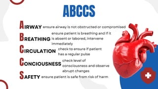
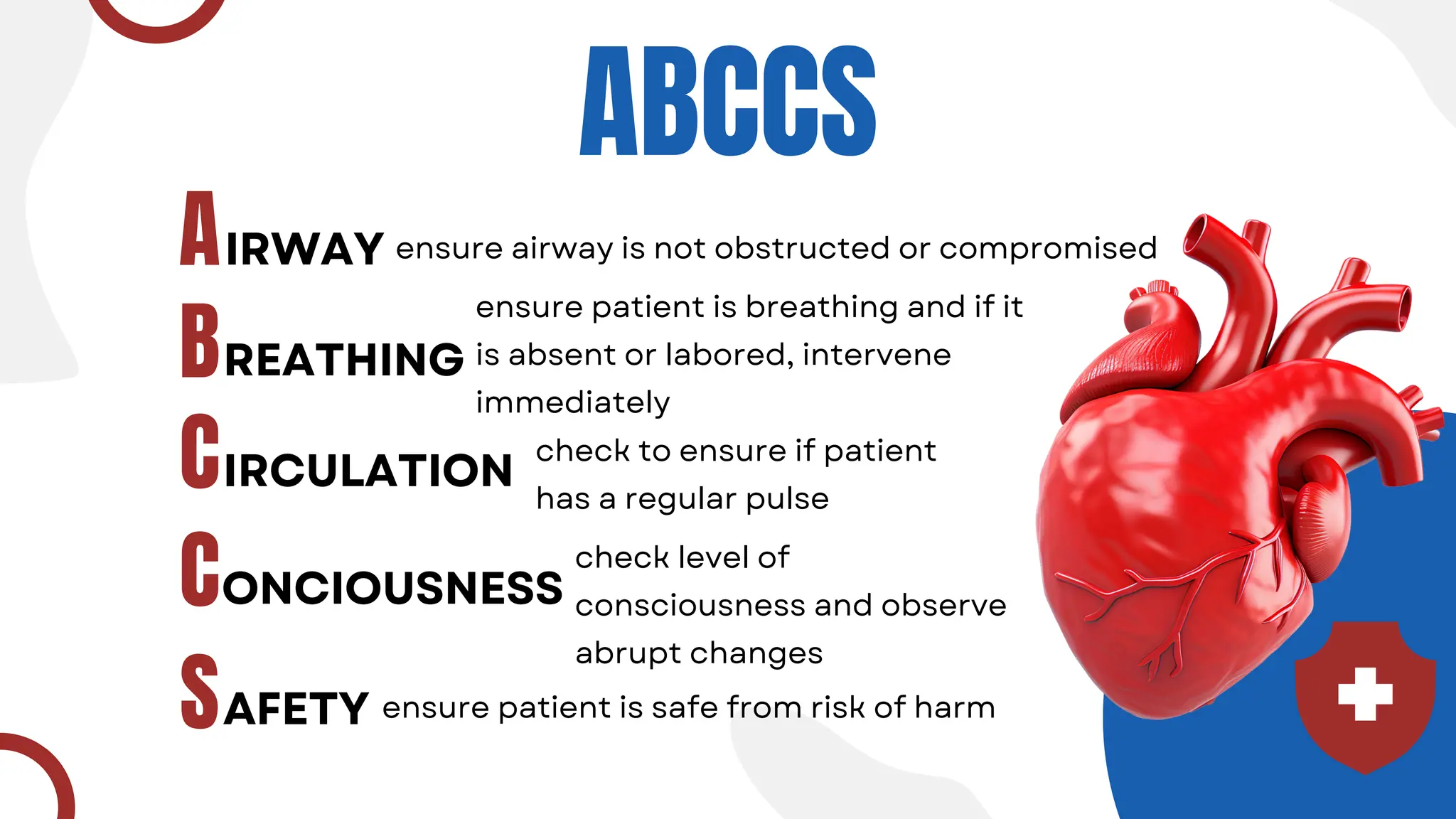
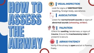
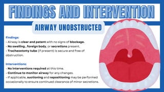
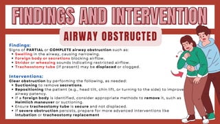
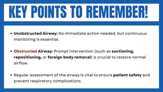
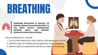
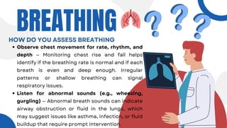
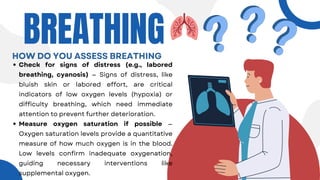
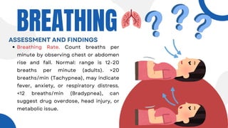
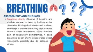
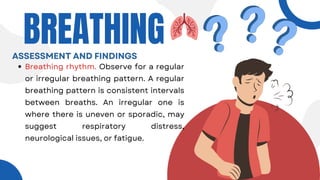
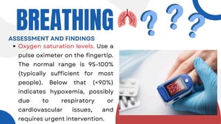
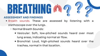
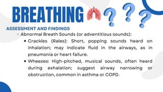
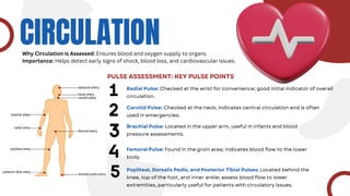
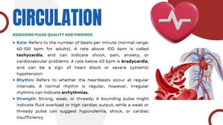
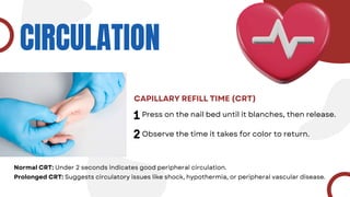
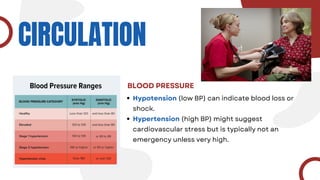
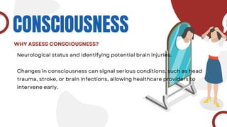
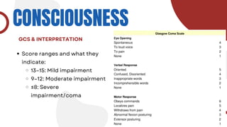
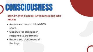
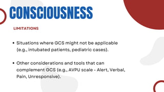
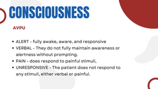
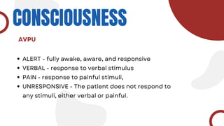
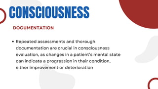
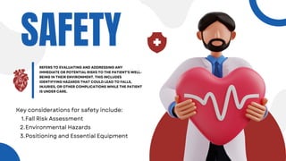
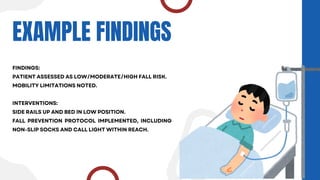
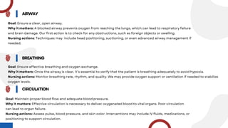
ABCCS
IRWAY
A
B
C
C
S
REATHING
IRCULATION
ONCIOUSNESS
AFETY
ensure airway is not obstructed or compromised
ensure patient is breathing and if it
is absent or labored, intervene
immediately
check to ensure if patient
has a regular pulse
check level of
consciousness and observe
abrupt changes
ensure patient is safe from risk of harm
CHECKLIST: INITAL AND EMERGENCY ASSESSMENT
AIRWAY Is the patient’s airway compromised?
Does the patient’s position need to be changed?
BREATHING
CIRCULATION
CONCIOUSNESS
SAFETY
Is the patient’s respiratory rate normal?
Does the patient breathe with ease or is it labored?
Is radial pulse present?
Is the patient’s skin color and temperature
normal?
Is the patient conscious?
What is patient’s level of consciousness?
Is patient free from any risk of harm or
injury?
AIRWAY
AIRWAY
ASSESSMENT
ASSESSMENT
OBJECTIVE
To assess the patient's airway for
patency and detect any obstruction
or complications affecting breathing.
HOW TO
HOW TO
ASSESS
ASSESS
THE
THE
AIRWAY
AIRWAY
VISUALINSPECTION
Look for signs of OBSTRUCTION
(swelling, foreign body, secretions).
AUSCULTATION
Listen for normal breath sounds or signs of
abnormal sounds (wheezing, stridor).
PALPATION
Check for swelling, tenderness, or signs of
trauma. Ensure the tracheostomy tube (if
present) is secure.
AIRWAYPATENCY
Verify if the airway is open and air is flowing
freely.
FINDINGS AND INTERVENTION
AIRWAY UNOBSTRUCTED
Findings:
- Airway is clear and patent with no signs of blockage.
- No swelling , foreign body, or secretions present.
- Tracheostomy tube (if present) is secure and free of
obstruction.
Interventions:
- No interventions required at this time.
- Continue to monitor airway for any changes.
- If applicable, suctioning and repositioning may be performed
occasionally to ensure continued clearance of minor secretions.
Findings:
Signs of PARTIAL or COMPLETE airway obstruction such as:
Swelling in the airway, causing narrowing.
Foreign body or secretions blocking airflow.
Stridor or wheezing sounds indicating restricted airflow.
Tracheostomy tube (if present) may be displaced or clogged.
Interventions:
Clear obstruction by performing the following, as needed:
Suctioning to remove secretions.
Repositioning the patient (e.g., head tilt, chin lift, or turning to the side) to improve
airway patency.
If a foreign body is identified, consider appropriate methods to remove it, such as
Heimlich maneuver or suctioning.
Ensure tracheostomy tube is secure and not displaced.
If severe obstruction persists, prepare for more advanced interventions like
intubation or tracheostomy replacement
FINDINGS AND INTERVENTION
AIRWAY OBSTRUCTED
KEY POINTS TO REMEMBER!
Unobstructed Airway: No immediate action needed, but continuous
monitoring is essential.
Obstructed Airway: Prompt intervention (such as suctioning,
repositioning, or foreign body removal) is crucial to restore normal
airflow.
Regular assessment of the airway is vital to ensure patient safety and
prevent respiratory complications.
BREATHING
ASSESSING BREATHING IS CRUCIAL TO
ENSURE ADEQUATE OXYGEN DELIVERY TO
THE BODY, DETECT RESPIRATORY
DISTRESS EARLY, AND PREVENT
COMPLICATIONS FROM HYPOXIA.
Key considerations include:
Ensure the respiratory rate, rhythm, and depth are normal.
1.
Identify signs of inadequate oxygenation (e.g., cyanosis).
2.
Detect signs of respiratory distress or labored breathing.
3.
Observe chest movement for rate, rhythm, and
depth — Monitoring chest rise and fall helps
identify if the breathing rate is normal and if each
breath is even and deep enough. Irregular
patterns or shallow breathing can signal
respiratory issues.
Listen for abnormal sounds (e.g., wheezing,
gurgling) — Abnormal breath sounds can indicate
airway obstruction or fluid in the lungs, which
may suggest issues like asthma, infection, or fluid
buildup that require prompt intervention.
BREATHING
HOW DO YOU ASSESS BREATHING
Check for signs of distress (e.g., labored
breathing, cyanosis) — Signs of distress, like
bluish skin or labored effort, are critical
indicators of low oxygen levels (hypoxia) or
difficulty breathing, which need immediate
attention to prevent further deterioration.
Measure oxygen saturation if possible —
Oxygen saturation levels provide a quantitative
measure of how much oxygen is in the blood.
Low levels confirm inadequate oxygenation,
guiding necessary interventions like
supplemental oxygen.
BREATHING
HOW DO YOU ASSESS BREATHING
BREATHING
Breathing Rate. Count breaths per
minute by observing chest or abdomen
rise and fall. Normal: range is 12-20
breaths per minute (adults). >20
breaths/min (Tachypnea), may indicate
fever, anxiety, or respiratory distress.
<12 breaths/min (Bradypnea), can
suggest drug overdose, head injury, or
metabolic issue.
ASSESSMENT AND FINDINGS
BREATHING
Breathing depth. Observe if breaths are
shallow, normal, or deep by looking at the
chest rise. Findings include normal, shallow,
and deep. A shallow breathing depth shows
minimal chest movement, could indicate
pain or respiratory compromise. A deep
breathing depth shows exaggerated chest
movement, possibly due to acidosis or
stress.
ASSESSMENT AND FINDINGS
Breathing rhythm. Observe for a regular
or irregular breathing pattern. A regular
breathing pattern is consistent intervals
between breaths. An irregular one is
where there is uneven or sporadic, may
suggest respiratory distress,
neurological issues, or fatigue.
BREATHING
ASSESSMENT AND FINDINGS
BREATHING
Oxygen saturation levels. Use a
pulse oximeter on the fingertip.
The normal range is 95-100%
(typically sufficient for most
people). Below that (<90%)
indicates hypoxemia, possibly
due to respiratory or
cardiovascular issues, and
requires urgent intervention.
ASSESSMENT AND FINDINGS
BREATHING
Breath sounds. These are assessed by listening with a
stethoscope over the lungs.
Normal Breath Sounds:
Vesicular: Soft, low-pitched sounds heard over most
lung areas, indicating normal air flow.
Bronchial: Loud, high-pitched sounds heard over the
trachea, normal in that location.
ASSESSMENT AND FINDINGS
BREATHING
Abnormal Breath Sounds (or adventitious sounds):
Crackles (Rales): Short, popping sounds heard on
inhalation; may indicate fluid in the airways, as in
pneumonia or heart failure.
Wheezes: High-pitched, musical sounds, often heard
during exhalation; suggest airway narrowing or
obstruction, common in asthma or COPD.
ASSESSMENT AND FINDINGS
BREATHING
Rhonchi: Low-pitched, snoring-like sounds, usually due
to secretions or obstruction in larger airways.
Stridor: Harsh, crowing sound on inhalation, usually
indicating an upper airway obstruction, often an
emergency.
Pleural Rub: Grating sound due to inflamed pleura
rubbing together, associated with pleuritis.
ASSESSMENT AND FINDINGS
CIRCULATION
PULSE ASSESSMENT: KEY PULSE POINTS
Why Circulation is Assessed: Ensures blood and oxygen supply to organs.
Importance: Helps detect early signs of shock, blood loss, and cardiovascular issues.
Radial Pulse: Checked at the wrist for convenience; good initial indicator of overall
circulation.
Carotid Pulse: Checked at the neck; indicates central circulation and is often
used in emergencies.
Brachial Pulse: Located in the upper arm, useful in infants and blood
pressure assessments.
Femoral Pulse: Found in the groin area; indicates blood flow to the lower
body.
Popliteal, Dorsalis Pedis, and Posterior Tibial Pulses: Located behind the
knee, top of the foot, and inner ankle; assess blood flow to lower
extremities, particularly useful for patients with circulatory issues.
CIRCULATION
ASSESSING PULSE QUALITY AND FINDINGS
Rate: Refers to the number of beats per minute (normal range:
60-100 bpm for adults). A rate above 100 bpm is called
tachycardia, and can indicate shock, pain, anxiety, or
cardiovascular problems. A rate below 60 bpm is bradycardia,
and can be a sign of heart block or severe systemic
hypotension
Rhythm: Refers to whether the heartbeats occur at regular
intervals. A normal rhythm is regular, however, irregular
rhythms can indicate arrhythmias.
Strength: Strong, weak, or thready; A bounding pulse might
indicate fluid overload or high cardiac output, while a weak or
thready pulse can suggest hypovolemia, shock, or cardiac
insufficiency
CIRCULATION
CAPILLARY REFILL TIME (CRT)
Press on the nail bed until it blanches, then release.
Normal CRT: Under 2 seconds indicates good peripheral circulation.
Prolonged CRT: Suggests circulatory issues like shock, hypothermia, or peripheral vascular disease.
Observe the time it takes for color to return.
CIRCULATION
SKIN COLOR
Pallor (paleness) can indicate poor circulation or
oxygenation.
Cyanosis (bluish tint) indicates low oxygen levels
and may be seen in conditions like severe hypoxia
or respiratory distress.
Erythema (redness) can indicate fever or localized
infection.
CIRCULATION
TEMPERATURE
Cool or clammy skin can suggest poor perfusion,
often a sign of shock.
Warm, flushed skin may indicate infection or
fever.
CIRCULATION
BLOOD PRESSURE
Hypotension (low BP) can indicate blood loss or
shock.
Hypertension (high BP) might suggest
cardiovascular stress but is typically not an
emergency unless very high.
CONSCIOUSNESS
WHAT IS CONSCIOUSNESS?
.Consciousness is the state of being awake and aware of oneself and the
environment.
arousal (being awake)
awareness (understanding and responding to surroundings).
Assessing consciousness helps determine a person’s mental and
neurological status.
CONSCIOUSNESS
WHY ASSESS CONSCIOUSNESS?
Neurological status and identifying potential brain injuries.
Changes in consciousness can signal serious conditions, such as head
trauma, stroke, or brain infections, allowing healthcare providers to
intervene early.
CONSCIOUSNESS
Score ranges and what they
indicate:
13–15: Mild impairment
9–12: Moderate impairment
≤8: Severe
impairment/coma
GCS & INTERPRETATION
CONSCIOUSNESS
Assess and record initial GCS
score.
Observe for changes in
response to treatment.
Report and document all
findings
STEP-BY-STEP GUIDE ON INTEGRATING GCS INTO
ABCCS:
CONSCIOUSNESS
Situations where GCS might not be applicable
(e.g., intubated patients, pediatric cases).
Other considerations and tools that can
complement GCS (e.g., AVPU scale - Alert, Verbal,
Pain, Unresponsive).
LIMITATIONS
CONSCIOUSNESS
ALERT - fully awake, aware, and responsive
VERBAL - They do not fully maintain awareness or
alertness without prompting.
PAIN - does respond to painful stimuli,
UNRESPONSIVE - The patient does not respond to
any stimuli, either verbal or painful.
AVPU
CONSCIOUSNESS
ALERT - fully awake, aware, and responsive
VERBAL - response to verbal stimulus
PAIN - response to painful stimuli,
UNRESPONSIVE - The patient does not respond to
any stimuli, either verbal or painful.
AVPU
CONSCIOUSNESS
Repeated assessments and thorough
documentation are crucial in consciousness
evaluation, as changes in a patient’s mental state
can indicate a progression in their condition,
either improvement or deterioration
DOCUMENTATION
SAFETY
REFERS TO EVALUATING AND ADDRESSING ANY
IMMEDIATE OR POTENTIAL RISKS TO THE PATIENT'S WELL-
BEING IN THEIR ENVIRONMENT. THIS INCLUDES
IDENTIFYING HAZARDS THAT COULD LEAD TO FALLS,
INJURIES, OR OTHER COMPLICATIONS WHILE THE PATIENT
IS UNDER CARE.
Key considerations for safety include:
Fall Risk Assessment
1.
Environmental Hazards
2.
Positioning and Essential Equipment
3.
EXAMPLE FINDINGS
FINDINGS:
PATIENT ASSESSED AS LOW/MODERATE/HIGH FALL RISK.
MOBILITY LIMITATIONS NOTED.
INTERVENTIONS:
SIDE RAILS UP AND BED IN LOW POSITION.
FALL PREVENTION PROTOCOL IMPLEMENTED, INCLUDING
NON-SLIP SOCKS AND CALL LIGHT WITHIN REACH.
SUMMARY OF
THE REPORT
AIRWAY
Goal: Ensure a clear, open airway.
Why it matters: A blocked airway prevents oxygen from reaching the lungs, which can lead to respiratory failure
and brain damage. Our first action is to check for any obstructions, such as foreign objects or swelling.
Nursing actions: Techniques may include head positioning, suctioning, or even advanced airway management if
needed.
BREATHING
Goal: Ensure effective breathing and oxygen exchange.
Why it matters: Once the airway is clear, it’s essential to verify that the patient is breathing adequately to avoid hypoxia.
Nursing actions: Monitor breathing rate, rhythm, and quality. We may provide oxygen support or ventilation if needed to stabilize
oxygen levels.
CIRCULATION
Goal: Maintain proper blood flow and adequate blood pressure.
Why it matters: Effective circulation is necessary to deliver oxygenated blood to vital organs. Poor circulation
can lead to organ failure.
Nursing actions: Assess pulse, blood pressure, and skin color. Interventions may include IV fluids, medications, or
positioning to support circulation.
CONSCIOUSNESS
Goal: Assess the patient’s level of alertness.
Why it matters: Consciousness reflects oxygenation and brain function. Changes in consciousness may
indicate neurological issues or inadequate oxygen supply.
Nursing actions: Use tools like AVPU (Alert, Voice, Pain, Unresponsive) to assess responsiveness and
track changes over time
SAFETY
Goal: Ensure a safe environment for the patient.
Why it matters: Patient safety helps prevent further injury or complications, particularly in critical settings.
Nursing actions: Implement measures to prevent falls, manage equipment safely, and provide emotional support to
reduce anxiety.
CONCLUSION
In summary, the ABCCS framework provides a structured, prioritized approach that guides us to focus
first on the most life-sustaining functions. For future nurses, mastering ABCCS means being prepared
to respond swiftly and effectively to emergencies, enhancing patient outcomes and delivering high-
quality care. This systematic approach will be a fundamental part of our daily practice.
REFERENCES
American Heart Association. (2020). Highlights of the 2020 American Heart
Association guidelines for cardiopulmonary resuscitation and emergency
cardiovascular care. https://doi.org/10.1161/CIR.0000000000000892
Australian Resuscitation Council. (2021). Guidelines for resuscitation.
https://resus.org.au/guidelines/
National Institutes of Health. (2019). Basic life support (BLS) training.
https://www.nhlbi.nih.gov/health-topics/basic-life-support
National Health Service. (2021). Basic life support guidelines.
https://www.resus.org.uk/pages/BLStraining.htm
World Health Organization. (2020). Basic emergency care: A guide for
trainers.https://www.who.int/publications/i/item/basic-emergency-care-a-
guide-for-trainers
REFERENCES
Jarvis, C. (2020). Physical Examination and Health Assessment (8th ed.)
Lewis, S. L., Dirksen, S. R., Heitkemper, M. M., & Bucher, L. (2017). Medical-Surgical
Nursing: Assessment and Management of Clinical Problems (10th ed.)
Seidel, H. M., Ball, J. W., Dains, J. E., Flynn, J. A., Solomon, B. S., & Stewart, R. W.
(2019). Mosby’s Guide to Physical Examination (8th ed.)
Bickley, L. S. (2016). Bates' Guide to Physical Examination and History Taking (12th
ed.)
Ignatavicius, D. D., & Workman, M. L. (2020). Medical-Surgical Nursing: Concepts
for Interprofessional Collaborative Care (9th ed.)
American Association of Respiratory Care (AARC) Clinical Practice Guidelines
THANK YOU
SOMUCH
Date
NOVEMBER07,2024
Presentationby
CUTIEGROUL

ABCCS-FRAMEWORK emergency nursing framework

  • 1.
    ABCCS IRWAY A B C C S REATHING IRCULATION ONCIOUSNESS AFETY ensure airway isnot obstructed or compromised ensure patient is breathing and if it is absent or labored, intervene immediately check to ensure if patient has a regular pulse check level of consciousness and observe abrupt changes ensure patient is safe from risk of harm
  • 2.
    CHECKLIST: INITAL ANDEMERGENCY ASSESSMENT AIRWAY Is the patient’s airway compromised? Does the patient’s position need to be changed? BREATHING CIRCULATION CONCIOUSNESS SAFETY Is the patient’s respiratory rate normal? Does the patient breathe with ease or is it labored? Is radial pulse present? Is the patient’s skin color and temperature normal? Is the patient conscious? What is patient’s level of consciousness? Is patient free from any risk of harm or injury?
  • 3.
    AIRWAY AIRWAY ASSESSMENT ASSESSMENT OBJECTIVE To assess thepatient's airway for patency and detect any obstruction or complications affecting breathing.
  • 4.
    HOW TO HOW TO ASSESS ASSESS THE THE AIRWAY AIRWAY VISUALINSPECTION Lookfor signs of OBSTRUCTION (swelling, foreign body, secretions). AUSCULTATION Listen for normal breath sounds or signs of abnormal sounds (wheezing, stridor). PALPATION Check for swelling, tenderness, or signs of trauma. Ensure the tracheostomy tube (if present) is secure. AIRWAYPATENCY Verify if the airway is open and air is flowing freely.
  • 5.
    FINDINGS AND INTERVENTION FINDINGSAND INTERVENTION AIRWAY UNOBSTRUCTED Findings: - Airway is clear and patent with no signs of blockage. - No swelling , foreign body, or secretions present. - Tracheostomy tube (if present) is secure and free of obstruction. Interventions: - No interventions required at this time. - Continue to monitor airway for any changes. - If applicable, suctioning and repositioning may be performed occasionally to ensure continued clearance of minor secretions.
  • 6.
    Findings: Signs of PARTIALor COMPLETE airway obstruction such as: Swelling in the airway, causing narrowing. Foreign body or secretions blocking airflow. Stridor or wheezing sounds indicating restricted airflow. Tracheostomy tube (if present) may be displaced or clogged. Interventions: Clear obstruction by performing the following, as needed: Suctioning to remove secretions. Repositioning the patient (e.g., head tilt, chin lift, or turning to the side) to improve airway patency. If a foreign body is identified, consider appropriate methods to remove it, such as Heimlich maneuver or suctioning. Ensure tracheostomy tube is secure and not displaced. If severe obstruction persists, prepare for more advanced interventions like intubation or tracheostomy replacement FINDINGS AND INTERVENTION FINDINGS AND INTERVENTION AIRWAY OBSTRUCTED
  • 7.
    KEY POINTS TOREMEMBER! Unobstructed Airway: No immediate action needed, but continuous monitoring is essential. Obstructed Airway: Prompt intervention (such as suctioning, repositioning, or foreign body removal) is crucial to restore normal airflow. Regular assessment of the airway is vital to ensure patient safety and prevent respiratory complications.
  • 8.
    BREATHING ASSESSING BREATHING ISCRUCIAL TO ENSURE ADEQUATE OXYGEN DELIVERY TO THE BODY, DETECT RESPIRATORY DISTRESS EARLY, AND PREVENT COMPLICATIONS FROM HYPOXIA. Key considerations include: Ensure the respiratory rate, rhythm, and depth are normal. 1. Identify signs of inadequate oxygenation (e.g., cyanosis). 2. Detect signs of respiratory distress or labored breathing. 3.
  • 9.
    Observe chest movementfor rate, rhythm, and depth — Monitoring chest rise and fall helps identify if the breathing rate is normal and if each breath is even and deep enough. Irregular patterns or shallow breathing can signal respiratory issues. Listen for abnormal sounds (e.g., wheezing, gurgling) — Abnormal breath sounds can indicate airway obstruction or fluid in the lungs, which may suggest issues like asthma, infection, or fluid buildup that require prompt intervention. BREATHING HOW DO YOU ASSESS BREATHING
  • 10.
    Check for signsof distress (e.g., labored breathing, cyanosis) — Signs of distress, like bluish skin or labored effort, are critical indicators of low oxygen levels (hypoxia) or difficulty breathing, which need immediate attention to prevent further deterioration. Measure oxygen saturation if possible — Oxygen saturation levels provide a quantitative measure of how much oxygen is in the blood. Low levels confirm inadequate oxygenation, guiding necessary interventions like supplemental oxygen. BREATHING HOW DO YOU ASSESS BREATHING
  • 11.
    BREATHING Breathing Rate. Countbreaths per minute by observing chest or abdomen rise and fall. Normal: range is 12-20 breaths per minute (adults). >20 breaths/min (Tachypnea), may indicate fever, anxiety, or respiratory distress. <12 breaths/min (Bradypnea), can suggest drug overdose, head injury, or metabolic issue. ASSESSMENT AND FINDINGS
  • 12.
    BREATHING Breathing depth. Observeif breaths are shallow, normal, or deep by looking at the chest rise. Findings include normal, shallow, and deep. A shallow breathing depth shows minimal chest movement, could indicate pain or respiratory compromise. A deep breathing depth shows exaggerated chest movement, possibly due to acidosis or stress. ASSESSMENT AND FINDINGS
  • 13.
    Breathing rhythm. Observefor a regular or irregular breathing pattern. A regular breathing pattern is consistent intervals between breaths. An irregular one is where there is uneven or sporadic, may suggest respiratory distress, neurological issues, or fatigue. BREATHING ASSESSMENT AND FINDINGS
  • 14.
    BREATHING Oxygen saturation levels.Use a pulse oximeter on the fingertip. The normal range is 95-100% (typically sufficient for most people). Below that (<90%) indicates hypoxemia, possibly due to respiratory or cardiovascular issues, and requires urgent intervention. ASSESSMENT AND FINDINGS
  • 15.
    BREATHING Breath sounds. Theseare assessed by listening with a stethoscope over the lungs. Normal Breath Sounds: Vesicular: Soft, low-pitched sounds heard over most lung areas, indicating normal air flow. Bronchial: Loud, high-pitched sounds heard over the trachea, normal in that location. ASSESSMENT AND FINDINGS
  • 16.
    BREATHING Abnormal Breath Sounds(or adventitious sounds): Crackles (Rales): Short, popping sounds heard on inhalation; may indicate fluid in the airways, as in pneumonia or heart failure. Wheezes: High-pitched, musical sounds, often heard during exhalation; suggest airway narrowing or obstruction, common in asthma or COPD. ASSESSMENT AND FINDINGS
  • 17.
    BREATHING Rhonchi: Low-pitched, snoring-likesounds, usually due to secretions or obstruction in larger airways. Stridor: Harsh, crowing sound on inhalation, usually indicating an upper airway obstruction, often an emergency. Pleural Rub: Grating sound due to inflamed pleura rubbing together, associated with pleuritis. ASSESSMENT AND FINDINGS
  • 18.
    CIRCULATION PULSE ASSESSMENT: KEYPULSE POINTS Why Circulation is Assessed: Ensures blood and oxygen supply to organs. Importance: Helps detect early signs of shock, blood loss, and cardiovascular issues. Radial Pulse: Checked at the wrist for convenience; good initial indicator of overall circulation. Carotid Pulse: Checked at the neck; indicates central circulation and is often used in emergencies. Brachial Pulse: Located in the upper arm, useful in infants and blood pressure assessments. Femoral Pulse: Found in the groin area; indicates blood flow to the lower body. Popliteal, Dorsalis Pedis, and Posterior Tibial Pulses: Located behind the knee, top of the foot, and inner ankle; assess blood flow to lower extremities, particularly useful for patients with circulatory issues.
  • 19.
    CIRCULATION ASSESSING PULSE QUALITYAND FINDINGS Rate: Refers to the number of beats per minute (normal range: 60-100 bpm for adults). A rate above 100 bpm is called tachycardia, and can indicate shock, pain, anxiety, or cardiovascular problems. A rate below 60 bpm is bradycardia, and can be a sign of heart block or severe systemic hypotension Rhythm: Refers to whether the heartbeats occur at regular intervals. A normal rhythm is regular, however, irregular rhythms can indicate arrhythmias. Strength: Strong, weak, or thready; A bounding pulse might indicate fluid overload or high cardiac output, while a weak or thready pulse can suggest hypovolemia, shock, or cardiac insufficiency
  • 20.
    CIRCULATION CAPILLARY REFILL TIME(CRT) Press on the nail bed until it blanches, then release. Normal CRT: Under 2 seconds indicates good peripheral circulation. Prolonged CRT: Suggests circulatory issues like shock, hypothermia, or peripheral vascular disease. Observe the time it takes for color to return.
  • 21.
    CIRCULATION SKIN COLOR Pallor (paleness)can indicate poor circulation or oxygenation. Cyanosis (bluish tint) indicates low oxygen levels and may be seen in conditions like severe hypoxia or respiratory distress. Erythema (redness) can indicate fever or localized infection.
  • 22.
    CIRCULATION TEMPERATURE Cool or clammyskin can suggest poor perfusion, often a sign of shock. Warm, flushed skin may indicate infection or fever.
  • 23.
    CIRCULATION BLOOD PRESSURE Hypotension (lowBP) can indicate blood loss or shock. Hypertension (high BP) might suggest cardiovascular stress but is typically not an emergency unless very high.
  • 24.
    CONSCIOUSNESS WHAT IS CONSCIOUSNESS? .Consciousnessis the state of being awake and aware of oneself and the environment. arousal (being awake) awareness (understanding and responding to surroundings). Assessing consciousness helps determine a person’s mental and neurological status.
  • 25.
    CONSCIOUSNESS WHY ASSESS CONSCIOUSNESS? Neurologicalstatus and identifying potential brain injuries. Changes in consciousness can signal serious conditions, such as head trauma, stroke, or brain infections, allowing healthcare providers to intervene early.
  • 26.
    CONSCIOUSNESS Score ranges andwhat they indicate: 13–15: Mild impairment 9–12: Moderate impairment ≤8: Severe impairment/coma GCS & INTERPRETATION
  • 27.
    CONSCIOUSNESS Assess and recordinitial GCS score. Observe for changes in response to treatment. Report and document all findings STEP-BY-STEP GUIDE ON INTEGRATING GCS INTO ABCCS:
  • 28.
    CONSCIOUSNESS Situations where GCSmight not be applicable (e.g., intubated patients, pediatric cases). Other considerations and tools that can complement GCS (e.g., AVPU scale - Alert, Verbal, Pain, Unresponsive). LIMITATIONS
  • 29.
    CONSCIOUSNESS ALERT - fullyawake, aware, and responsive VERBAL - They do not fully maintain awareness or alertness without prompting. PAIN - does respond to painful stimuli, UNRESPONSIVE - The patient does not respond to any stimuli, either verbal or painful. AVPU
  • 30.
    CONSCIOUSNESS ALERT - fullyawake, aware, and responsive VERBAL - response to verbal stimulus PAIN - response to painful stimuli, UNRESPONSIVE - The patient does not respond to any stimuli, either verbal or painful. AVPU
  • 31.
    CONSCIOUSNESS Repeated assessments andthorough documentation are crucial in consciousness evaluation, as changes in a patient’s mental state can indicate a progression in their condition, either improvement or deterioration DOCUMENTATION
  • 32.
    SAFETY REFERS TO EVALUATINGAND ADDRESSING ANY IMMEDIATE OR POTENTIAL RISKS TO THE PATIENT'S WELL- BEING IN THEIR ENVIRONMENT. THIS INCLUDES IDENTIFYING HAZARDS THAT COULD LEAD TO FALLS, INJURIES, OR OTHER COMPLICATIONS WHILE THE PATIENT IS UNDER CARE. Key considerations for safety include: Fall Risk Assessment 1. Environmental Hazards 2. Positioning and Essential Equipment 3.
  • 33.
    EXAMPLE FINDINGS FINDINGS: PATIENT ASSESSEDAS LOW/MODERATE/HIGH FALL RISK. MOBILITY LIMITATIONS NOTED. INTERVENTIONS: SIDE RAILS UP AND BED IN LOW POSITION. FALL PREVENTION PROTOCOL IMPLEMENTED, INCLUDING NON-SLIP SOCKS AND CALL LIGHT WITHIN REACH.
  • 34.
  • 35.
    AIRWAY Goal: Ensure aclear, open airway. Why it matters: A blocked airway prevents oxygen from reaching the lungs, which can lead to respiratory failure and brain damage. Our first action is to check for any obstructions, such as foreign objects or swelling. Nursing actions: Techniques may include head positioning, suctioning, or even advanced airway management if needed. BREATHING Goal: Ensure effective breathing and oxygen exchange. Why it matters: Once the airway is clear, it’s essential to verify that the patient is breathing adequately to avoid hypoxia. Nursing actions: Monitor breathing rate, rhythm, and quality. We may provide oxygen support or ventilation if needed to stabilize oxygen levels. CIRCULATION Goal: Maintain proper blood flow and adequate blood pressure. Why it matters: Effective circulation is necessary to deliver oxygenated blood to vital organs. Poor circulation can lead to organ failure. Nursing actions: Assess pulse, blood pressure, and skin color. Interventions may include IV fluids, medications, or positioning to support circulation.
  • 36.
    CONSCIOUSNESS Goal: Assess thepatient’s level of alertness. Why it matters: Consciousness reflects oxygenation and brain function. Changes in consciousness may indicate neurological issues or inadequate oxygen supply. Nursing actions: Use tools like AVPU (Alert, Voice, Pain, Unresponsive) to assess responsiveness and track changes over time SAFETY Goal: Ensure a safe environment for the patient. Why it matters: Patient safety helps prevent further injury or complications, particularly in critical settings. Nursing actions: Implement measures to prevent falls, manage equipment safely, and provide emotional support to reduce anxiety. CONCLUSION In summary, the ABCCS framework provides a structured, prioritized approach that guides us to focus first on the most life-sustaining functions. For future nurses, mastering ABCCS means being prepared to respond swiftly and effectively to emergencies, enhancing patient outcomes and delivering high- quality care. This systematic approach will be a fundamental part of our daily practice.
  • 37.
    REFERENCES American Heart Association.(2020). Highlights of the 2020 American Heart Association guidelines for cardiopulmonary resuscitation and emergency cardiovascular care. https://doi.org/10.1161/CIR.0000000000000892 Australian Resuscitation Council. (2021). Guidelines for resuscitation. https://resus.org.au/guidelines/ National Institutes of Health. (2019). Basic life support (BLS) training. https://www.nhlbi.nih.gov/health-topics/basic-life-support National Health Service. (2021). Basic life support guidelines. https://www.resus.org.uk/pages/BLStraining.htm World Health Organization. (2020). Basic emergency care: A guide for trainers.https://www.who.int/publications/i/item/basic-emergency-care-a- guide-for-trainers
  • 38.
    REFERENCES Jarvis, C. (2020).Physical Examination and Health Assessment (8th ed.) Lewis, S. L., Dirksen, S. R., Heitkemper, M. M., & Bucher, L. (2017). Medical-Surgical Nursing: Assessment and Management of Clinical Problems (10th ed.) Seidel, H. M., Ball, J. W., Dains, J. E., Flynn, J. A., Solomon, B. S., & Stewart, R. W. (2019). Mosby’s Guide to Physical Examination (8th ed.) Bickley, L. S. (2016). Bates' Guide to Physical Examination and History Taking (12th ed.) Ignatavicius, D. D., & Workman, M. L. (2020). Medical-Surgical Nursing: Concepts for Interprofessional Collaborative Care (9th ed.) American Association of Respiratory Care (AARC) Clinical Practice Guidelines
  • 39.