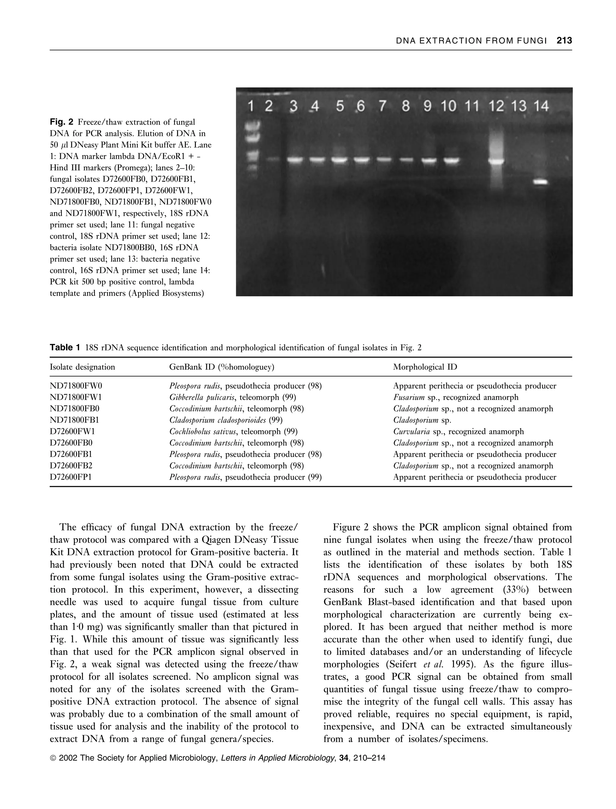
An improved method for genomic dna extraction from strawberry leaves
A rapid and efficient assay for extracting dna from fungi
1. Letters in Applied Microbiology 2002, 34, 210–214
A rapid and efficient assay for extracting DNA from fungi
D.W. Griffin, C.A. Kellogg, K.K. Peak and E.A. Shinn
United States Geological Survey, Center for Coastal and Regional Marine Studies, St Petersburg, FL, USA
2001/218: received 4 October 2001 and accepted 18 December 2001
D . W . G R I F F I N , C . A . K E L L O G G , K . K . P E A K A N D E . A . S H I N N . 2002.
Aims: A method for the rapid extraction of fungal DNA from small quantities of tissue in a
batch-processing format was investigated.
Methods and Results: Tissue (< 3Æ0 mg) was scraped from freshly-grown fungal isolates.
The tissue was suspended in buffer AP1 and subjected to seven rounds of freeze/thaw using a
crushed dry ice/ethanol bath and a boiling water bath. After a 30 min boiling step, the tissue
was quickly ground against the wall of the microfuge tube using a sterile pipette tip. The
Qiagen DNeasy Plant Tissue Kit protocol was then used to purify the DNA for PCR/
sequencing applications.
Conclusions: The method allowed batch DNA extraction from multiple fungal isolates using a
simple yet rapid and reliable assay.
Significance and Impact of the Study: Use of this assay will allow researchers to obtain
DNA from fungi quickly for use in molecular assays that previously required specialized
instrumentation, was time-consuming or was not conducive to batch processing.
use of isopropanol and ethanol to elute and purify DNA
INTRODUCTION
from samples (Elsas et al. 2000). The drawback to this type
A method for efficient extraction of DNA from the various of assay is that it is time-consuming. Homogenization is
suites of microbes has been the focus of research in another method used to extract fungal DNA, incorporating
laboratories around the planet. One of the central problems the use of glass-bead-beating or a similar type of cell lysis
faced by microbiologists using PCR to study microbial matrix (Smit et al. 1999; Borneman et al. 2000). While some
occurrence or microbial genetics is to determine the most of these protocols show promise, they require the purchase
efficient (speed and sensitivity) method for isolating small of specialized instrumentation, and the ability to isolate
quantities of DNA from a limited number of cells. This is of DNA from a limited number of cells is questionable.
particular concern when attempting to extract DNA from Freezing of cells in liquid nitrogen followed by grinding
cell types that possess rigid cell walls and resist lysis with a mortar and pestle is another method used to extract
techniques commonly used for most species of micro- DNA (Smith et al. 1996). This has proven to be a reliable
organisms. Several protocols have been used to extract DNA method, but the limitation is the inability to process
from fungi for genetic analysis, but most are time-consu- multiple isolates simultaneously due to method timing
ming or require mass quantities of tissue. requirements and the need for a matching number of mortar
A classic method is to compromise the integrity of the and pestle sets to prevent sample cross contamination.
fungal cell wall and membrane, followed by the use of A study which compared various commercial DNA
phenol/chloroform to isolate and purify the DNA from cell extraction kits found that the QIAamp Tissue Kit (Qiagen,
and environmental debris (Bever et al. 2000). The drawback Valencia, CA, USA) was the most efficient kit of those
to this method is that loss of DNA can occur during the analysed (Loffler et al. 1997). The Qiagen RNeasy and
purification step, which is particularly important when DNeasy kits have been used successfully for a number of
attempting to isolate DNA from a small number of fungal environmental PCR-based assay studies with outstanding
cells. A similar type of extraction protocol incorporates the results (Griffin et al. 1999, 2001). It has also been found that
a previously published freeze/thaw protocol is useful in
Correspondence to: Dr D.W. Griffin, United States Geological Survey, Center
extracting DNA from microbes, which are typically resistant
for Coastal and Regional Marine Studies, 600 4th St South, St Petersburg, to most of the standard cell lysis protocols (Johnson 1995).
Florida 33701, USA (e-mail: dgriffin@usgs.gov). In this paper, an assay is presented for extracting DNA from
ª 2002 The Society for Applied Microbiology
2. DNA EXTRACTION FROM FUNGI 211
a broad range of air-borne fungal isolates by combining the up. Filters were incubated in the dark at room temperature
freeze/thaw protocol with the Qiagen DNeasy Plant Mini and monitored for growth over a 2 week period. Fungal
Kit protocol which has proven to be rapid, efficient and colonies were isolated from each other by isolation streaking
reproducible for PCR-based studies. on fresh plates of R2A. Once isolated, colonies were grown
overnight in Tryptic Soy Broth (Fisher Scientific) and the
following day, 1 ml of culture was transferred to a sterile
MATERIALS AND METHODS
cryogenic storage tube containing 200 ll sterile glycerol.
Air samples for isolation of fungi These isolated colonies were then stored at )70°C for
cataloguing.
Pre-sterilized filter housings containing 47 mm diameter
analytical test filters with a pore size of 0Æ2 lm were
obtained from Fisher Scientific (Atlanta, GA, USA). To Freeze-thaw extraction of fungal DNA
take the air sample, the filters were removed from their
Isolates from )70°C storage were streaked out onto plates of
respective sterile bags, placed on an analytical filter mani-
R2A agar and grown for 2 days at room temperature.
fold, the lids removed and a vacuum applied using a vacuum
Approximately 2Æ5 mg of fungus tissue from each isolate (see
pump for a set period of time. Airflow rates through the
Fig. 1) were placed in a sterile 1Æ5 ml microcentrifuge tube.
filters were 9Æ3 l min)1 for 15–29 min sampling)1. To
To each tube, 400 ll AP1 buffer (DNeasy Plant Mini Kit,
control for handling contamination, an additional filter was
Qiagen) and 4 ll RNase A (supplied with the kit) were
removed from its bag, the filter placed on the manifold and
added. Freeze/thaw was used to lyse fungal cells. This
allowed to sit without removing the lid. Both filters were
process was repeated seven times using crushed dry ice/
then removed from the manifold, the lids sealed with
ethanol and a boiling water bath. After the last thaw step, the
parafilm, replaced in their respective bags, sealed with tape
samples were boiled for 30 min. A sterile 1 ml micropipette
and refrigerated (4°C) until shipment. Once the filters were
tip was then used to grind any visible tissue in the tubes
received at the United States Geological Survey (USGS)
briefly (5 s) between the tip and conical bottom of the
microbiological laboratory in St Petersburg, Florida, they
microcentrifuge tube. The DNeasy Plant Mini Kit ‘Protocol
were refrigerated (4°C) until analysis. All analyses was
for Isolation of DNA from Plant Tissue with the DNeasy
conducted within a horizontal laminar airflow cabinet using
Plant Mini Kit’ procedure starting with Step 4 (add 130 ll of
sterile technique. R2A agar (Fisher Scientific) was used for
Buffer AP2…) was then followed. The DNA was eluted in 50
fungal analysis. One quarter to one half of each filter was cut
ll buffer AE and 5 ll of eluted DNA was used for PCR.
using sterile scissors and placed on R2A agar, sample side
Fig. 1 Example of the amount of fungal
tissue used for extraction of DNA for PCR
applications. Ruler ¼ centimetre scale
ª 2002 The Society for Applied Microbiology, Letters in Applied Microbiology, 34, 210–214
3. 212 D . W . G R I F F I N ET AL.
Bead-beating extraction of fungal DNA Preparation of isolates for morphological
characterization
Isolates from )70°C storage were streaked onto plates of
R2A agar and grown for 2 days at room temperature. Isolates were grown for whole thallus and microscopic
Approximately 2Æ5 mg of fungus tissue from each isolate characterization on R2A agar at room temperature. Isolate
were then transferred to sterile 2Æ0 ml cryogenic/microcen- purity was achieved by streak-plating fruiting bodies on
trifuge tubes equipped with an O-ring. To each fungus R2A. After 1–3 days of development at room temperature,
tissue sample, 400 ll AP1 buffer (DNeasy Plant Mini Kit) single, well-isolated microthalli were visualized with a hand
and 4 ll RNase A (supplied with the kit) were added. To lens or dissecting microscope and re-plated on R2A to assure
each tube, a volume of sterile glass beads (0Æ1 mm diameter, isolate purity. Thalli were screened with a dissecting
BioSpec Products, Inc., Bartlesville, OK, USA) approxi- microscope or hand lens at 2–3 day intervals; once evident,
mately equal to 100 ll was also added. Tubes were loaded in fruiting bodies were examined microscopically using tape
a Mini-BeadBeater-8 (BioSpec Products). Samples were mounts, tease mounts (McGinnis 1980) or coverslip pre-
bead-beaten for 2 min at maximum speed. They were then parations in Lactophenol Cotton Blue mounting fluid. In a
chilled on ice for 5 min. The bead-beating/cooling steps variation on the technique of Mitchell and Britt (1981),
were repeated twice. The samples were then centrifuged for coverslip preparations were made by streak-plating a
10 min at 14 000 rev min)1 in a microcentrifuge. The microthallus to an R2A plate and then embedding three
supernatant fluid from each tube was transferred to a sterile sterile coverslips (at a 45° angle) into the agar, at the point of
1Æ5 ml microcentrifuge tube. The DNeasy Plant Mini Kit heaviest inoculum. Developing aerial mycelia and fruiting
‘Protocol for Isolation of DNA from Plant Tissue with the bodies adhered to the embedded coverslips. The coverslips
DNeasy Plant Mini Kit’ procedure starting with Step 4 (add were mounted successively, at two to several day intervals,
130 ll of Buffer AP2…) was then followed. The DNA was allowing for a ‘time-lapsed’ study of progressive fruiting
eluted in 50 ll buffer AE and 5 ll of eluted DNA was used body development.
for PCR.
RESULTS AND DISCUSSION
Genetic identification of microbial isolates
Early laboratory experiments, which investigated a number
PCR was used for 18S rDNA amplification using a of methods for efficient extraction of fungal DNA in a
universal fungal primer set [EF3 and EF4]. The PCR batch-processing format, indicated the need for an alternat-
master mix recipe per reaction was: 10 ll GeneAmp 10· ive protocol. To address the applicability of freeze/thaw in
PCR buffer (Applied Biosystems), 12 ll 25 mmol l)1 extracting fungal DNA, several experiments were conduc-
MgCl2 (Applied Biosystems), 2 ll 10 mmol l)1 dNTP ted, with stepwise modifications to each, in an effort to
mix (Promega), 0Æ5 ll 5 U ll)1 Taq polymerase determine the most reliable assay. Once an assay demon-
(Promega), 1 ll each of 10 nmol l)1 upstream and down- strated potential, it was compared with a bead-beating
stream primer (synthesized by Operon Technologies, Inc.) extraction assay. In this experiment, no PCR amplicon was
and 69 ll 0Æ02 lm filter-sterilized autoclaved H2O. The detected using bead beating, and a light to heavy PCR
PCR amplification profile used was: one cycle for 2 min at amplicon signal was detected using freeze/thaw (three
94°C, 40 cycles of [30 s at 94°C, 30 s at 45°C, 2 min at specimens ¼ Sclerotium sp., Aspergillus versicolor and
72°C], one cycle of 10 min at 72°C, and hold at 4°C. After Coccodinium bartschii). In this experiment, the DNA
PCR, amplicon was cleaned and eluted using a QIAquick captured in the Qiagen Plant Tissue Kit spin column was
PCR Purification Kit (Qiagen). Amplicon was cloned into eluted in 50 ll of water for PCR analysis. In later
a plasmid vector using a TOPO TA Cloning Kit experiments it was found that elution into the Qiagen Plant
(Invitrogen Corp., Carlsabad, CA, USA). Plasmid was Tissue Kit buffer AE (as recommended) resulted in an
isolated from the TOPO TA cloning Kit host using a enhanced PCR signal. One of the more widely used
WizardÒ Plus SV Miniprep Kit (Promega). Clones were protocols for extracting fungal DNA is the use of liquid
verified using EcoR I digestion (Promega), following the nitrogen and a mortar and pestle, as stated in the introduc-
manufacturers directions, and electrophoresis. Plasmid tion. The Qiagen DNeasy Plant Tissue Kit recommends the
insert (PCR amplicon) was sequenced (single strand, one use of this approach prior to using the kit to purify and elute
reaction, approximately 750 bases) by the University of the DNA. The drawbacks of this protocol, as stated, are the
Florida DNA Sequencing Core Laboratory (Gainesville, limitation on batch processing and precautions needed for
FL, USA). GenBank Blast search (http://www.ncbi. use of liquid nitrogen. It should be stated that liquid
nlm.nih.gov/BLAST/) was used for amplicon/isolate nitrogen could be used in place of a crushed dry ice/ethanol
identification. bath for freezing samples in the freeze/thaw protocol.
ª 2002 The Society for Applied Microbiology, Letters in Applied Microbiology, 34, 210–214
4. DNA EXTRACTION FROM FUNGI 213
Fig. 2 Freeze/thaw extraction of fungal
DNA for PCR analysis. Elution of DNA in
50 ll DNeasy Plant Mini Kit buffer AE. Lane
1: DNA marker lambda DNA/EcoR1 + -
Hind III markers (Promega); lanes 2–10:
fungal isolates D72600FB0, D72600FB1,
D72600FB2, D72600FP1, D72600FW1,
ND71800FB0, ND71800FB1, ND71800FW0
and ND71800FW1, respectively, 18S rDNA
primer set used; lane 11: fungal negative
control, 18S rDNA primer set used; lane 12:
bacteria isolate ND71800BB0, 16S rDNA
primer set used; lane 13: bacteria negative
control, 16S rDNA primer set used; lane 14:
PCR kit 500 bp positive control, lambda
template and primers (Applied Biosystems)
Table 1 18S rDNA sequence identification and morphological identification of fungal isolates in Fig. 2
Isolate designation GenBank ID (%homologuey) Morphological ID
ND71800FW0 Pleospora rudis, pseudothecia producer (98) Apparent perithecia or pseudothecia producer
ND71800FW1 Gibberella pulicaris, teleomorph (99) Fusarium sp., recognized anamorph
ND71800FB0 Coccodinium bartschii, teleomorph (98) Cladosporium sp., not a recognized anamorph
ND71800FB1 Cladosporium cladosporioides (99) Cladosporium sp.
D72600FW1 Cochliobolus sativus, teleomorph (99) Curvularia sp., recognized anamorph
D72600FB0 Coccodinium bartschii, teleomorph (98) Cladosporium sp., not a recognized anamorph
D72600FB1 Pleospora rudis, pseudothecia producer (98) Apparent perithecia or pseudothecia producer
D72600FB2 Coccodinium bartschii, teleomorph (98) Cladosporium sp., not a recognized anamorph
D72600FP1 Pleospora rudis, pseudothecia producer (99) Apparent perithecia or pseudothecia producer
The efficacy of fungal DNA extraction by the freeze/ Figure 2 shows the PCR amplicon signal obtained from
thaw protocol was compared with a Qiagen DNeasy Tissue nine fungal isolates when using the freeze/thaw protocol
Kit DNA extraction protocol for Gram-positive bacteria. It as outlined in the material and methods section. Table 1
had previously been noted that DNA could be extracted lists the identification of these isolates by both 18S
from some fungal isolates using the Gram-positive extrac- rDNA sequences and morphological observations. The
tion protocol. In this experiment, however, a dissecting reasons for such a low agreement (33%) between
needle was used to acquire fungal tissue from culture GenBank Blast-based identification and that based upon
plates, and the amount of tissue used (estimated at less morphological characterization are currently being ex-
than 1Æ0 mg) was significantly smaller than that pictured in plored. It has been argued that neither method is more
Fig. 1. While this amount of tissue was significantly less accurate than the other when used to identify fungi, due
than that used for the PCR amplicon signal observed in to limited databases and/or an understanding of lifecycle
Fig. 2, a weak signal was detected using the freeze/thaw morphologies (Seifert et al. 1995). As the figure illus-
protocol for all isolates screened. No amplicon signal was trates, a good PCR signal can be obtained from small
noted for any of the isolates screened with the Gram- quantities of fungal tissue using freeze/thaw to compro-
positive DNA extraction protocol. The absence of signal mise the integrity of the fungal cell walls. This assay has
was probably due to a combination of the small amount of proved reliable, requires no special equipment, is rapid,
tissue used for analysis and the inability of the protocol to inexpensive, and DNA can be extracted simultaneously
extract DNA from a range of fungal genera/species. from a number of isolates/specimens.
ª 2002 The Society for Applied Microbiology, Letters in Applied Microbiology, 34, 210–214
5. 214 D . W . G R I F F I N ET AL.
Cryptosporidium oocysts in water samples. Applied and Environmental
REFERENCES
Microbiology 61, 3849–3855.
Bever, R.J., Jr, Couch, L.H., Sutherland, J.B. et al. (2000) DNA Loffler, J., Hebart, H., Schumacher, U., Reitze, H. and Einsele, H.
adduct formation by Fusarium culture extracts: lack of role of fusarin (1997) Comparison of different methods for extraction of DNA of
C. Chemico–Biological Interactions 128, 141–157. fungal pathogens from cultures and blood. Journal of Clinical
Borneman, J. and Hartin, R.J. (2000) PCR primers that amplify fungal Microbiology 35, 3311–3312.
rRNA genes from environmental samples. Applied and Environmental McGinnis, M. (1980) Laboratory Handbook of Medical Mycology.
Microbiology 66, 4356–4360. Academic Press.
Elsas, J.D.V., Duarte, G.F., Keijzer-Wolters, A. and Smit, E. (2000) Mitchell, J.L. and Britt, E.M. (1981) A coverslip culture technique for
Analysis of the dynamics of fungal communities in soil via fungal- preparing permanent fungus mounts. Mycopathologia 76, 23–25.
specific PCR of soil DNA followed by denaturing gradient gel Seifert, K.A., Wingfield, B.D. and Wingfield, M.J. (1995) A critique of
electrophoresis. Journal of Microbiological Methods 43, 133–151. DNA sequence analysis in the taxonomy of filamentous ascomycetes
Griffin, D.W., Garrison, V.H., Herman, J.R. and Shinn, E.A. (2001) and ascomycetous anamorphs. Canadian Journal of Botany 73, S760–
African desert dust in the Caribbean atmosphere: microbiology and S767.
public health. Aerobiologia 17, 203–213. Smit, E., Leeflang, P., Glandorg, B., Elsas, J.D.V. and Wernars, K.
Griffin, D., Gibson, C.J., III, Lipp, E.K., Riley, K., Paul, J.H. and (1999) Analysis of fungal diversity in the wheat rhizosphere by
Rose, J.B. (1999) Detection of viral pathogens by reverse transcrip- sequencing of cloned PCR-amplified genes encoding 18S rRNA and
tase PCR and of microbial indicators by standard methods in the temperature gradient gel electrophoresis. Applied and Environmental
canals of the Florida Keys. Applied and Environmental Microbiology Microbiology 65, 2614–2621.
65, 4118–4125. Smith, G.W., Ives, L.D., Nagelkerken, I.A. and Ritchie, K.B. (1996)
Johnson, D.W., Pieniazek, N.J., Griffin, D.W., Misener, L. and Rose, Caribbean sea-fan mortalities. Nature 383, 487.
J.B. (1995) Development of a PCR protocol for sensitive detection of
ª 2002 The Society for Applied Microbiology, Letters in Applied Microbiology, 34, 210–214