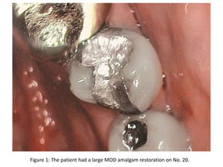
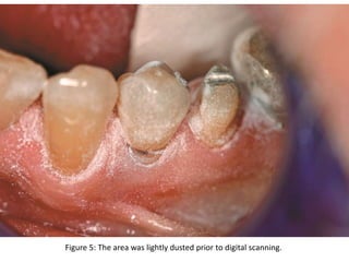
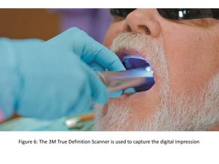
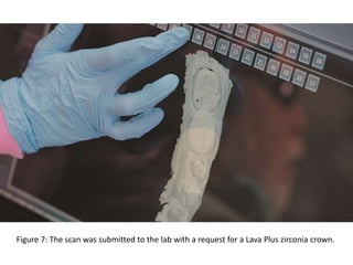
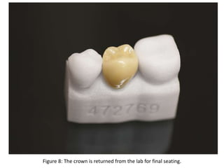
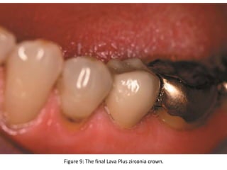
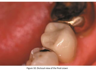
Figure 1: The patient had a large MOD amalgam restoration on No. 20.
Figure 2: Buccal view of the tooth.
Figure 3: The prep prior to placement of the cord.
Figure 4: A hemostatic agent was applied and 00 cord was placed
Figure 5: The area was lightly dusted prior to digital scanning.
Figure 6: The 3M True Definition Scanner is used to capture the digital impression
Figure 7: The scan was submitted to the lab with a request for a Lava Plus zirconia crown.
Figure 8: The crown is returned from the lab for final seating.
Figure 9: The final Lava Plus zirconia crown.
Figure 10: Occlusal view of the final crown
3M™ ESPE™ Lava™ Plus High Translucency Zirconia
• Designed to deliver the esthetics of a layered
restoration in a monolithic all-zirconia
• Unique formulation provides higher translucency
• High strength allows for a tooth-preserving preparation

3M true definition scanner

  • 1.
    Figure 1: Thepatient had a large MOD amalgam restoration on No. 20.
  • 2.
    Figure 2: Buccalview of the tooth.
  • 3.
    Figure 3: Theprep prior to placement of the cord.
  • 4.
    Figure 4: Ahemostatic agent was applied and 00 cord was placed
  • 5.
    Figure 5: Thearea was lightly dusted prior to digital scanning.
  • 6.
    Figure 6: The3M True Definition Scanner is used to capture the digital impression
  • 7.
    Figure 7: Thescan was submitted to the lab with a request for a Lava Plus zirconia crown.
  • 8.
    Figure 8: Thecrown is returned from the lab for final seating.
  • 9.
    Figure 9: Thefinal Lava Plus zirconia crown.
  • 10.
    Figure 10: Occlusalview of the final crown
  • 11.
    3M™ ESPE™ Lava™Plus High Translucency Zirconia • Designed to deliver the esthetics of a layered restoration in a monolithic all-zirconia • Unique formulation provides higher translucency • High strength allows for a tooth-preserving preparation