SlideShare a Scribd company logo
1
Characterizing steric limitations of the heme pocket in the gas-binding Tt H-NOX protein
using site-specific incorporation of unnatural amino acids.
Lukasz T. Olenginski and Christine M. Phillips-Piro
Franklin & Marshall College, Department of Chemistry, Lancaster, PA 17604-3003
ABSTRACT
Heme Nitric Oxide and/or Oxygen (H-NOX) binding proteins are bacterial O2 and/or NO
gas-sensing proteins involved in signaling a variety of functions to the cell. The heme-binding
pocket in Thermoanaerobacter tencongensis H-NOX (Tt H-NOX) has been characterized using
site-directed mutagenesis with the 20 naturally occurring amino acids. The present study aims to
further characterize the heme-binding pocket of Tt H-NOX by incorporating unnatural amino
acids (UAAs) into the H-NOX scaffold, shedding light on both ligand discrimination and the
tuning of ligand affinity. Currently, we are working to understand the steric limitations in this
pocket by incorporating halogenated phenylalanine residues (pClF, pBrF, pIF) at the Y140 site.
Interestingly, the level of expression decreased as the size of the halogen constituent increased,
suggesting a steric limitation to incorporating larger UAAs. This result will inform our choice of
UAAs to incorporate into the protein and any further mutations needed to accommodate the
desired UAAs. Future work is focused on the incorporation of UAAs at other sites in Tt H-NOX
in order to assess the spectroscopic and structural properties of these proteins as well as assessing
enzyme and aggregation kinetics activity upon UAA incorporation in T4 Lysozyme.
INTRODUCTION
The genetically encoded method for incorporating unnatural amino acids (UAAs) has
allowed the thoughtful addition of a multitude of functionalities site-specifically in proteins with
a variety of applications.1-6 While successful incorporation of UAAs has proven difficult,
Franklin and Marshall College has witnessed significant success in incorporating both
commercially available UAAs and novel UAAs synthesized in-house.4-10 With this local
expertise, we have tremendous potential on incorporating UAAs into the protein systems
described herein. Further, structural studies on proteins containing UAAs have been elusory,
illustrating the importance of a systematic structure-function study of UAA-incorporated
proteins.
Heme-based sensors are a diverse group of signal transducing proteins that respond to
gases like nitric oxide (NO), oxygen (O2), and carbon monoxide (CO).11 The past 20 years has
witnessed an explosion in the number of known sensor proteins, from just two recognized
members, FixL and soluble guanylate cyclase (sGC), to four distinct families comprising more
than 50 sensors.12 Heme-based sensors now feature four diverse heme-binding motifs: the heme-
binding PAS domain, globin-coupled sensor (GCs), CooA, and Heme Nitric Oxide and/or
Oxygen (H-NOX) binding domains.11-13 Containing both a PAS-like domain and an H-NOX
domain, the signal transducing protein sGC responds to nitric oxide (NO), a potent modulator of
cardiovascular physiology in mammals.14 A group of prokaryotic proteins have been found to be
related in sequence to the NO-binding sGCs.13,15-17 In facultative aerobes, these domains are
predicted to contain ~190 residues and are found in an operon with a histidine kinase (HK).11
Homologous domains are found in obligate anaerobes, where they fuse through membrane-
2
spanning region to a predicted methyl-accepting chemotaxis protein (MCP) domain.11 Similar to
the sGCs, the heme domains from facultative aerobes bind NO, but not O2. In contrast, the heme
domain bound to and MCP from the obligate anaerobe Thermoanaerobacter tengcongensis binds
O2, NO, and CO.13 In light of this discovered specificity for oxygen in some of these domains
they have also been referred to as Heme Nitric Oxide and/or Oxygen (H-NOX) binding domains.
Pellicena et al. (2004) reported the crystal structure of the O2-bound H-NOX domain
from T. tengcongensis (Tt H-NOX). Structural analysis reveals the H-NOX family to have
evolved a novel protein fold consisting of seven -helices and a four-stranded anti-parallel -
sheet (Figure 1). A major finding from the structure of the Tt H-NOX domain and critical to
ligand discrimination is the hydrogen bonding (H-bonding) network that surrounds the bound O2
molecule.11,15,18-21 Y140 is involved in a 2.7 Å H-bond to O2 as well as to N74 and W9 are
(Figure 1).11 H-NOX sequence alignments strongly suggest that all three of these residues are
unique to members of the H-NOX family that bind O2.11
Gas ligand affinity to the heme of H-NOX proteins has been studied using site-directed
mutagenesis (SDM) with the 20 naturally occurring amino acids.18,20,22-30 For instance, P115, a
conserved residue in the H-NOX family, was mutated to an alanine and used to demonstrate that
decreasing heme distortion increases affinity for oxygen, providing a clear link between the
heme conformation and Tt H-NOX structure.19 Y140L provided further evidence that the distal
Figure 1. The heme binding pocket of Tt H-NOX with O2 bound (PDB ID: 1U55). The heme and
some residues shown in orange sticks. Y140 and H-bonding network known to be crucial for tight O2
binding affinity.
3
pocket tyrosine not only stabilizes the O2 complex but also discriminates between NO and O2
using a kinetic selection.18 Furthermore, the double mutant F78Y/Y140L demonstrated that O2
binding could be rescued as long as there was a distal pocket tyrosine.11,18
The protein has been cloned into the appropriate expression vector, all necessary
mutagenesis has been completed, the expression protocol has been optimized, and the
purification protocol was troubleshot. Most significantly, our SDS-PAGE gels have shown
evidence of steric limitation in incorporating larger UAAs (Figure 4, 5). Thus, we initially aimed
to further characterize the size limitations in the heme pocket by using the halogenated
phenylalanine UAAs. However, based on the nontrivial nature of Tt H-NOX purification, we
decided to expand our study, focusing on the incorporation of UAAs at other sites in Tt H-NOX
in order to assess the spectroscopic and structural properties of these proteins as well as assessing
enzyme activity and aggregation kinetics upon UAA incorporation in T4 Lysozyme (Figure 2).
The study of Tt H-NOX allows the following three questions to be explored: how does
UAA incorporation alter (1) protein environment (2) protein structure (3) protein function. The
present work with the halogenated phenylalanine UAAs has informed our decision to further
mutate the heme pocket in order to accommodate UAAs of interest and investigate question (3).
However, the other two questions (1, 2) can be addressed via assessing the spectroscopic and
structural properties of Tt H-NOX proteins with UAAs incorporated at other sites. The study of
Y24
Y18
F67
F153
Figure 2. Structure of T4 Lysozyme (PDB ID 148L) with a substrate mimic bound and shown in
magenta sticks and colored by atom type. Sites for UAA incorporation are shown in yellow sticks and
colored by atom type.
4
T4 Lysozyme allows a different approach to answering question (3), as well as introducing a new
question: (4) how does UAA incorporation alter protein aggregation kinetics.
MATERIALS AND METHODS
Cloning Tt H-NOX into appropriate expression vector. Tt H-NOX was cloned out of the pCW
vector (Appendix A) and digested with the restriction enzymes NcoI and XhoI (Appendix B).
Simultaneously, the pBAD vector containing sfGFP was digested with the same restrictions
enzymes and the Tt H-NOX insert was ligated into the empty pBAD vector using the following
plasmid/insert ratios – 1:1, 1:2, 2:1 (Appendix B).
SDM to create Tt H-NOX Amber (TAG) mutants. Successful cloning of Tt H-NOX out of pCW
and into pBAD required the second residue (K) to be mutated to glutamate, and thus once it was
in pBAD it was mutated back to the native lysine using the QuikChange® protocol (Appendix
C). The C terminus His6-tag was removed (necessary for successful crystallization) by the
insertion of a TAA stop codon after the protein sequence and before the His6-tag (Appenix A).
Lastly, the amber (TAG) codon was inserted at the Y140 position in order for the pDULE vector
– containing both the tRNA and the aminoacyl-tRNA synthetase – to add UAAs (Appendix B).
The end result of the multiple rounds of SDM was the non-His6-tagged version of Tt H-NOX
with an amber (TAG) codon at the Y140 site. All other Amber mutants (F52, F78, Y85, F151,
F169, F183, Y185) were created in the same manner – using the QuikChange® protocol
(Appendix C).
SDM to create T4Lys Amber (TAG) mutants. Successful cloning of T4Lys out of pHS1403 and
into pBAD required the second residue (N) to be mutated to aspartic acid and thus once it was in
pBAD it was mutated back to the native asparagine using the QuikChange® protocol (Appendix
C). The C terminus His6-tag was removed (necessary for successful crystallization) by the
insertion of a TAA stop codon after the protein sequence and before the His6-tag (Appenix C).
Lastly, the amber (TAG) codon was inserted at the following sites – Y18, Y24, F67, and F153.
Tt H-NOX Expression. Appropriate Tt H-NOX expression construct and desired pDULE
synthetase construct were dual-transformed into chemically competent DH10B E. coli cells in
the afternoon and then plated on LB/Ampicillin/Tetracycline Agar plates and incubated at 37 C
overnight (Appenidx D). The next morning, the plates were taken out of the incubator,
parafilmed, and stored in the refrigerator at 4 C. Later that afternoon, the plates were taken out
of the refrigerator and used to make starter cultures in non-inducing media (Appendix E). The
next morning, auto-induction media was prepared (Appendix E). Expression volume was 250 ml
in a 500 ml baffled flask. Expression cultures were inoculated with starter culture (1ml:1 L) and
grown up at 37 C and 250 rpm for 30-36 hrs. Directly following incubation of the expression
cultures, the UAA solutions were prepared (Appendix E). Approximately 1 hour after the start of
incubation, the UAA solution (1 uM) and heme-precursor, 5-aminolevulinic acid (1 uM), were
added to the cultures. After 30-36 hrs, the cultures were spun down at 5000 rpm for 10 min and
the cell pellets were flash frozen and stored at -80 C.
Expressions were also performed in a yeast extract media. The Tt H-NOX expression
construct was transformed into the same cell line as described above. However, when making
starter cultures, LB broth was used rather than non-inducing media. The yeast extract media was
prepared the day before the expression. Expression volume was 1 L in a 2 L baffled flask; thus
5
45 g yeast extract, 10 ml glycerol, and 900 ml ddH2O were added to each flask and autoclaved.
Before inoculation 100 ml of a 170 mM KH2PO4, 720 mM K2HPO4 phosphate buffer was added
to each expression culture. Cultures were inoculated as described above and allowed to grow at
37 C and 250 rpm. Once the OD600 reached ~ 0.7-0 .8 the incubation temperature was dropped
to 18 C and the cultures were induced with the same amount of 20% arabinose as prescribed in
the auto-induction media (2.5 ml/L). UAA and ALA solution were added in the same manner as
before (1 hr after induction). Cultures grew at 18 C and 250 rpm overnight and the next
morning the cell pellet was collected in the same fashion as mentioned above.
Tt H-NOX Purification. Round 1 – Cell pellet was thawed on ice, re-suspended in either 10 ml
(250 ml culture) or 40 ml (1 L culture) Lysis Buffer (50 mM TEA, pH 8.5, 20 mM NaCl), and
lysed by the addition of Lysozyme (0.25 mg/ml). PMSF (0.5 mM) and DNAse (3.75 mM) were
also added in order to inhibit endogenous proteases from degrading our protein and breaking
down the DNA, respectively. Lysis solution was then sonicated at 40 % amplitude for 2 min (2 s
pulse on, 2 s pulse off) and boiled at 70 C for 30 min. Samples were then spun down at 20,000
rpm for 45 min and the supernatant was flash frozen and stored at -80 C. Purity and presence of
protein was analyzed by SDS-PAGE, with each gel containing a re-suspension, pellet, and
supernatant sample.
Round 2 – The supernatant (~50 ml) was thawed and loaded onto a ~80-100 ml Q-650
column (anion exchange) pre-equilibrated with ~150 ml Lysis Buffer. Protein was run through
column using the Lysis Buffer at 1.5 ml/min. Red fractions (~ 30 ml) were collected and
concentrated to 2.5 ml. The protein was then run over a pre-packed PD10 column pre-
equilibrated with 25 ml Buffer B (50 mM HEPES, pH 6.5, 5% glycerol). After Tt H-NOX
protein was loaded, 3.5 ml Buffer B was added to make protein elute. Again, the red fractions
were collected. Then, the protein was loaded onto a ~ 80-100 ml CM-650 column (cation
exchange) pre-equilibrated with ~150 ml Buffer B and run through at 1.5 ml/min. All red colored
fractions were collected and analyzed with SDS-PAGE to inform which fractions should be
combined, concentrated, and ran over the S75 column. The CM-650 fractions that included Tt H-
NOX protein were combined (~25 ml) and concentrated down to ~ 4 ml. Protein was then
filtered through syringe filter, leaving 3 ml Tt H-NOX protein. Proteins was then loaded onto the
S75 column (size exclusion) pre-equilibrated with Buffer C (50 mM TEA, pH 7.5, 50 mM NaCl,
5% glycerol) and run at 0.20 ml/min. with the max pressure set at 0.27 MPa. Fractions near
peaks corresponding to the correct MW (~ 22 kDa) were analyzed by SDS-PAGE. Fractions
containing Tt H-NOX protein were combined and concentrated down to ~500 ul and
concentration was assessed by a Wave Scan.
RESULTS
6
Addition of ALA aids expression of Tt H-NOX. Early expression attempts in auto-induction
media with both pCNF and mNO2Y resulted in low expression yields. Further, expression
cultures lacked the red color, indicative of heme-incorporation. Thus, we added the heme
precursor, 5-aminolevulinic acid (ALA), to the expression cultures and not only saw redder
cultures but increased expression as well (Figure 3).
Steric limitations involved in the heme pocket of Tt H-NOX. Despite the addition of ALA
aiding the expression of Tt H-NOX, consistent expression with pCNF and mNO2Y was not
observed. This led us to ponder whether our expression protocol was flawed. Thus, we set up a 5
ml test expression in auto-induction media with both Wt Tt H-NOX_His6 and Tt H-
NOX_Y140_His6 with the following UAAs – pCNF, pClF, pBrF, pIF, pNH2F, and mNO2Y – to
tease out whether the problems encountered were a result of our expression protocol or simply
the UAA incorporation. The His6 constructs were used due to their simpler purification protocol
(in case further characterization was necessary). Interestingly, we saw expression of both the Wt
Tt H-NOX_His6 and the Tt H-NOX_Y140_His6 with the halogenated phenylalanine UAAs,
suggesting that neither our expression protocol or our method of incorporating the UAA is
flawed, but rather the UAA itself is what is limiting (Figure 4). In other words, these data suggest
that there is a steric limitation to what UAAs can be incorporated into the heme pocket.
Figure 3. Impact of addition of ALA on Tt H-NOX_Y140 + pCNF expression. Parallel 250 ml
expressions were setup in 500 ml baffled flasks both with and without addition of ALA. (A) Expression
cultures after 30-36 hr growth period. (B) Pellet following growth period. (C) Purification supernatant.
(D) Purification pellet. (E) SDS-PAGE gel of Tt H-NOX_Y140 + pCNF post round 1 purification with
(lanes 3-5) and without (lanes 6-8) addition of ALA.
7
Still, we were not completely convinced by the data for multiple reasons – the expression
volumes were very small and we had yet to see consistent results with the auto-induction media
in the first place. Thus, we decided to try the expression of the Tt H-NOX_Y140 with the
halogenated phenylalanine UAAs in a media that we had recent success with on the Wt Tt H-
NOX – the yeast extract media. We saw the same trend of the larger the size of the halogen
constituent at the para-position of the phenylalanine ring, the smaller the level of expression
Further, we were able to show that we could express the halogenated phenylalanine UAAs with
larger culture sizes (Figure 4). Thus, the data in Figure 4 not only support the steric hypothesis
but also highight that we can express Tt H-NOX_Y140 constructs with UAAs liters worth at a
time, which ultimately allows us to set crystal trays and elucidate the structural consequences of
UAA incorporation into the H-NOX scaffold.
Figure 4. SDS-PAGE gel of Tt H-NOX_His6 + various UAA constructs following round 1 of
purification. Samples were run on a self-poured 12 % SDS-PAGE gel with Precision Plus Protein™
Kaleidoscope™ (BIORAD, #161-0375) and 2x Laemmli Sample Buffer dye (BIO-RAD, #161-0737)
at 200 V for 45 min. As indicated by lane 2 on each gel Tt H-NOX protein should appear ~ 22 kDa.
(A) As seen in lane 4, 6, 8, and 10 only the halogenated phenylalanine constructs seemed to express
well. (B) None of the UAA containing Tt H-NOXconstructs expressed.
8
DISCUSSION
The first half of the fall semester was plagued by unsuccessful purification attempts with
Tt H-NOX. Despite the tremendous progress in cloning the appropriate expression constructs and
expressing Tt H-NOX with halogenated phenylalanine UAAs, we could not acquire sufficient
protein yield for crystallographic work. The reasoning for the small yields seemed to be the
purification protocol, and thus minor changes were made. Notably, following use of the Q-650
anion exchange column the protein was concentrated and loaded right onto the S75 size
exclusion column, skipping the CM-650 cation exchange column completely. This modification
saved time but did not sacrifice protein purity as concluded from SDS-PAGE analysis of a gel
containing Tt H-NOX protein post anion exchange and post cation exchange. Despite saving
time with skipping the cation exchange column, a lot of difficulty was encountered with the S75
Figure 5. Size limitations in the heme pocket of Tt H-NOX. (A) side and (B) top view of
heme pocket in Tt H-NOX (PDB ID: IU55) using surface representation. (C) SDS-PAGE
gel of both Wt Tt H-NOX and Tt H-NOX_Y140 with halogenated phenylalanine UAAs
following round 1 of purification. Samples were run on a self-poured 12 % SDS-PAGE gel
with Precision Plus Protein™ Kaleidoscope™ (BIORAD, #161-0375) and 2x Laemmli
Sample Buffer dye (BIO-RAD, #161-0737) at 200 V for 45 min. As indicated by lane 2 Tt
H-NOXprotein should appear ~ 22 kDa. We see successfulexpression of all constructs.
9
size exclusion column on the FPLC. Specifically, during the semester it malfunctioned several of
times and crippled our efforts to move the project forward and purify protein. We did, however,
purify Tt H-NOX + pIF (~1.5 ml, ~7 mg/ml), but again this amount was insufficient for
crystallographic work.
As mentioned, there are a variety of future directions for next semester. To begin, we will
further mutate the heme pocket in an attempt to accommodate larger UAAs. Molecular analysis
of the heme pocket reveals the phenyl ring of F78 to be in very close proximity to Y140 (Figure
6). Thus, we propose to mutate F78 to alanine, freeing up space for the meta and para positions
on the phenyl ring of Y140. With the extra space in the pocket, we plan to incorporate unnatural
tyrosine analogs with either withdrawing groups (EWG) or electron donating groups (EDG) at
the para-position on the phenyl ring to either make the tyrosine a better or poorer hydrogen bond
donor and thus a stronger or weaker O2 binder, respectively (Figure 7). Ultimately, we will
structurally characterize the mutant H-NOX proteins and assess their O2 binding affinity to relate
the H-bond donating ability of the tyrosine to the O2 affinity of the protein and any structural
alterations that may have occurred. Thus, this study addresses question (3) discussed earlier.
Further, we are not limited to solely incorporating UAAs at the Y140 site. We plan to
express Tt H-NOX at a variety of sites with different protein environments (Appendix F). We
chose to start with five sites on the protein, all of which are natively tyrosine or phenylalanine
residues to ensure a most conservative replacement. We have chosen sites that are fully buried
(Y52, Y85), partially buried (F151, F169, F183), and solvent exposed (Y185) (Figure 8). These
sites offer the potential to report on protein environment and protein structure upon UAA
incorporation, and thus addresses questions (1, 2) discussed earlier. The former can be
Y140
F78
Y140
A78
Figure 6. Heme pocket of Tt H-NOX (PDB ID: 1U55). Protein shown in surface representation
with van der Waal radii of the atoms in Y140 and F78 shown. Left image shows F78 modeled in
whereas the left image shows the propsed F78 mutant modeled in.
10
accomplished with vibrational reporter UAAs and the later can be accomplished with X-ray
crystallography.
Figure 7. Method of tuning the H-bonding environment above the heme pocket in Tt H-NOX.
Figure 6. Tt H-NOX showed in cartoon representation (PDB ID: IU55) with select tyrosine and
phenylalanine residues shown in yellow sticks and colored by atom.
11
As mentioned, if we encounter similar problems with purifying Tt H-NOX, the focus of
the study might shift towards T4 Lysozyme. Much like in Tt H-NOX, we have chosen a variety
of sites with different protein environments – sites lining the substrate-binding pocket (Y18,
Y24) and two sites far from the substrate-binding pocket (F67, F153) (Figure 2). Again, these
sites offer the potential to report on protein environment and protein structure upon UAA
incorporation, and thus addresses questions (1, 2) discussed earlier. Additionally, study of T4
Lysozyme addresses question (3), but in a different way than Tt H-NOX. Rather than assessing
altered gas binding, T4 Lysozyme sheds light on whether the incorporation of UAAs directly
impacts enzyme catalysis. Once purified mutant T4 Lysozyme protein is obtained, we can begin
to look at its activity, which is straightforward due to the variety of commercially available
enzyme assays for this protein. Lastly, due to this protein’s propensity to aggregate we can
explore whether incorporation of UAAs alters aggregation kinetics, addressing question (4)
discussed earlier. The scope of this project has become quite large, however we are interested in
very specific questions (1-4) and have developed thoughtful ways of investigating them.
At this point, the mutagenesis to create all of the Tt H-NOX and T4 Lysozyme Amber
mutants described herein has been completed. Further, they have been dually transformed into
chemically competent DH10B E. coli cells with the pCNF synthetase. Thus, we have what we
need in order to start expressing new protein and progress this project forward.
REFERENCES
(1) Seyedsayamdost, M. R., Yee, C. S., and Stubbe, J. (2007) Site-specific incorporation of
fluorotyrosines into the R2 subunit of E. coli ribonucleotide reductase by expressed protein
ligation. Nat Protoc 2, 1225–1235.
(2) Wang, L., Xie, J., Deniz, A. A., and Schultz, P. G. (2003) Unnatural amino acid mutagenesis
of green fluorescent protein. J. Org. Chem. 68, 174–176.
(3) Groff, D., Wang, F., Jockusch, S., Turro, N. J., and Schultz, P. G. (2010) A New Strategy to
Photoactivate Green Fluorescent Protein. Angew. Chem. Int. Ed. 49, 7677–7679.
(4) Miyake-Stoner, S. J., Miller, A. M., Hammill, J. T., Peeler, J. C., Hess, K. R., Mehl, R. A.,
and Brewer, S. H. (2009) Probing protein folding using site-specifically encoded unnatural
amino acids as FRET donors with tryptophan. Biochemistry 48, 5953–5962.
(5) Bazewicz, C. G., Lipkin, J. S., Smith, E. E., Liskov, M. T., and Brewer, S. H. (2012)
Expanding the utility of 4-cyano-L-phenylalanine as a vibrational reporter of protein
environments. J Phys Chem B 116, 10824–10831.
(6) Bazewicz, C. G., Liskov, M. T., Hines, K. J., and Brewer, S. H. (2013) Sensitive, site-
specific, and stable vibrational probe of local protein environments: 4-azidomethyl-L-
phenylalanine. J Phys Chem B 117, 8987–8993.
(7) Pavic, K., Rios, P., Dzeyk, K., Koehler, C., Lemke, E. A., and Köhn, M. (2014) Unnatural
Amino Acid Mutagenesis Reveals Dimerization As a Negative Regulatory Mechanism of VHR's
Phosphatase Activity. ACS Chem. Biol.
(8) Jackson, J. C., Duffy, S. P., Hess, K. R., and Mehl, R. A. (2006) Improving nature's enzyme
active site with genetically encoded unnatural amino acids. J. Am. Chem. Soc. 128, 11124–
11127.
(9) Taskent-Sezgin, H., Chung, J., Patsalo, V., Miyake-Stoner, S. J., Miller, A. M., Brewer, S.
H., Mehl, R. A., Green, D. F., Raleigh, D. P., and Carrico, I. (2009) Interpretation of p-
12
cyanophenylalanine fluorescence in proteins in terms of solvent exposure and contribution of
side-chain quenchers: a combined fluorescence, IR and molecular dynamics study. Biochemistry
48, 9040–9046.
(10) Smith, E. E., Linderman, B. Y., Luskin, A. C., and Brewer, S. H. (2011) Probing local
environments with the infrared probe: L-4-nitrophenylalanine. J Phys Chem B 115, 2380–2385.
(11) Pellicena, P., Karow, D. S., Boon, E. M., Marletta, M. A., and Kuriyan, J. (2004) Crystal
structure of an oxygen-binding heme domain related to soluble guanylate cyclases. Proc. Natl.
Acad. Sci. 101, 12854-12859.
(12) Gilles-Gonzalez, M. A., and Gonzalez, G. (2005) Heme-based sensors: defining
characteristics, recent developments, and regulatory hypotheses. J. Inorg. Biochem. 99, 1-22.
(13) Karow, D. S., Pan, D., Tran, R., Pellicena, P., Presley, A., Mathies, R. A., and Marletta,
M.A. (2004) Spectroscopic characterization of the soluble guanylate cyclase-like heme domains
from Vibrio cholera and Thermoanaerobacter tengcongensis. Biochemistry. 43, 10203-10211.
(14) Denninger, J. W. and Marletta, M. A. (1999) Guanylate cyclase and the NO/cGMP
signaling pathway. Biochim. Biophy. Acta. 1411, 334-350.
(15) Schmidt, P. M., Schramm, M., Schröder, H., Wunder, F., and Stasch, J. P. (2004)
Identification of residues crucially involved in the binding of heme moiety of soluble guanylate
cyclase. J. Biol. Chem. 279, 3025-3032.
(16) Winger, J. A., Derbyshire, E. R., and Marletta, M. A. (2007) Dissociation of nitric oxide
from soluble guanylate cyclase and heme-nitric oxide/oxygen binding domain constructs. J. Biol.
Chem. 282, 897-907.
(17) 12. Boon, E. M., Davis, J. H., Karow, D .S., Huang, S. H., Tran, R., Miazgowicz, M. M.,
Mathies, R., and Marletta, M. A. (2006) Nitric oxide binding to prokaryotic homologs of the
soluble guanylate cyclase b1 H-NOX domain. J. Biol. Chem. 281, 21892-902.
(18) Boon, E. M., Huang, S. H., and Marletta, M. A. (2005) A molecular basis for NO selectivity
in soluble guanylate cyclases. Nat. Chem. Biol. 1, 53-59.
(19) Olea, C., Boon, E. M., Pellicena, P., Kuriyan, J., and Marletta, M. A. (2008) Probing the
function of heme distortion in the H-NOX family. ACS Chem. Biol. 3, 703-710.
(20) Weinert, E. E., Plate, L., Whited, C. A., Olea, C. Jr., and Marletta, M. A. (2010)
Determinants of ligand affinity and heme reactivity in H-NOX domains. Angew. Chem., Int. Ed.
49, 720-723.
(21) Martin, E., Berka, V., Bogatenkova, E., Murad, F., and Tsai, A. (2006) Ligand selectivity of
guanylyl cyclase: effect of the hydrogen-binding tyrosine in the distal heme pocket on binding of
oxygen, nitric oxide, and carbon monoxide. J. Biol. Chem. 281, 27836-27845.
(22) Weinert, E. E., Phillips-Piro, C. M., Tran, R., Mathies, R. A., and Marletta, M. A. (2011)
Controlling conformational flexibility of an O2-binding H-NOX domain. Biochemistry. 50, 6832-
6840.
(23) Weinert, E. E., Phillips-Piro, C. M., and Marletta, M. A. (2013) Porphyrin π-stacking in a
heme protein scaffold tunes gas ligand affinity. J. Inorg. Biochem. 127, 7–12.
(24) Olea, C., Jr., Kuriyan, J., and Marletta, M. A. (2010) Modulating Heme Redox Potential
through Protein-Induced Porphyrin Distortion. J. Am. Chem. Soc. 132, 12794–12795.
(25) Olea, C., Boon, E. M., Pellicena, P., Kuriyan, J., and Marletta, M. A. (2008) Probing the
Function of Heme Distortion in the H-NOX Family. ACS Chem. Biol. 3, 703–710.
(26) Olea, C., Jr., Herzik, M. A., Jr., Kuriyan, J., and Marletta, M. A. (2010) Structural insights
into the molecular mechanism of H-NOX activation. Protein Science 19, 881–887.
13
(27) Derbyshire E. R., Deng S., and Marletta M. A. (2010) Incorporation of tyrosine and
glutamine residues into the soluble guanylate cyclase heme distal pocket alters NO and O2
binding. J Biol Chem. 285, 17471-8.
(28) Tran R., Boon E. M., Marletta M. A., and Mathies R. A. (2009) Resonance raman spectra of
an O2-binding H-NOX domain reveal heme relaxation upon mutation. Biochemistry. 48, 8568-
77.
(29) Kosowicz, J. G. and Boon, E. M. (2013) Insights into the distal pocket of H-NOX using
fluoride as a probe for H-bonding interactions. J Inorg. Biochem. 126, 91-5.
(30) Dai, Z. and Boon, E. M. (2011) Probing the local electronic and geometric properties of the
heme iron center in an O2-binding Heme-Nitric oxide and/or Oxygen binding domain. J Inorg.
Biochem. 105, 784-792.
14
APPENDIX
A. Cloning Tt H-NOX out of pCW
NcoI – GAG – residue 3 – F
5’ – GC CC ATG GAG GGG ACA ATC GTC GGG ACA TGG ATA AAG ACC C – 3’
Tt H-NOX – STOP – XhoI - R
5’ – CG CTCGAG TTA ATT TTT CTT ATA CTC AAA AAC GGG G – 3’
Cloning Reaction
Volume (ul)
Tt H-NOX in pCW (25 ng) 0.425
Primer – F (10 uM) 2
Primer – R (10 uM) 2
10 X Buffer 5
ddH2O 38.35
dNTP mix (10 mM) 1.25
Pfu II Turbo Polymerase 1
50 ul Total Reaction
Ran PCR program “PIRO PCR” (subdirectory 1, program 1)
1. 95 C for 6 min.
2. 95 C for 30 s.
3. 55 C for 30 s.
4. 72 C for 2 min, 30 s.
5. Repeat steps 2-4 (34x)
6. 72 C for 10 min.
7. 4 C for 10 min.
8. 10 C forever
B. Digestion and Ligation Protocols
Single Digestion Reaction
Volume (ul)
XhoI/NcoI 1
pBAD-GFP (1 ug) 6
10 X Cutsmart Buffer 5
ddH2O 38
50 ul Total Reaction
15
Double Digestion Reaction
Volume (ul)
XhoI
NcoI
1
1
pBAD-GFP (1 ug) 6
10 X Cutsmart Buffer 5
ddH2O 37
50 ul Total Reaction
Ligation Reactions
1:1 Reaction (ul) 2:1 Reaction (ul) 1:2 Reaction (ul)
10 X DNA Ligase Buffer 2 2 2
pBAD + NcoI + XhoI 2 4 2
Tt H-NOX_Y140 insert 2 2 4
Nuclease free water 13 11 11
T4 DNA Ligase 1 1 1
20 ul Total 20 ul Total 20 ul Total
Sequencing results of Tt H-NOX_Y140 in pBAD
Y140 construct exhibits incorrect second residue and nonsense codon at position Y140,
indicative of successful entry into pBAD
16
C. SDM to create appropriate expression constructs
Construct Forward primer (5’-3’) Reverse Primer (5’-3’)
Tt H-NOX-Y140-E2K GGGCTAACAGGAGGAATTAACCAT
GAAGGGGACAATCGTCGGGACATG
GATAAAGACCC
GGGTCTTTATCCATGTCCCGACGAT
TGTCCCCTTCATGGTTAATTCCTCC
TGTTAGCCC
Tt H-NOX-F52Amb GAGGTTAGGAGAATTTAGGCTAAG
GTGAGTGAAAAAACT
AGTTTTTTCACTCACCTTAGCCTAA
ATTCTCCTAACCTC
Tt H-NOX_F78Amb GGCAGAACATAAAAACTTAGAGCG
AATGGTTTCCCTCC
GGAGGGAAACCATTCGCTCTAAGTT
TTTATGTTCTGCC
Tt H-NOX-F78A GGCAGAACATAAAAACTGCCAGCG
AATGGTTTCCCTCC
GGAGGGAAACCATTCGCTGGCAGTT
TTTATGTTCTGCC
Tt H-NOX-Y85Amb CGAATGGTTTCCCTCCTAGTTTGC
AGGGAGAAGGCTAGTG
CACTAGCCTTCTCCCTGCAAACTAG
GAGGGAAACCATTCG
Tt H-NOX-F151Amb ATAGAGGGTAGTTCTAAATAGTTC
AAGGAAGAAATTTCAG
CTGAAATTTCTTCCTTGAACTATTT
AGAACTACCCTCTAT
Tt H-NOX-F169Amb CGAAAGAGGCGAAAAAGATGGCTA
GTCAAGGCTAAAAGTC
GACTTTTAGCCTTGACTAGCCATCT
TTTTCGCCTCTTTCG
Tt H-NOX-F183Amb AAATTTAAAAACCCCGTTTAGGAG
TATAAGAAAAATTAAC
GTTAATTTTTCTTATACTCCTAAAC
GGGGTTTTTAAATTT
Tt H-NOX-Y185Amb CCCCGTTTTTGAGTAGAAGAAAAA
TTAACTCGAGATCTGC
GCAGATCTCGAGTTAATTTTTCTTC
TACTCAAAAACGGGG
T4Lys-STOP GGCACTTGGGACGCGTATAAAAATCTA
AGCCATCATCATCATCATC
GATGATGATGATGATGGCTTAGATTTTT
ATACGCGTCCCAAGTGCC
T4Lys-Y18Amb GAAGGTCTTAGACTTAAAATCTAGAAA
GACACAGAAGGCTATTACAC
GTGTAATAGCCTTCTGTGTCTTTCTAGA
TTTTAAGTCTAAGACCTTC
T4Lys-Y24Amb CTATAAAGACACAGAAGGCTAGTACAC
TATTGGCATCGGTCATTTGC
GCAAATGACCGATGCCAATAGTGTACTA
GCCTTCTGTGTCTTTATAG
T4Lys-F67Amb GATGAGGCTGAAAAACTCTAGAATCAG
GATGTTGATGCTGC
GCAGCATCAACATCCTGATTCTAGAGTT
TTTCAGCCTCATC
QuikChange® SDM Protocol
10 ng plasmid reaction (ul) 50 ng plasmid reaction (ul)
E2K-primer-F (1 uM) 6.8 6.8
E2K-primer-R (1 uM) 6.96 6.96
pBAD_Tt H-NOXY140 0.648 3.24
Pfu Ultra II 10 X Buffer 5 5
dNTPs (10 mM) 1 1
ddH2O 28.6 (29.6 for control) 26 (27 for control)
Pfu Ultra II Polymerase 1 (0 for control) 1 (0 for control)
50 ul Total Reaction 50 ul Total Reaction
Ran PCR program “PIRO-SDM” (subdirectory 1, program 3)
1. 95 C for 30 s.
2. 95 C for 30 s.
3. 55 C for 30 s.
4. 68 C for 4 min, 30 s.
5. Repeat steps 2-4 (18x)
6. 10 C forever
17
Folowing PCR - Add 1 ul DpnI and incubate (37 C) for 1 hr and then transform SDM product
Sequencing results following SDM reacions
Tt H-NOX-Y140-E2K
Tt H-NOX-F52Amb
18
Tt H-NOX-Y85Amb
Tt H-NOX-F151Amb
19
Tt H-NOX-F169Amb
Tt H-NOX-F183Amb
20
Tt H-NOX-Y185Amb
T4Lys-D2N
21
T4Lys-Hist2STOP
T4Lys-Y18Amb
22
T4Lys-Y24Amb
T4Lys-F67Amb
23
T4Lys-F153Amb
D. Dual-transformation of pBAD and pDule constructs
Dual-transformation
1. Thaw chemically competent DH10B E. coli cells on ice
2. Pipet 50 ul DH10B cells into 15 ml culture tube
3. 1 ul pBAD_TtY140 and 1 ul pDULE construct pipetted into cells
4. Allow cells to incubate on ice for 20 min.
5. Heat shock cells in water bath (42 C) for 45 s.
6. Put cells back on ice
7. Rescue cells by adding 200 ul SOC media
8. Place 15 ml culture tubes in incubator (37 C, 250 rpm) for 1 hr.
9. While cells are incubating warm up LB/Amp/Tet plates
a. Place plates right-side up with lid cock-eyed for 5-10 min.
b. Put lid on and turn plates over for remainder of incubation.
10. Plate 50 ul of cells and place in 37 C bench-top incubator
a. Place plates right-side up with lid cock-eyed for 5-10 min.
b. Put lid on and turn plates over and leave overnight
11. Next morning take plates out of incubator, parafilm, and store in 4 C refrigerator
(Perform under sterile conditions)
24
E. Auto-induction Expression Protocol
Non-inducing Media – 50 ml
5 % Aspartate (pH 7.5) 2.5 ml (autoclave)
25 X M salts 2 ml (autoclave)
18 AA mix (25 X, 4 C) 2 ml
40 % Glucose 625 ul (autoclave)
1 M MgSO4 100 ul (autoclave)
Trace metals (1000 X) 10 ul
Leucine (4 mg/ml, pH 7.5) 500 ul (autoclave)
Sterile ddH2O Dilute to 50 ml (autoclave)
(Prepare under sterile conditions)
Preparing starter cultures for pBAD/pDULE dual-constructs
1. Add to a 15 ml culture tube: 5 ml non-inducing media
5 ul ampicillin
5 ul tetracycline
2. With a P2 pipet - gently scoop up a single colony from transformation LB/Amp/Tet plate and
dispense into culture tube.
3. Incubate (37 C, 250 rpm) overnight
Auto-induction Media – 1 L
5 % Aspartate (pH 7.5) 50 ml (autoclave)
10 % Glycerol 50 ml (autoclave)
18 AA mix (25 X, 4 C) 40 ml
25 X M salts 40 ml (autoclave)
Leucine (4 mg/ml, pH 7.5) 10 ml (autoclave)
20 % Arabinose 2.5 ml (sterile-filtered)
1 M MgSO4 2 ml (autoclave)
40 % Glucose 1.25 ml (autoclave)
Trace metals (1000 X) 1 ml
Sterile ddH2O Dilute to 1 L (autoclave)
Expression protocol
1. Prepare necessary quantity of auto-induction media
2. Add necessary antibiotics (Amp/Tet) each with 1000 X final concentration
3. Aliquot 250 ml auto-induction media into 500 ml baffled flasks
4. Inoculate expression culture with 2.5 ml of non-inducing starter culture
5. Incubate (37 C, 250 rpm) for 30-36 hrs.
6. 1 hr after start of growth period, add both the UAA solution and ALA solution.
a. UAA solution is prepared as follows:
i. UAA will have 1 mM final concentration.
ii. Weigh out 1/4th of the MW of desired UAA in mgs.
25
iii. Add 1 ml sterile ddH2O and 8 M NaOH (dropwise) to help solution
dissolve.
iv. Add entire solution to expression culture.
b. A 1 M ALA solution is made and then 2.5 ml are added to expression culture
7. Expression cultures continue to grow for remainder of 30-36 hrs.
F. UAA incorporation at other sites in Tt H-NOX
Residue Total Apolar Backbone Sidechain Ratio In/out
Y17 32.67 18.13 23.77 8.90 4.6 in
F52 1.34 1.34 0.00 1.34 0.7 in
F78 34.66 34.66 0.00 34.66 19.2 in
F82 6.52 6.52 1.34 5.17 2.9 in
Y85 27.54 25.75 0.38 27.16 14.1 in
F86 16.48 11.03 5.44 11.03 6.1 in
F94 7.55 7.55 0.00 7.55 4.2 in
Y131 7.31 6.38 0.00 7.31 3.8 in
Y138 19.25 18.28 0.55 18.70 9.7 in
Y140 19.42 19.18 0.00 19.42 10.1 in
F141 9.41 9.40 1.82 7.59 4.2 in
F151 81.78 71.86 18.02 63.76 35.4
F152 28.71 16.21 18.12 10.60 5.9 in
F169 84.71 84.64 0.14 84.58 47.0
F178 5.33 4.90 0.51 4.83 2.7 in
F183 54.81 50.40 12.52 42.29 23.5
Y185 184.83 133.69 26.98 157.84 81.7 out
Table I. Solvent accessibility data for all tyrosine and phenylalanine sites in Tt H-NOX from
GETAREA software. Sites to explore highlighted in yellow. Buried sites: Y52, Y85, Y140 (which we
already have); Partially buried sites: F151, F169, F183; Solvent exposed sites: Y185.

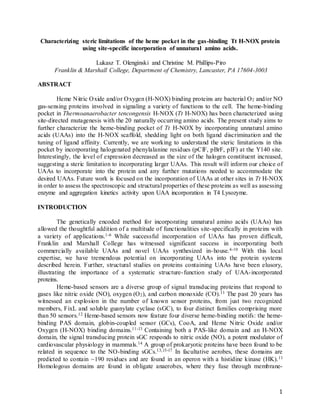
More Related Content

Similar to 141221-Fall490Final

150202-Biophysical Society Poster2
150202-Biophysical Society Poster2150202-Biophysical Society Poster2
150202-Biophysical Society Poster2Lukasz Olenginski
 
Dissecting NOS electronics_Biochem J 2013
Dissecting NOS electronics_Biochem J 2013Dissecting NOS electronics_Biochem J 2013
Dissecting NOS electronics_Biochem J 2013Luciana Hannibal, Ph.D.
 
caspase6 FMK paper BMCL2011
caspase6 FMK paper BMCL2011caspase6 FMK paper BMCL2011
caspase6 FMK paper BMCL2011Alex Kiselyov
 
Catalytic Antibodies_JACS 1987 Powell et. al.
Catalytic Antibodies_JACS 1987 Powell et. al.Catalytic Antibodies_JACS 1987 Powell et. al.
Catalytic Antibodies_JACS 1987 Powell et. al.Michael Powell
 
Carbon−Heteroatom Coupling Using Pd-PEPPSI Complexes (1)
Carbon−Heteroatom Coupling Using Pd-PEPPSI Complexes (1)Carbon−Heteroatom Coupling Using Pd-PEPPSI Complexes (1)
Carbon−Heteroatom Coupling Using Pd-PEPPSI Complexes (1)
DrMAdamSayah
 
Gonzalez et al., JACS 2011, 133, 5500-5507
Gonzalez et al., JACS 2011, 133, 5500-5507Gonzalez et al., JACS 2011, 133, 5500-5507
Gonzalez et al., JACS 2011, 133, 5500-5507Vincent M
 
J. biol. chem. 1986-fushitani-8414-23
J. biol. chem. 1986-fushitani-8414-23J. biol. chem. 1986-fushitani-8414-23
J. biol. chem. 1986-fushitani-8414-23guestfebc32d
 
tuning the pH Response of i-Motif DNA Oligonucleotides_Lannes_et_al-2015-Chem...
tuning the pH Response of i-Motif DNA Oligonucleotides_Lannes_et_al-2015-Chem...tuning the pH Response of i-Motif DNA Oligonucleotides_Lannes_et_al-2015-Chem...
tuning the pH Response of i-Motif DNA Oligonucleotides_Lannes_et_al-2015-Chem...saheli halder
 
Lysozyme
LysozymeLysozyme
Lysozyme
RGCL
 
Guest Molecule-Responsive Functional CaPhosphonates proton conductivity (JACS)
Guest Molecule-Responsive Functional CaPhosphonates proton conductivity (JACS)Guest Molecule-Responsive Functional CaPhosphonates proton conductivity (JACS)
Guest Molecule-Responsive Functional CaPhosphonates proton conductivity (JACS)maria papadaki
 
A STUDY OF NCOA4 EXPRESSION AND BINDING TO FERRITINS
A STUDY OF NCOA4 EXPRESSION AND BINDING TO FERRITINSA STUDY OF NCOA4 EXPRESSION AND BINDING TO FERRITINS
A STUDY OF NCOA4 EXPRESSION AND BINDING TO FERRITINS
Fernando Carmona Rodriguez-Acosta, PhD
 
8-glycolysis.ppt
8-glycolysis.ppt8-glycolysis.ppt
8-glycolysis.ppt
MuhammadRizwan863722
 
Pharmacology Of Pain Essay
Pharmacology Of Pain EssayPharmacology Of Pain Essay
Pharmacology Of Pain Essay
Leslie Lee
 

Similar to 141221-Fall490Final (20)

150202-Biophysical Society Poster2
150202-Biophysical Society Poster2150202-Biophysical Society Poster2
150202-Biophysical Society Poster2
 
150426-490 Presentation
150426-490 Presentation150426-490 Presentation
150426-490 Presentation
 
Dissecting NOS electronics_Biochem J 2013
Dissecting NOS electronics_Biochem J 2013Dissecting NOS electronics_Biochem J 2013
Dissecting NOS electronics_Biochem J 2013
 
CcP2APX_Biochem_2008
CcP2APX_Biochem_2008CcP2APX_Biochem_2008
CcP2APX_Biochem_2008
 
caspase6 FMK paper BMCL2011
caspase6 FMK paper BMCL2011caspase6 FMK paper BMCL2011
caspase6 FMK paper BMCL2011
 
Catalytic Antibodies_JACS 1987 Powell et. al.
Catalytic Antibodies_JACS 1987 Powell et. al.Catalytic Antibodies_JACS 1987 Powell et. al.
Catalytic Antibodies_JACS 1987 Powell et. al.
 
MBHPurificationJBC
MBHPurificationJBCMBHPurificationJBC
MBHPurificationJBC
 
ol902123h
ol902123hol902123h
ol902123h
 
ol902123h
ol902123hol902123h
ol902123h
 
Carbon−Heteroatom Coupling Using Pd-PEPPSI Complexes (1)
Carbon−Heteroatom Coupling Using Pd-PEPPSI Complexes (1)Carbon−Heteroatom Coupling Using Pd-PEPPSI Complexes (1)
Carbon−Heteroatom Coupling Using Pd-PEPPSI Complexes (1)
 
Gonzalez et al., JACS 2011, 133, 5500-5507
Gonzalez et al., JACS 2011, 133, 5500-5507Gonzalez et al., JACS 2011, 133, 5500-5507
Gonzalez et al., JACS 2011, 133, 5500-5507
 
J. biol. chem. 1986-fushitani-8414-23
J. biol. chem. 1986-fushitani-8414-23J. biol. chem. 1986-fushitani-8414-23
J. biol. chem. 1986-fushitani-8414-23
 
tuning the pH Response of i-Motif DNA Oligonucleotides_Lannes_et_al-2015-Chem...
tuning the pH Response of i-Motif DNA Oligonucleotides_Lannes_et_al-2015-Chem...tuning the pH Response of i-Motif DNA Oligonucleotides_Lannes_et_al-2015-Chem...
tuning the pH Response of i-Motif DNA Oligonucleotides_Lannes_et_al-2015-Chem...
 
Lysozyme
LysozymeLysozyme
Lysozyme
 
Guest Molecule-Responsive Functional CaPhosphonates proton conductivity (JACS)
Guest Molecule-Responsive Functional CaPhosphonates proton conductivity (JACS)Guest Molecule-Responsive Functional CaPhosphonates proton conductivity (JACS)
Guest Molecule-Responsive Functional CaPhosphonates proton conductivity (JACS)
 
A STUDY OF NCOA4 EXPRESSION AND BINDING TO FERRITINS
A STUDY OF NCOA4 EXPRESSION AND BINDING TO FERRITINSA STUDY OF NCOA4 EXPRESSION AND BINDING TO FERRITINS
A STUDY OF NCOA4 EXPRESSION AND BINDING TO FERRITINS
 
8-glycolysis.ppt
8-glycolysis.ppt8-glycolysis.ppt
8-glycolysis.ppt
 
Pharmacology Of Pain Essay
Pharmacology Of Pain EssayPharmacology Of Pain Essay
Pharmacology Of Pain Essay
 
C4DT02257B
C4DT02257BC4DT02257B
C4DT02257B
 
CcP_IJQC_2001
CcP_IJQC_2001CcP_IJQC_2001
CcP_IJQC_2001
 

141221-Fall490Final

  • 1. 1 Characterizing steric limitations of the heme pocket in the gas-binding Tt H-NOX protein using site-specific incorporation of unnatural amino acids. Lukasz T. Olenginski and Christine M. Phillips-Piro Franklin & Marshall College, Department of Chemistry, Lancaster, PA 17604-3003 ABSTRACT Heme Nitric Oxide and/or Oxygen (H-NOX) binding proteins are bacterial O2 and/or NO gas-sensing proteins involved in signaling a variety of functions to the cell. The heme-binding pocket in Thermoanaerobacter tencongensis H-NOX (Tt H-NOX) has been characterized using site-directed mutagenesis with the 20 naturally occurring amino acids. The present study aims to further characterize the heme-binding pocket of Tt H-NOX by incorporating unnatural amino acids (UAAs) into the H-NOX scaffold, shedding light on both ligand discrimination and the tuning of ligand affinity. Currently, we are working to understand the steric limitations in this pocket by incorporating halogenated phenylalanine residues (pClF, pBrF, pIF) at the Y140 site. Interestingly, the level of expression decreased as the size of the halogen constituent increased, suggesting a steric limitation to incorporating larger UAAs. This result will inform our choice of UAAs to incorporate into the protein and any further mutations needed to accommodate the desired UAAs. Future work is focused on the incorporation of UAAs at other sites in Tt H-NOX in order to assess the spectroscopic and structural properties of these proteins as well as assessing enzyme and aggregation kinetics activity upon UAA incorporation in T4 Lysozyme. INTRODUCTION The genetically encoded method for incorporating unnatural amino acids (UAAs) has allowed the thoughtful addition of a multitude of functionalities site-specifically in proteins with a variety of applications.1-6 While successful incorporation of UAAs has proven difficult, Franklin and Marshall College has witnessed significant success in incorporating both commercially available UAAs and novel UAAs synthesized in-house.4-10 With this local expertise, we have tremendous potential on incorporating UAAs into the protein systems described herein. Further, structural studies on proteins containing UAAs have been elusory, illustrating the importance of a systematic structure-function study of UAA-incorporated proteins. Heme-based sensors are a diverse group of signal transducing proteins that respond to gases like nitric oxide (NO), oxygen (O2), and carbon monoxide (CO).11 The past 20 years has witnessed an explosion in the number of known sensor proteins, from just two recognized members, FixL and soluble guanylate cyclase (sGC), to four distinct families comprising more than 50 sensors.12 Heme-based sensors now feature four diverse heme-binding motifs: the heme- binding PAS domain, globin-coupled sensor (GCs), CooA, and Heme Nitric Oxide and/or Oxygen (H-NOX) binding domains.11-13 Containing both a PAS-like domain and an H-NOX domain, the signal transducing protein sGC responds to nitric oxide (NO), a potent modulator of cardiovascular physiology in mammals.14 A group of prokaryotic proteins have been found to be related in sequence to the NO-binding sGCs.13,15-17 In facultative aerobes, these domains are predicted to contain ~190 residues and are found in an operon with a histidine kinase (HK).11 Homologous domains are found in obligate anaerobes, where they fuse through membrane-
  • 2. 2 spanning region to a predicted methyl-accepting chemotaxis protein (MCP) domain.11 Similar to the sGCs, the heme domains from facultative aerobes bind NO, but not O2. In contrast, the heme domain bound to and MCP from the obligate anaerobe Thermoanaerobacter tengcongensis binds O2, NO, and CO.13 In light of this discovered specificity for oxygen in some of these domains they have also been referred to as Heme Nitric Oxide and/or Oxygen (H-NOX) binding domains. Pellicena et al. (2004) reported the crystal structure of the O2-bound H-NOX domain from T. tengcongensis (Tt H-NOX). Structural analysis reveals the H-NOX family to have evolved a novel protein fold consisting of seven -helices and a four-stranded anti-parallel - sheet (Figure 1). A major finding from the structure of the Tt H-NOX domain and critical to ligand discrimination is the hydrogen bonding (H-bonding) network that surrounds the bound O2 molecule.11,15,18-21 Y140 is involved in a 2.7 Å H-bond to O2 as well as to N74 and W9 are (Figure 1).11 H-NOX sequence alignments strongly suggest that all three of these residues are unique to members of the H-NOX family that bind O2.11 Gas ligand affinity to the heme of H-NOX proteins has been studied using site-directed mutagenesis (SDM) with the 20 naturally occurring amino acids.18,20,22-30 For instance, P115, a conserved residue in the H-NOX family, was mutated to an alanine and used to demonstrate that decreasing heme distortion increases affinity for oxygen, providing a clear link between the heme conformation and Tt H-NOX structure.19 Y140L provided further evidence that the distal Figure 1. The heme binding pocket of Tt H-NOX with O2 bound (PDB ID: 1U55). The heme and some residues shown in orange sticks. Y140 and H-bonding network known to be crucial for tight O2 binding affinity.
  • 3. 3 pocket tyrosine not only stabilizes the O2 complex but also discriminates between NO and O2 using a kinetic selection.18 Furthermore, the double mutant F78Y/Y140L demonstrated that O2 binding could be rescued as long as there was a distal pocket tyrosine.11,18 The protein has been cloned into the appropriate expression vector, all necessary mutagenesis has been completed, the expression protocol has been optimized, and the purification protocol was troubleshot. Most significantly, our SDS-PAGE gels have shown evidence of steric limitation in incorporating larger UAAs (Figure 4, 5). Thus, we initially aimed to further characterize the size limitations in the heme pocket by using the halogenated phenylalanine UAAs. However, based on the nontrivial nature of Tt H-NOX purification, we decided to expand our study, focusing on the incorporation of UAAs at other sites in Tt H-NOX in order to assess the spectroscopic and structural properties of these proteins as well as assessing enzyme activity and aggregation kinetics upon UAA incorporation in T4 Lysozyme (Figure 2). The study of Tt H-NOX allows the following three questions to be explored: how does UAA incorporation alter (1) protein environment (2) protein structure (3) protein function. The present work with the halogenated phenylalanine UAAs has informed our decision to further mutate the heme pocket in order to accommodate UAAs of interest and investigate question (3). However, the other two questions (1, 2) can be addressed via assessing the spectroscopic and structural properties of Tt H-NOX proteins with UAAs incorporated at other sites. The study of Y24 Y18 F67 F153 Figure 2. Structure of T4 Lysozyme (PDB ID 148L) with a substrate mimic bound and shown in magenta sticks and colored by atom type. Sites for UAA incorporation are shown in yellow sticks and colored by atom type.
  • 4. 4 T4 Lysozyme allows a different approach to answering question (3), as well as introducing a new question: (4) how does UAA incorporation alter protein aggregation kinetics. MATERIALS AND METHODS Cloning Tt H-NOX into appropriate expression vector. Tt H-NOX was cloned out of the pCW vector (Appendix A) and digested with the restriction enzymes NcoI and XhoI (Appendix B). Simultaneously, the pBAD vector containing sfGFP was digested with the same restrictions enzymes and the Tt H-NOX insert was ligated into the empty pBAD vector using the following plasmid/insert ratios – 1:1, 1:2, 2:1 (Appendix B). SDM to create Tt H-NOX Amber (TAG) mutants. Successful cloning of Tt H-NOX out of pCW and into pBAD required the second residue (K) to be mutated to glutamate, and thus once it was in pBAD it was mutated back to the native lysine using the QuikChange® protocol (Appendix C). The C terminus His6-tag was removed (necessary for successful crystallization) by the insertion of a TAA stop codon after the protein sequence and before the His6-tag (Appenix A). Lastly, the amber (TAG) codon was inserted at the Y140 position in order for the pDULE vector – containing both the tRNA and the aminoacyl-tRNA synthetase – to add UAAs (Appendix B). The end result of the multiple rounds of SDM was the non-His6-tagged version of Tt H-NOX with an amber (TAG) codon at the Y140 site. All other Amber mutants (F52, F78, Y85, F151, F169, F183, Y185) were created in the same manner – using the QuikChange® protocol (Appendix C). SDM to create T4Lys Amber (TAG) mutants. Successful cloning of T4Lys out of pHS1403 and into pBAD required the second residue (N) to be mutated to aspartic acid and thus once it was in pBAD it was mutated back to the native asparagine using the QuikChange® protocol (Appendix C). The C terminus His6-tag was removed (necessary for successful crystallization) by the insertion of a TAA stop codon after the protein sequence and before the His6-tag (Appenix C). Lastly, the amber (TAG) codon was inserted at the following sites – Y18, Y24, F67, and F153. Tt H-NOX Expression. Appropriate Tt H-NOX expression construct and desired pDULE synthetase construct were dual-transformed into chemically competent DH10B E. coli cells in the afternoon and then plated on LB/Ampicillin/Tetracycline Agar plates and incubated at 37 C overnight (Appenidx D). The next morning, the plates were taken out of the incubator, parafilmed, and stored in the refrigerator at 4 C. Later that afternoon, the plates were taken out of the refrigerator and used to make starter cultures in non-inducing media (Appendix E). The next morning, auto-induction media was prepared (Appendix E). Expression volume was 250 ml in a 500 ml baffled flask. Expression cultures were inoculated with starter culture (1ml:1 L) and grown up at 37 C and 250 rpm for 30-36 hrs. Directly following incubation of the expression cultures, the UAA solutions were prepared (Appendix E). Approximately 1 hour after the start of incubation, the UAA solution (1 uM) and heme-precursor, 5-aminolevulinic acid (1 uM), were added to the cultures. After 30-36 hrs, the cultures were spun down at 5000 rpm for 10 min and the cell pellets were flash frozen and stored at -80 C. Expressions were also performed in a yeast extract media. The Tt H-NOX expression construct was transformed into the same cell line as described above. However, when making starter cultures, LB broth was used rather than non-inducing media. The yeast extract media was prepared the day before the expression. Expression volume was 1 L in a 2 L baffled flask; thus
  • 5. 5 45 g yeast extract, 10 ml glycerol, and 900 ml ddH2O were added to each flask and autoclaved. Before inoculation 100 ml of a 170 mM KH2PO4, 720 mM K2HPO4 phosphate buffer was added to each expression culture. Cultures were inoculated as described above and allowed to grow at 37 C and 250 rpm. Once the OD600 reached ~ 0.7-0 .8 the incubation temperature was dropped to 18 C and the cultures were induced with the same amount of 20% arabinose as prescribed in the auto-induction media (2.5 ml/L). UAA and ALA solution were added in the same manner as before (1 hr after induction). Cultures grew at 18 C and 250 rpm overnight and the next morning the cell pellet was collected in the same fashion as mentioned above. Tt H-NOX Purification. Round 1 – Cell pellet was thawed on ice, re-suspended in either 10 ml (250 ml culture) or 40 ml (1 L culture) Lysis Buffer (50 mM TEA, pH 8.5, 20 mM NaCl), and lysed by the addition of Lysozyme (0.25 mg/ml). PMSF (0.5 mM) and DNAse (3.75 mM) were also added in order to inhibit endogenous proteases from degrading our protein and breaking down the DNA, respectively. Lysis solution was then sonicated at 40 % amplitude for 2 min (2 s pulse on, 2 s pulse off) and boiled at 70 C for 30 min. Samples were then spun down at 20,000 rpm for 45 min and the supernatant was flash frozen and stored at -80 C. Purity and presence of protein was analyzed by SDS-PAGE, with each gel containing a re-suspension, pellet, and supernatant sample. Round 2 – The supernatant (~50 ml) was thawed and loaded onto a ~80-100 ml Q-650 column (anion exchange) pre-equilibrated with ~150 ml Lysis Buffer. Protein was run through column using the Lysis Buffer at 1.5 ml/min. Red fractions (~ 30 ml) were collected and concentrated to 2.5 ml. The protein was then run over a pre-packed PD10 column pre- equilibrated with 25 ml Buffer B (50 mM HEPES, pH 6.5, 5% glycerol). After Tt H-NOX protein was loaded, 3.5 ml Buffer B was added to make protein elute. Again, the red fractions were collected. Then, the protein was loaded onto a ~ 80-100 ml CM-650 column (cation exchange) pre-equilibrated with ~150 ml Buffer B and run through at 1.5 ml/min. All red colored fractions were collected and analyzed with SDS-PAGE to inform which fractions should be combined, concentrated, and ran over the S75 column. The CM-650 fractions that included Tt H- NOX protein were combined (~25 ml) and concentrated down to ~ 4 ml. Protein was then filtered through syringe filter, leaving 3 ml Tt H-NOX protein. Proteins was then loaded onto the S75 column (size exclusion) pre-equilibrated with Buffer C (50 mM TEA, pH 7.5, 50 mM NaCl, 5% glycerol) and run at 0.20 ml/min. with the max pressure set at 0.27 MPa. Fractions near peaks corresponding to the correct MW (~ 22 kDa) were analyzed by SDS-PAGE. Fractions containing Tt H-NOX protein were combined and concentrated down to ~500 ul and concentration was assessed by a Wave Scan. RESULTS
  • 6. 6 Addition of ALA aids expression of Tt H-NOX. Early expression attempts in auto-induction media with both pCNF and mNO2Y resulted in low expression yields. Further, expression cultures lacked the red color, indicative of heme-incorporation. Thus, we added the heme precursor, 5-aminolevulinic acid (ALA), to the expression cultures and not only saw redder cultures but increased expression as well (Figure 3). Steric limitations involved in the heme pocket of Tt H-NOX. Despite the addition of ALA aiding the expression of Tt H-NOX, consistent expression with pCNF and mNO2Y was not observed. This led us to ponder whether our expression protocol was flawed. Thus, we set up a 5 ml test expression in auto-induction media with both Wt Tt H-NOX_His6 and Tt H- NOX_Y140_His6 with the following UAAs – pCNF, pClF, pBrF, pIF, pNH2F, and mNO2Y – to tease out whether the problems encountered were a result of our expression protocol or simply the UAA incorporation. The His6 constructs were used due to their simpler purification protocol (in case further characterization was necessary). Interestingly, we saw expression of both the Wt Tt H-NOX_His6 and the Tt H-NOX_Y140_His6 with the halogenated phenylalanine UAAs, suggesting that neither our expression protocol or our method of incorporating the UAA is flawed, but rather the UAA itself is what is limiting (Figure 4). In other words, these data suggest that there is a steric limitation to what UAAs can be incorporated into the heme pocket. Figure 3. Impact of addition of ALA on Tt H-NOX_Y140 + pCNF expression. Parallel 250 ml expressions were setup in 500 ml baffled flasks both with and without addition of ALA. (A) Expression cultures after 30-36 hr growth period. (B) Pellet following growth period. (C) Purification supernatant. (D) Purification pellet. (E) SDS-PAGE gel of Tt H-NOX_Y140 + pCNF post round 1 purification with (lanes 3-5) and without (lanes 6-8) addition of ALA.
  • 7. 7 Still, we were not completely convinced by the data for multiple reasons – the expression volumes were very small and we had yet to see consistent results with the auto-induction media in the first place. Thus, we decided to try the expression of the Tt H-NOX_Y140 with the halogenated phenylalanine UAAs in a media that we had recent success with on the Wt Tt H- NOX – the yeast extract media. We saw the same trend of the larger the size of the halogen constituent at the para-position of the phenylalanine ring, the smaller the level of expression Further, we were able to show that we could express the halogenated phenylalanine UAAs with larger culture sizes (Figure 4). Thus, the data in Figure 4 not only support the steric hypothesis but also highight that we can express Tt H-NOX_Y140 constructs with UAAs liters worth at a time, which ultimately allows us to set crystal trays and elucidate the structural consequences of UAA incorporation into the H-NOX scaffold. Figure 4. SDS-PAGE gel of Tt H-NOX_His6 + various UAA constructs following round 1 of purification. Samples were run on a self-poured 12 % SDS-PAGE gel with Precision Plus Protein™ Kaleidoscope™ (BIORAD, #161-0375) and 2x Laemmli Sample Buffer dye (BIO-RAD, #161-0737) at 200 V for 45 min. As indicated by lane 2 on each gel Tt H-NOX protein should appear ~ 22 kDa. (A) As seen in lane 4, 6, 8, and 10 only the halogenated phenylalanine constructs seemed to express well. (B) None of the UAA containing Tt H-NOXconstructs expressed.
  • 8. 8 DISCUSSION The first half of the fall semester was plagued by unsuccessful purification attempts with Tt H-NOX. Despite the tremendous progress in cloning the appropriate expression constructs and expressing Tt H-NOX with halogenated phenylalanine UAAs, we could not acquire sufficient protein yield for crystallographic work. The reasoning for the small yields seemed to be the purification protocol, and thus minor changes were made. Notably, following use of the Q-650 anion exchange column the protein was concentrated and loaded right onto the S75 size exclusion column, skipping the CM-650 cation exchange column completely. This modification saved time but did not sacrifice protein purity as concluded from SDS-PAGE analysis of a gel containing Tt H-NOX protein post anion exchange and post cation exchange. Despite saving time with skipping the cation exchange column, a lot of difficulty was encountered with the S75 Figure 5. Size limitations in the heme pocket of Tt H-NOX. (A) side and (B) top view of heme pocket in Tt H-NOX (PDB ID: IU55) using surface representation. (C) SDS-PAGE gel of both Wt Tt H-NOX and Tt H-NOX_Y140 with halogenated phenylalanine UAAs following round 1 of purification. Samples were run on a self-poured 12 % SDS-PAGE gel with Precision Plus Protein™ Kaleidoscope™ (BIORAD, #161-0375) and 2x Laemmli Sample Buffer dye (BIO-RAD, #161-0737) at 200 V for 45 min. As indicated by lane 2 Tt H-NOXprotein should appear ~ 22 kDa. We see successfulexpression of all constructs.
  • 9. 9 size exclusion column on the FPLC. Specifically, during the semester it malfunctioned several of times and crippled our efforts to move the project forward and purify protein. We did, however, purify Tt H-NOX + pIF (~1.5 ml, ~7 mg/ml), but again this amount was insufficient for crystallographic work. As mentioned, there are a variety of future directions for next semester. To begin, we will further mutate the heme pocket in an attempt to accommodate larger UAAs. Molecular analysis of the heme pocket reveals the phenyl ring of F78 to be in very close proximity to Y140 (Figure 6). Thus, we propose to mutate F78 to alanine, freeing up space for the meta and para positions on the phenyl ring of Y140. With the extra space in the pocket, we plan to incorporate unnatural tyrosine analogs with either withdrawing groups (EWG) or electron donating groups (EDG) at the para-position on the phenyl ring to either make the tyrosine a better or poorer hydrogen bond donor and thus a stronger or weaker O2 binder, respectively (Figure 7). Ultimately, we will structurally characterize the mutant H-NOX proteins and assess their O2 binding affinity to relate the H-bond donating ability of the tyrosine to the O2 affinity of the protein and any structural alterations that may have occurred. Thus, this study addresses question (3) discussed earlier. Further, we are not limited to solely incorporating UAAs at the Y140 site. We plan to express Tt H-NOX at a variety of sites with different protein environments (Appendix F). We chose to start with five sites on the protein, all of which are natively tyrosine or phenylalanine residues to ensure a most conservative replacement. We have chosen sites that are fully buried (Y52, Y85), partially buried (F151, F169, F183), and solvent exposed (Y185) (Figure 8). These sites offer the potential to report on protein environment and protein structure upon UAA incorporation, and thus addresses questions (1, 2) discussed earlier. The former can be Y140 F78 Y140 A78 Figure 6. Heme pocket of Tt H-NOX (PDB ID: 1U55). Protein shown in surface representation with van der Waal radii of the atoms in Y140 and F78 shown. Left image shows F78 modeled in whereas the left image shows the propsed F78 mutant modeled in.
  • 10. 10 accomplished with vibrational reporter UAAs and the later can be accomplished with X-ray crystallography. Figure 7. Method of tuning the H-bonding environment above the heme pocket in Tt H-NOX. Figure 6. Tt H-NOX showed in cartoon representation (PDB ID: IU55) with select tyrosine and phenylalanine residues shown in yellow sticks and colored by atom.
  • 11. 11 As mentioned, if we encounter similar problems with purifying Tt H-NOX, the focus of the study might shift towards T4 Lysozyme. Much like in Tt H-NOX, we have chosen a variety of sites with different protein environments – sites lining the substrate-binding pocket (Y18, Y24) and two sites far from the substrate-binding pocket (F67, F153) (Figure 2). Again, these sites offer the potential to report on protein environment and protein structure upon UAA incorporation, and thus addresses questions (1, 2) discussed earlier. Additionally, study of T4 Lysozyme addresses question (3), but in a different way than Tt H-NOX. Rather than assessing altered gas binding, T4 Lysozyme sheds light on whether the incorporation of UAAs directly impacts enzyme catalysis. Once purified mutant T4 Lysozyme protein is obtained, we can begin to look at its activity, which is straightforward due to the variety of commercially available enzyme assays for this protein. Lastly, due to this protein’s propensity to aggregate we can explore whether incorporation of UAAs alters aggregation kinetics, addressing question (4) discussed earlier. The scope of this project has become quite large, however we are interested in very specific questions (1-4) and have developed thoughtful ways of investigating them. At this point, the mutagenesis to create all of the Tt H-NOX and T4 Lysozyme Amber mutants described herein has been completed. Further, they have been dually transformed into chemically competent DH10B E. coli cells with the pCNF synthetase. Thus, we have what we need in order to start expressing new protein and progress this project forward. REFERENCES (1) Seyedsayamdost, M. R., Yee, C. S., and Stubbe, J. (2007) Site-specific incorporation of fluorotyrosines into the R2 subunit of E. coli ribonucleotide reductase by expressed protein ligation. Nat Protoc 2, 1225–1235. (2) Wang, L., Xie, J., Deniz, A. A., and Schultz, P. G. (2003) Unnatural amino acid mutagenesis of green fluorescent protein. J. Org. Chem. 68, 174–176. (3) Groff, D., Wang, F., Jockusch, S., Turro, N. J., and Schultz, P. G. (2010) A New Strategy to Photoactivate Green Fluorescent Protein. Angew. Chem. Int. Ed. 49, 7677–7679. (4) Miyake-Stoner, S. J., Miller, A. M., Hammill, J. T., Peeler, J. C., Hess, K. R., Mehl, R. A., and Brewer, S. H. (2009) Probing protein folding using site-specifically encoded unnatural amino acids as FRET donors with tryptophan. Biochemistry 48, 5953–5962. (5) Bazewicz, C. G., Lipkin, J. S., Smith, E. E., Liskov, M. T., and Brewer, S. H. (2012) Expanding the utility of 4-cyano-L-phenylalanine as a vibrational reporter of protein environments. J Phys Chem B 116, 10824–10831. (6) Bazewicz, C. G., Liskov, M. T., Hines, K. J., and Brewer, S. H. (2013) Sensitive, site- specific, and stable vibrational probe of local protein environments: 4-azidomethyl-L- phenylalanine. J Phys Chem B 117, 8987–8993. (7) Pavic, K., Rios, P., Dzeyk, K., Koehler, C., Lemke, E. A., and Köhn, M. (2014) Unnatural Amino Acid Mutagenesis Reveals Dimerization As a Negative Regulatory Mechanism of VHR's Phosphatase Activity. ACS Chem. Biol. (8) Jackson, J. C., Duffy, S. P., Hess, K. R., and Mehl, R. A. (2006) Improving nature's enzyme active site with genetically encoded unnatural amino acids. J. Am. Chem. Soc. 128, 11124– 11127. (9) Taskent-Sezgin, H., Chung, J., Patsalo, V., Miyake-Stoner, S. J., Miller, A. M., Brewer, S. H., Mehl, R. A., Green, D. F., Raleigh, D. P., and Carrico, I. (2009) Interpretation of p-
  • 12. 12 cyanophenylalanine fluorescence in proteins in terms of solvent exposure and contribution of side-chain quenchers: a combined fluorescence, IR and molecular dynamics study. Biochemistry 48, 9040–9046. (10) Smith, E. E., Linderman, B. Y., Luskin, A. C., and Brewer, S. H. (2011) Probing local environments with the infrared probe: L-4-nitrophenylalanine. J Phys Chem B 115, 2380–2385. (11) Pellicena, P., Karow, D. S., Boon, E. M., Marletta, M. A., and Kuriyan, J. (2004) Crystal structure of an oxygen-binding heme domain related to soluble guanylate cyclases. Proc. Natl. Acad. Sci. 101, 12854-12859. (12) Gilles-Gonzalez, M. A., and Gonzalez, G. (2005) Heme-based sensors: defining characteristics, recent developments, and regulatory hypotheses. J. Inorg. Biochem. 99, 1-22. (13) Karow, D. S., Pan, D., Tran, R., Pellicena, P., Presley, A., Mathies, R. A., and Marletta, M.A. (2004) Spectroscopic characterization of the soluble guanylate cyclase-like heme domains from Vibrio cholera and Thermoanaerobacter tengcongensis. Biochemistry. 43, 10203-10211. (14) Denninger, J. W. and Marletta, M. A. (1999) Guanylate cyclase and the NO/cGMP signaling pathway. Biochim. Biophy. Acta. 1411, 334-350. (15) Schmidt, P. M., Schramm, M., Schröder, H., Wunder, F., and Stasch, J. P. (2004) Identification of residues crucially involved in the binding of heme moiety of soluble guanylate cyclase. J. Biol. Chem. 279, 3025-3032. (16) Winger, J. A., Derbyshire, E. R., and Marletta, M. A. (2007) Dissociation of nitric oxide from soluble guanylate cyclase and heme-nitric oxide/oxygen binding domain constructs. J. Biol. Chem. 282, 897-907. (17) 12. Boon, E. M., Davis, J. H., Karow, D .S., Huang, S. H., Tran, R., Miazgowicz, M. M., Mathies, R., and Marletta, M. A. (2006) Nitric oxide binding to prokaryotic homologs of the soluble guanylate cyclase b1 H-NOX domain. J. Biol. Chem. 281, 21892-902. (18) Boon, E. M., Huang, S. H., and Marletta, M. A. (2005) A molecular basis for NO selectivity in soluble guanylate cyclases. Nat. Chem. Biol. 1, 53-59. (19) Olea, C., Boon, E. M., Pellicena, P., Kuriyan, J., and Marletta, M. A. (2008) Probing the function of heme distortion in the H-NOX family. ACS Chem. Biol. 3, 703-710. (20) Weinert, E. E., Plate, L., Whited, C. A., Olea, C. Jr., and Marletta, M. A. (2010) Determinants of ligand affinity and heme reactivity in H-NOX domains. Angew. Chem., Int. Ed. 49, 720-723. (21) Martin, E., Berka, V., Bogatenkova, E., Murad, F., and Tsai, A. (2006) Ligand selectivity of guanylyl cyclase: effect of the hydrogen-binding tyrosine in the distal heme pocket on binding of oxygen, nitric oxide, and carbon monoxide. J. Biol. Chem. 281, 27836-27845. (22) Weinert, E. E., Phillips-Piro, C. M., Tran, R., Mathies, R. A., and Marletta, M. A. (2011) Controlling conformational flexibility of an O2-binding H-NOX domain. Biochemistry. 50, 6832- 6840. (23) Weinert, E. E., Phillips-Piro, C. M., and Marletta, M. A. (2013) Porphyrin π-stacking in a heme protein scaffold tunes gas ligand affinity. J. Inorg. Biochem. 127, 7–12. (24) Olea, C., Jr., Kuriyan, J., and Marletta, M. A. (2010) Modulating Heme Redox Potential through Protein-Induced Porphyrin Distortion. J. Am. Chem. Soc. 132, 12794–12795. (25) Olea, C., Boon, E. M., Pellicena, P., Kuriyan, J., and Marletta, M. A. (2008) Probing the Function of Heme Distortion in the H-NOX Family. ACS Chem. Biol. 3, 703–710. (26) Olea, C., Jr., Herzik, M. A., Jr., Kuriyan, J., and Marletta, M. A. (2010) Structural insights into the molecular mechanism of H-NOX activation. Protein Science 19, 881–887.
  • 13. 13 (27) Derbyshire E. R., Deng S., and Marletta M. A. (2010) Incorporation of tyrosine and glutamine residues into the soluble guanylate cyclase heme distal pocket alters NO and O2 binding. J Biol Chem. 285, 17471-8. (28) Tran R., Boon E. M., Marletta M. A., and Mathies R. A. (2009) Resonance raman spectra of an O2-binding H-NOX domain reveal heme relaxation upon mutation. Biochemistry. 48, 8568- 77. (29) Kosowicz, J. G. and Boon, E. M. (2013) Insights into the distal pocket of H-NOX using fluoride as a probe for H-bonding interactions. J Inorg. Biochem. 126, 91-5. (30) Dai, Z. and Boon, E. M. (2011) Probing the local electronic and geometric properties of the heme iron center in an O2-binding Heme-Nitric oxide and/or Oxygen binding domain. J Inorg. Biochem. 105, 784-792.
  • 14. 14 APPENDIX A. Cloning Tt H-NOX out of pCW NcoI – GAG – residue 3 – F 5’ – GC CC ATG GAG GGG ACA ATC GTC GGG ACA TGG ATA AAG ACC C – 3’ Tt H-NOX – STOP – XhoI - R 5’ – CG CTCGAG TTA ATT TTT CTT ATA CTC AAA AAC GGG G – 3’ Cloning Reaction Volume (ul) Tt H-NOX in pCW (25 ng) 0.425 Primer – F (10 uM) 2 Primer – R (10 uM) 2 10 X Buffer 5 ddH2O 38.35 dNTP mix (10 mM) 1.25 Pfu II Turbo Polymerase 1 50 ul Total Reaction Ran PCR program “PIRO PCR” (subdirectory 1, program 1) 1. 95 C for 6 min. 2. 95 C for 30 s. 3. 55 C for 30 s. 4. 72 C for 2 min, 30 s. 5. Repeat steps 2-4 (34x) 6. 72 C for 10 min. 7. 4 C for 10 min. 8. 10 C forever B. Digestion and Ligation Protocols Single Digestion Reaction Volume (ul) XhoI/NcoI 1 pBAD-GFP (1 ug) 6 10 X Cutsmart Buffer 5 ddH2O 38 50 ul Total Reaction
  • 15. 15 Double Digestion Reaction Volume (ul) XhoI NcoI 1 1 pBAD-GFP (1 ug) 6 10 X Cutsmart Buffer 5 ddH2O 37 50 ul Total Reaction Ligation Reactions 1:1 Reaction (ul) 2:1 Reaction (ul) 1:2 Reaction (ul) 10 X DNA Ligase Buffer 2 2 2 pBAD + NcoI + XhoI 2 4 2 Tt H-NOX_Y140 insert 2 2 4 Nuclease free water 13 11 11 T4 DNA Ligase 1 1 1 20 ul Total 20 ul Total 20 ul Total Sequencing results of Tt H-NOX_Y140 in pBAD Y140 construct exhibits incorrect second residue and nonsense codon at position Y140, indicative of successful entry into pBAD
  • 16. 16 C. SDM to create appropriate expression constructs Construct Forward primer (5’-3’) Reverse Primer (5’-3’) Tt H-NOX-Y140-E2K GGGCTAACAGGAGGAATTAACCAT GAAGGGGACAATCGTCGGGACATG GATAAAGACCC GGGTCTTTATCCATGTCCCGACGAT TGTCCCCTTCATGGTTAATTCCTCC TGTTAGCCC Tt H-NOX-F52Amb GAGGTTAGGAGAATTTAGGCTAAG GTGAGTGAAAAAACT AGTTTTTTCACTCACCTTAGCCTAA ATTCTCCTAACCTC Tt H-NOX_F78Amb GGCAGAACATAAAAACTTAGAGCG AATGGTTTCCCTCC GGAGGGAAACCATTCGCTCTAAGTT TTTATGTTCTGCC Tt H-NOX-F78A GGCAGAACATAAAAACTGCCAGCG AATGGTTTCCCTCC GGAGGGAAACCATTCGCTGGCAGTT TTTATGTTCTGCC Tt H-NOX-Y85Amb CGAATGGTTTCCCTCCTAGTTTGC AGGGAGAAGGCTAGTG CACTAGCCTTCTCCCTGCAAACTAG GAGGGAAACCATTCG Tt H-NOX-F151Amb ATAGAGGGTAGTTCTAAATAGTTC AAGGAAGAAATTTCAG CTGAAATTTCTTCCTTGAACTATTT AGAACTACCCTCTAT Tt H-NOX-F169Amb CGAAAGAGGCGAAAAAGATGGCTA GTCAAGGCTAAAAGTC GACTTTTAGCCTTGACTAGCCATCT TTTTCGCCTCTTTCG Tt H-NOX-F183Amb AAATTTAAAAACCCCGTTTAGGAG TATAAGAAAAATTAAC GTTAATTTTTCTTATACTCCTAAAC GGGGTTTTTAAATTT Tt H-NOX-Y185Amb CCCCGTTTTTGAGTAGAAGAAAAA TTAACTCGAGATCTGC GCAGATCTCGAGTTAATTTTTCTTC TACTCAAAAACGGGG T4Lys-STOP GGCACTTGGGACGCGTATAAAAATCTA AGCCATCATCATCATCATC GATGATGATGATGATGGCTTAGATTTTT ATACGCGTCCCAAGTGCC T4Lys-Y18Amb GAAGGTCTTAGACTTAAAATCTAGAAA GACACAGAAGGCTATTACAC GTGTAATAGCCTTCTGTGTCTTTCTAGA TTTTAAGTCTAAGACCTTC T4Lys-Y24Amb CTATAAAGACACAGAAGGCTAGTACAC TATTGGCATCGGTCATTTGC GCAAATGACCGATGCCAATAGTGTACTA GCCTTCTGTGTCTTTATAG T4Lys-F67Amb GATGAGGCTGAAAAACTCTAGAATCAG GATGTTGATGCTGC GCAGCATCAACATCCTGATTCTAGAGTT TTTCAGCCTCATC QuikChange® SDM Protocol 10 ng plasmid reaction (ul) 50 ng plasmid reaction (ul) E2K-primer-F (1 uM) 6.8 6.8 E2K-primer-R (1 uM) 6.96 6.96 pBAD_Tt H-NOXY140 0.648 3.24 Pfu Ultra II 10 X Buffer 5 5 dNTPs (10 mM) 1 1 ddH2O 28.6 (29.6 for control) 26 (27 for control) Pfu Ultra II Polymerase 1 (0 for control) 1 (0 for control) 50 ul Total Reaction 50 ul Total Reaction Ran PCR program “PIRO-SDM” (subdirectory 1, program 3) 1. 95 C for 30 s. 2. 95 C for 30 s. 3. 55 C for 30 s. 4. 68 C for 4 min, 30 s. 5. Repeat steps 2-4 (18x) 6. 10 C forever
  • 17. 17 Folowing PCR - Add 1 ul DpnI and incubate (37 C) for 1 hr and then transform SDM product Sequencing results following SDM reacions Tt H-NOX-Y140-E2K Tt H-NOX-F52Amb
  • 23. 23 T4Lys-F153Amb D. Dual-transformation of pBAD and pDule constructs Dual-transformation 1. Thaw chemically competent DH10B E. coli cells on ice 2. Pipet 50 ul DH10B cells into 15 ml culture tube 3. 1 ul pBAD_TtY140 and 1 ul pDULE construct pipetted into cells 4. Allow cells to incubate on ice for 20 min. 5. Heat shock cells in water bath (42 C) for 45 s. 6. Put cells back on ice 7. Rescue cells by adding 200 ul SOC media 8. Place 15 ml culture tubes in incubator (37 C, 250 rpm) for 1 hr. 9. While cells are incubating warm up LB/Amp/Tet plates a. Place plates right-side up with lid cock-eyed for 5-10 min. b. Put lid on and turn plates over for remainder of incubation. 10. Plate 50 ul of cells and place in 37 C bench-top incubator a. Place plates right-side up with lid cock-eyed for 5-10 min. b. Put lid on and turn plates over and leave overnight 11. Next morning take plates out of incubator, parafilm, and store in 4 C refrigerator (Perform under sterile conditions)
  • 24. 24 E. Auto-induction Expression Protocol Non-inducing Media – 50 ml 5 % Aspartate (pH 7.5) 2.5 ml (autoclave) 25 X M salts 2 ml (autoclave) 18 AA mix (25 X, 4 C) 2 ml 40 % Glucose 625 ul (autoclave) 1 M MgSO4 100 ul (autoclave) Trace metals (1000 X) 10 ul Leucine (4 mg/ml, pH 7.5) 500 ul (autoclave) Sterile ddH2O Dilute to 50 ml (autoclave) (Prepare under sterile conditions) Preparing starter cultures for pBAD/pDULE dual-constructs 1. Add to a 15 ml culture tube: 5 ml non-inducing media 5 ul ampicillin 5 ul tetracycline 2. With a P2 pipet - gently scoop up a single colony from transformation LB/Amp/Tet plate and dispense into culture tube. 3. Incubate (37 C, 250 rpm) overnight Auto-induction Media – 1 L 5 % Aspartate (pH 7.5) 50 ml (autoclave) 10 % Glycerol 50 ml (autoclave) 18 AA mix (25 X, 4 C) 40 ml 25 X M salts 40 ml (autoclave) Leucine (4 mg/ml, pH 7.5) 10 ml (autoclave) 20 % Arabinose 2.5 ml (sterile-filtered) 1 M MgSO4 2 ml (autoclave) 40 % Glucose 1.25 ml (autoclave) Trace metals (1000 X) 1 ml Sterile ddH2O Dilute to 1 L (autoclave) Expression protocol 1. Prepare necessary quantity of auto-induction media 2. Add necessary antibiotics (Amp/Tet) each with 1000 X final concentration 3. Aliquot 250 ml auto-induction media into 500 ml baffled flasks 4. Inoculate expression culture with 2.5 ml of non-inducing starter culture 5. Incubate (37 C, 250 rpm) for 30-36 hrs. 6. 1 hr after start of growth period, add both the UAA solution and ALA solution. a. UAA solution is prepared as follows: i. UAA will have 1 mM final concentration. ii. Weigh out 1/4th of the MW of desired UAA in mgs.
  • 25. 25 iii. Add 1 ml sterile ddH2O and 8 M NaOH (dropwise) to help solution dissolve. iv. Add entire solution to expression culture. b. A 1 M ALA solution is made and then 2.5 ml are added to expression culture 7. Expression cultures continue to grow for remainder of 30-36 hrs. F. UAA incorporation at other sites in Tt H-NOX Residue Total Apolar Backbone Sidechain Ratio In/out Y17 32.67 18.13 23.77 8.90 4.6 in F52 1.34 1.34 0.00 1.34 0.7 in F78 34.66 34.66 0.00 34.66 19.2 in F82 6.52 6.52 1.34 5.17 2.9 in Y85 27.54 25.75 0.38 27.16 14.1 in F86 16.48 11.03 5.44 11.03 6.1 in F94 7.55 7.55 0.00 7.55 4.2 in Y131 7.31 6.38 0.00 7.31 3.8 in Y138 19.25 18.28 0.55 18.70 9.7 in Y140 19.42 19.18 0.00 19.42 10.1 in F141 9.41 9.40 1.82 7.59 4.2 in F151 81.78 71.86 18.02 63.76 35.4 F152 28.71 16.21 18.12 10.60 5.9 in F169 84.71 84.64 0.14 84.58 47.0 F178 5.33 4.90 0.51 4.83 2.7 in F183 54.81 50.40 12.52 42.29 23.5 Y185 184.83 133.69 26.98 157.84 81.7 out Table I. Solvent accessibility data for all tyrosine and phenylalanine sites in Tt H-NOX from GETAREA software. Sites to explore highlighted in yellow. Buried sites: Y52, Y85, Y140 (which we already have); Partially buried sites: F151, F169, F183; Solvent exposed sites: Y185.