2. 2 Journal of Parenteral and Enteral Nutrition XX(X)
dependent on parenteral nutrition (PN).1
PNALD had been con-
sidered multifactorial, with the main risk factors including pre-
term birth, low birth weight, surgical resection, lack of enteral
feeding, prolonged use of PN, and recurrent sepsis from central
venous catheter–related bloodstream infections.2,3
In the past
decade, the use of soy-based parenteral lipid emulsion has
emerged as a major risk factor of cholestasis and hepatic injury.
The long-chain polyunsaturated fatty acids (PUFAs) in lipid
emulsions can undergo peroxidation and produce free radical
peroxides that are believed to contribute to the liver damage
observed in PNALD.4,5
Vitamin E is added to lipid emulsions to
reduce the risk of such peroxidation and potentially can also
provide increased antioxidant delivery, which may benefit the
patient, protecting host cellular membranes from lipid peroxi-
dation.6
Conventional soybean oil–based lipid emulsions (eg,
Intralipid; Fresenius Kabi, Bad Homburg, Germany) contain
relatively low amounts of vitamin E. In contrast, the third-gen-
eration lipid emulsions, containing fish oil, have substantially
higher α-tocopherol, the most active antioxidant form of vita-
min E, with content in the range of 8–11 times that of the con-
ventional lipid.7
Therefore, it is unclear if the benefit of treating
PNALD that has been observed with fish oil–based emulsions
is due to the PUFA content directly or to the added vitamin E.
Recent data from preterm piglets suggest that vitamin E may be
the main beneficial factor.8
Certainly, supplementing conven-
tional parenteral lipid with vitamin E would be a cost-effective
preventative strategy for PNALD. Furthermore, the mecha-
nisms of this beneficial effect need to be explored.
The aim of this research is to further investigate the role of
supplemental α-tocopherol in the prevention of PNALD. We
hypothesized adding α-tocopherol to conventional soy-based
lipid would improve bile flow and liver chemistry in associa-
tion with reduction of oxidative stress during PN therapy.
Materials and Methods
Experiments were conducted in accordance with the guidelines
of the Canadian Council of Animal Care with the approval of
the Animal Policy and Welfare Committee, University of
Alberta.
Sixteen female Large/Landrace White and Duroc cross pig-
lets (Hypor, Regina, SK, Canada) aged 2–5 days and weighing
1.8–2.5 kg were obtained from the University of Alberta Swine
Research and Technology Centre (SRTC). The animals were
randomized into 2 groups: parenteral nutrition (PN) with soy
lipid (Intralipid; Fresenius Kabi) (SO, n = 8) or the same lipid
plus vitamin E (SO+E, n = 8).
Piglets underwent general anesthesia and received a
5-French (Fr) central venous catheter in the left external jugu-
lar vein for PN delivery. Immediately after the catheter inser-
tion, piglets were housed in metabolic cages individually,
secured by a tether-swivel system (Alice King Chatman
Medical Arts, Los Angeles, CA) to allow free movement. The
room temperature was maintained at 25°C with the aid of a
heat lamp, and lighting followed a 12-hour light/dark cycle.
Broad-spectrum antibiotics, ampicillin (Sandoz, Boucherville,
Quebec, Canada) and trimethoprim-sulfadoxine (Merck
Animal Health, Kirkland, Quebec, Canada), were administered
from days 0–4 and 8–12 to prevent catheter sepsis.
PN was started at 50% of the target volume of 324 mL/kg/d
(13.5 mL/kg/h) and advanced by 25% up to 100%, every 12
hours. Final target nutrient intakes were 282 kcal/kg/d, 18 g amino
acids/kg, and 10 g fat/kg; these are based on maintaining growth
and protein accretion for PN-fed piglets in comparison to sow-fed
full-term newborn piglets.9
The PN solutions were prepared as an
all-in-one admixture under sterile conditions in our laboratory as
previously described.10
All infusion bags were light protected to
reduce lipid peroxidation. In the vitamin E–supplemented group
(SO+E), an extra 0.5 mg DL–α-tocopherol acetate (Ephynal;
Bayer Hispania, S.L., Barcelona, Spain) was added per gram of
lipid from Intralipid, immediately prior to the PN infusion com-
mencing. This represents 0.67 mg α-tocopherol equivalents or
0.34 mg α-tocopherol per gram of Intralipid. Considering its
γ-tocopherol content but converting it to α-tocopherol equivalents,
Intralipid has a natural vitamin E content of 0.25–0.40 mg
α-tocopherol equivalents/g of lipid. Accordingly, the final total
concentration of vitamin E after supplementation was 120–150
mg/L α-tocopherol equivalent (α-TE).5,7
This amount is in the
equivalent range of the third-generation fish oil emulsions:
Omegaven (approximately 170 mg/L α-TE; Fresenius Kabi) and
SMOF lipid (130 mg/L α-TE; Fresenius Kabi).7
To provide a normative range for all data, we also studied 8
healthy sow-fed newborn piglets at an equivalent age (CON)
for all the measured outcomes. Raised under standard farm
conditions at the SRTC, they represent the gold standard for
postnatal growth and development of preweaned piglets.
Serum Vitamin E
To confirm supplemental vitamin E, the total serum vitamin E
levels were measured in all groups on day 17, analyzed by high-
performance liquid chromatography (Michigan State University,
Diagnostic Center for Population and Animal Health).
Bile Flow and Liver Chemistry
On day 17, as a primary outcome of liver function, bile flow
was measured as previously reported.11,12
Briefly, with the gall-
bladder emptied and the cystic duct ligated, a 7-Fr polyure-
thane catheter was cannulated into the common bile duct. After
a 5-minute normalization period, under body temperature–con-
trolled conditions, bile samples were collected into preweighed
vials for 10 minutes, with a 5-minute rest period between col-
lections. Bile flow was deemed representative once there was
<10% different between 3 collections or 6 individual collec-
tions had been performed. Following humane euthanasia with
pentobarbital sodium (Schering, Pointe-Claire, Québec,
Canada), the liver was excised and weighed.
Blood samples were collected on day 17 from direct veni-
puncture of the superior vena cava. Serum bile acids, total
at Kagoshima University on October 19, 2015pen.sagepub.comDownloaded from
3. Muto et al 3
bilirubin, γ-glutamyltransferase (GGT), alkaline phosphatase
(ALP), and alanine aminotransferase (ALT) were measured by
automated procedures at a veterinary laboratory (IDEXX
Reference Laboratories Ltd, Edmonton, Alberta, Canada).
Bile Acid Metabolism
Quantitative real-time polymerase chain reaction (qPCR) was
performed on frozen liver samples. Hepatic expression of genes
involved in BAsynthesis: cholesterol 7-hydroxylase (CYP7A1);
BA sensing: farnesoid X receptor (FXR), small heterodimer
partner (SHP); BA transporter in enterohepatic circulation:
organic solute transporter alpha (OSTα); BA uptake mediator
from blood into liver: Na+
-taurocholate cotransporting polypep-
tide (NTCP); BA efflux mediator from liver into bile canaliculi:
bile salt export pump (BSEP); BAtransporter into the bile along
with cholesterol: multidrug resistance–associated proteins 2
and 3 (MRP2, MRP3) were assessed as previously reported13
(for primers see Suppl. Table S1).
Bile Acid Composition
Bile samples collected at the time of measuring bile flow
were pooled and stored at –80°C. Analysis of bile composi-
tion was undertaken using a 20-µL bile sample with addition
of 200 µL internal standard solution (GCA-d4, 1 ppm) using
a solid phase extraction method followed by a liquid chromatog-
raphy/tandem mass spectrometry (LC-MS/MS) analysis, as we
have described previously,13
with modifications (see Suppl. Table
S2). Cholic acid (CA), chenodeoxycholic acid (CDCA), litho-
cholic acid (LCA), taurocholic acid (TCA), hyocholic acid
(HCA), taurolithocholic acid (TLCA), glycolithocholic acid
(GLCA), hyodeoxycholic acid (HDCA), and taurohyocholic acid
(THCA) were all accurately quantified using the authentic stan-
dards. Taurochenodeoxycholic acid (TCDCA) and taurohyode-
oxycholic acid (THDCA) were estimated using the calibration
curve of taurodeoxycholic acid (TDCA); glycochenodeoxycho-
lic acid (GCDCA) and glycohyodeoxycholic acid (GHDCA)
were estimated using the calibration curves of glycolithocholic
acid (GDCA) and glycoursodeoxycholic acid (GUDCA),
respectively.
Inflammation and Oxidative Stress
To assess systemic inflammation, serum C-reactive protein
(CRP) was measured using an enzyme-linked immunosorbent
assay (ELISA) test kit (Genway, San Diego, CA). To evaluate
oxidative stress, 2 plasma measurements were undertaken:
plasma 8-isoprostane, a biomarker of free radical production
specifically from PUFAs, and plasma nitrates/nitrites, a bio-
marker of nitrous oxide free radical production. Plasma 8-iso-
prostane was measured using a standard EIA Kit (Cayman
Chemical Company, Ann Arbor, MI). Plasma nitrates/nitrites
were measured using a calometric assay kit, which measures
nitrates after first converting to nitrites and then measures total
nitrites using a photometric absorbance method (Cayman
Chemical Company). Blood samples were stored at –80°C
until they were analyzed according to the manufacturer’s
instructions. Due to the need to batch samples and store for
only a limited time before final analysis, only 4 control sam-
ples were available for these analyses.
Statistical Analysis
Data for treated animals are presented as mean value ± stan-
dard deviation (SD). Data for controls are provided as a range,
with minimum to maximum values. The statistical analysis is
between the 2 treatment groups (SO vs SO+E) and used the
Student t test. All data, with the exception of plasma nitrates,
quantified bile acids, and PCR data, were normally distributed.
For these outcomes, nonparametric statistics (Mann-Whitney
U test) were undertaken, and those P values are reported.
Statistics were performed using SPSS (version 21; SPSS, Inc,
an IBM Company, Chicago, IL) and considered statistically
significant when P values were <.05.
Results
All piglets remained healthy and had no clinical evidence of
sepsis during the experimental period. One piglet in SO+E
pulled its catheter out of the vein and so was excluded from the
results. Final piglet numbers were SO (n = 8) and SO+E (n =
7). Both groups had equivalent weight gain while on trial, with
no differences in baseline weight (2.27 vs 2.16 kg; P = .23),
final total body weights (5.17 vs 5.05 kg; P = .34), or weight
gain per day (181 vs 181 g; P = .99). However, consistent with
prior studies in our laboratory, final body weights were lower
than healthy sow-fed controls (range, 4.3–7.8 kg).
As expected, vitamin E concentrations at the end of trial
were significantly lower in the SO group compared with the
SO+E group (2.66 vs 7.61 µg/mL; P = .001). The CON values
ranged from 3.9–6.0 µg/mL. In 5 cases, the plasma values for
SO+E were higher than the CON upper limit of normal, and in
all cases, the plasma values for SO were below the CON lower
limit of normal (see Table 1).
Bile Flow and Liver Chemistry
There was no significant difference in the bile flow between
SO and SO+E groups (5.91 vs 5.54 µL/g liver; P = .83).
PN-treated animals had lower bile flow compared with CON
values (8.20–14.44 µL/g), suggestive of early onset of PNALD
in the PN groups, but no differences between vitamin E
treatments.
No differences were found between PN-treated groups in
serum bile acids (P = .12), total bilirubin (P = .56), and GGT
(P = .34). See Table 2 for liver chemistry results, compared
with CON reference values. Compared with the CON, the PN
at Kagoshima University on October 19, 2015pen.sagepub.comDownloaded from
4. 4 Journal of Parenteral and Enteral Nutrition XX(X)
animals had serum bilirubin, bile acids, and GGT values all
elevated outside of the normal range, while ALT was lower
than CON. Again, this suggests that liver disease was observed
in the PN animals.
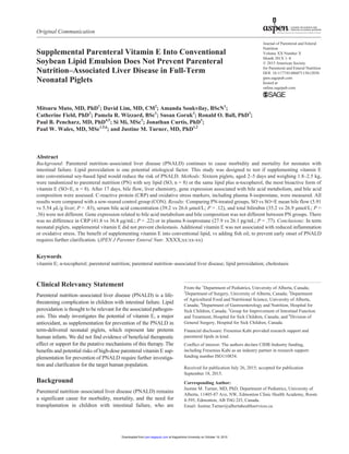
Bile Acid Metabolism
Hepatic gene expressions associated with bile acid metabolism
are shown in Figure 1. No differences were found between
PN-treated groups in CYP7A1 (P = .49), FXR (P = .13), SHP
(P = .10), OSTα (P = .91), NTCP (P = .91), BSEP (P = .64),
MRP2 (P = .64), and MRP3 (P = .36).Additional vitamin E did
not affect gene expressions in the liver, regulating bile acid
synthesis, sensing, and transporting (see Figure 1).
Bile Acid Composition
Bile acids quantification is shown in Table 3. There was no dif-
ference between SO and SO+E groups in any of the identified
bile acids. Amounts of measured bile acids in PN-treated pig-
lets were lower than CON, consistent with a restricted bile acid
pool with PN. The only exception was LCA, which was in the
range of CON values.
Inflammation and Oxidative Stress
There was no difference in CRP levels between SO and SO+E
groups (41.8 vs 36.8 µg/mL; P = .22). The CON CRP values
(n = 6) ranged from 26.4–46.5 µg/mL, after exclusion of 2 pig-
lets with extreme outlying values, above the mean by more
than 3 times the interquartile range. We suspect these piglets
may have been subject to inflammation in the barn; certainly,
treated animals did not have CRP values outside the normal
range. Plasma 8-isoprostane levels were not different between
SO and SO+E groups (27.9 vs 26.1 pg/mL; P = .77) and were
in the range of CON values (11.6–24.1 pg/mL). Plasma nitrates
were also not different between SO and SO+E groups (12.8 vs
25.7 µM; P = .56; CON range, 9.4–27.7 µM). So in summary,
both biomarkers consistently showed no differences between
PN-treated groups, and no evidence of enhanced oxidative
stress, by 2 different pathways, was observed (see Table 1).
Discussion
Parenteral lipid emulsions, specifically soy-based emulsions,
are now recognized as one of the major associated risk factors
for PNALD.4,5
Conventional soy-based lipid emulsions are
predominant in ω-6 PUFAs, implicated for proinflammatory
eicosanoid production.6
They are also abundant in phytoster-
ols, which may be a factor in PNALD as they have been shown
to alter bile acid transport and metabolic function.14,15
Finally,
they are low in vitamin E, a major lipid-soluble antioxidant. In
this study, we focused on vitamin E as a potential therapy to
prevent PNALD. Parenteral PUFAs are prone to peroxidation
even if the emulsion is covered during treatments, and they
enhance oxidative species free radical production.16
This likely
Table 1. Vitamin E, C-Reactive Protein, and Oxidative Stress Markers.
Measurements SO (n = 7) SO+E (n = 7) P Value Control Range (n = 4)
Vitamin E, µg/mL 2.7 ± 0.4 7.6 ± 2.4 .001 3.9–6.0
CRP, µg/mL 41.8 ± 6.7a
36.8 ± 8.0b
.22 26.4–46.5
8-Isoprostane, pg/mL 27.9 ± 12.5a
26.1 ± 10.3 .77 11.6–24.1
Nitrates, µM 12.8 ± 13.0a
25.7 ± 23.2 .56 9.4–27.7
CRP, C-reactive protein; SO, soy lipid without added vitamin E; SO+E, soy lipid plus vitamin E. Data are expressed as the means ± standard deviation.
Animal numbers as stated except the following: a
SO, n = 8; b
control, n = 6. P value refers to between-group comparison (SO vs SO+E) by Student t tests,
with the exception of nitrates, which used the Mann-Whitney U test. Control range of values is considered to represent the normal reference range for
age.
Table 2. Markers of Liver Disease.
Measurements SO (n = 8) SO+E (n = 7) P Value Control Range (n = 8)
Bile flow, µL/g liver 5.91 ± 3.90 5.54 ± 2.23 .83 8.20–14.40
Total bilirubin, µmol/L 35.2 ± 29.3 26.9 ± 24.1 .56 2.5–7.9
Bile acid, µmol/L 39.2 ± 16.6 26.6 ± 11.9 .12 5.4–12.7
GGT, IU/L 141.6 ± 94.8 185.9 ± 88.4 .37 14.0–38.0
ALP, IU/L 1033.0 ± 278.1 1144.4 ± 364.6 .52 606.0–1163.0
ALT, IU/L 11.8 ± 3.5 13.0 ± 1.6 .38 20.0–32.0
ALP, alkaline phosphatase; ALT, alanine aminotransferase; GGT, γ-glutamyltranspeptidase; SO, soy lipid without added vitamin E; SO+E, soy lipid
plus vitamin E. Data are expressed as the means ± standard deviation. P value refers to between-group comparison, SO vs SO+E. Control range of values
is considered to represent the normal reference range for age.
at Kagoshima University on October 19, 2015pen.sagepub.comDownloaded from
5. Muto et al 5
translates to oxidative stress, which has been suggested as one
of the putative mechanisms for PNALD.16,17
We hypothesized
that supplementing soy-based lipid with additional vitamin E
could reduce oxidative stress and prevent liver damage during
PN therapy. Furthermore, recent investigation in preterm neo-
natal piglets suggested that the addition of vitamin E into soy-
based lipid may prevent early onset of PNALD, perhaps by
altering the molecular mechanisms of bile acid transport.8
The
aim of this study was to explore the role of supplemental vita-
min E in prevention of PNALD using term-delivered piglets.
Investigation in term-delivered neonatal piglets has relevance
as many infants with intestinal failure are born late preterm, for
which the term neonatal piglet is a recognized model, due to
similarities in anatomy and physiology,10,18,19
while having
delayed gastrointestinal ontogeny.20,21
We used DL–α-tocopherol acetate, the synthetic form of the
most bioactive vitamin E isoform. Comparing α-tocopherol
content in conventional soy-based lipid to that of third-genera-
tion lipids, containing fish oil, the latter contains 8–11 times
higher α-tocopherol.7
Fish oil–based lipid emulsion has been
suggested to play an important role in both prevention and
reversal of PNALD.22
However, it remains unclear if the ben-
efit is given by the PUFA content, by lack of phytosterols con-
tent, or by abundant antioxidant agent, vitamin E.
Contrary to our hypothesis, vitamin E supplementation did
not reduce the risk of developing cholestasis in term piglets. As
Figure 1. Hepatic expression of genes associated with bile acid synthesis (CYP7A1), sensing (FXR, SHP), and transport (OSTα,
NTCP, BSEP, MRP2, MRP3). Data are expressed in fold change scale with the means of target gene messenger RNA (mRNA)/
hypoxanthine phosphoribosyltransferase 1 (HPRT1) ± standard deviation. Between-group comparisons used Mann-Whitney U tests.
No differences were found between PN-treated groups (SO, n = 8; SO+E, n = 7); expected values are represented by CON data (n =
8). BSEP, bile salt export pump; CON, sow-fed control; CYP7A1, cholesterol 7α-hydroxylase; FXR, farnesoid X receptor; MRP2,
multidrug resistance–associated protein 2; MRP3, multidrug resistance–associated protein 3; NTCP, Na+-taurocholate cotransporting
polypeptide; OSTα, organic solute transporter α; SHP, small heterodimer partner; SO, soy lipid without added vitamin E; SO+E, soy
lipid plus vitamin E.
at Kagoshima University on October 19, 2015pen.sagepub.comDownloaded from
6. 6 Journal of Parenteral and Enteral Nutrition XX(X)
we have observed in prior studies, the conventional lipid
(Intralipid) was associated with a reduction in bile flow and
increase in markers of cholestasis (total bilirubin, bile acids,
and GGT).13,18
As would be expected with PN feeding, the total
bile acid pool was markedly restricted compared with healthy
controls, which had quantitatively more of every individual
bile acid (with the exception of LCA, which was in the range
of CON). Furthermore, as we have also recently shown, the
composition of the bile was altered.13
We noted a shift toward
more LCA, TCDCA, and GLCDCA as a percentage of the total
bile acid pool in the PN piglets. These tend to be the more
hydrophobic bile acids in pig bile. LCA is the most hepatotoxic
of all bile acids, hence why it represents only a small amount
of both pig and human bile. For the PN piglets, there was also
less HCA, the most hydrophilic of the bile acids in pig bile.
Altogether, we suspect this represents a shift toward a more
hepatotoxic bile composition with PN feeding. Regardless, the
compositional shift was very consistent between both PN
groups, with and without added vitamin E.
Not finding an improvement in bile flow with vitamin E
treatment contrasts with our findings and those of others,
where bile flow is improved by use of parenteral lipids contain-
ing ω-3 fatty acids in this same animal model.11,18
Furthermore,
no evidence was shown for supplemental vitamin E decreasing
oxidative stress, arguably best indicated by considering the
most specific biomarker for free radical production from
PUFA, 8-isoprostane.23
In fact, we found no evidence for
increased oxidative stress through enhanced free radical pro-
duction from either PUFA or the nitrous oxide pathway. Hence,
our study does not support a clearly defined physiological
mechanism for vitamin E supplementation improving
PNALD.8
Finally, no difference was found in hepatic gene
expression associated with bile acid synthesis (CYP7A1),
sensing (FXR, SHP), or transport (OSTα, NTCP, BSEP, MRP2,
MRP3) between PN-treated groups. Therefore, in our opinion,
considering our prior research findings, where bile flow was a
primary outcome strongly associated with other measures of
cholestasis,10,18,24
we do not believe that the abundant vitamin
E is the dominant factor conferring clinical benefit in PNALD
when using fish oil–based lipid emulsions. Other disadvan-
tages of conventional soy-based lipid, such as the lack of key
ω-3 long-chain PUFAs during neonatal development, remain
reasons to consider third-generation lipids that include fish oil
for infants at risk of PNALD.
We do speculate that beneficial effects of supplementary
vitamin E may be enhanced with increasing prematurity,
although additional vitamin E may not be necessary in more
mature infants. Unlike our study in term piglets, Ng et al8
sup-
plemented preterm piglets with vitamin E (d–α-tocopherol
added into Intralipid) and found significantly lower levels of
bile acids, total bilirubin, and GGT compared with piglets that
were not treated with vitamin E. The total amount of
α-tocopherols delivered (7.5–8.3 mg α-TE/kg/d) was similar to
our own (6.8–8.3 mg α-TE/kg/d). Hence, the major difference
was the maturity of the piglets. The antioxidant enzyme system
is believed to be upregulated during the last 15% of gestation.25
Therefore, as is reported in preterm human infants,26,27
it is pre-
sumed that the more preterm, the piglet the more immature the
defense system against oxygen-derived free radicals. Vitamin
E supplementation may be effective in such animals. Further
studies should seek to clarify by what potential mechanisms
additional vitamin E may prevent the early onset of PNALD in
preterm piglets.
Admittedly, a limitation of the present study is that, unlike
Ng et al,8
we did not measure the tissue content of vitamin E
in the liver. Therefore, we cannot be sure the delivered
Table 3. Composition of Bile.
Bile Acid SO (n = 8) SO+E (n = 7) P Value Control Range (n = 8)
CA, µg/mL 0.34 ± 0.18 0.24 ± 0.13 .36 0.80–3.90
LCA, µg/mL 0.39 ± 0.04 0.41 ± 0.13 .88 0.30–0.70
TCA, µg/mL 14.01 ± 16.30 18.27 ± 16.54 .72 7.10–120.90
HCA, µg/mL 0.96 ± 1.08 0.63 ± 0.67 .56 9.50–50.00
TLCA, µg/mL 0.06 ± 0.07 0.06 ± 0.07 .72 0.30–6.10
GLCA, µg/mL 0.13 ± 0.18 0.10 ± 0.09 .60 0.60–5.00
HDCA, µg/mL 0.49 ± 0.24 0.46 ± 0.18 .95 0.40–2.10
THCA, µg/mL 104.10 ± 75.77 106.27 ± 70.66 1.0 572.90–2547.00
TCDCA, µg/mL 46.48 ± 35.10 64.04 ± 36.26 .17 103.50–609.00
GCDCA, µg/mL 129.94 ± 110.57 159.14 ± 137.13 .49 164.40–1342.40
THDCA, µg/mL 6.08 ± 5.33 6.80 ± 3.25 .56 108.30–986.90
GHDCA, µg/mL 73.21 ± 69.46 80.90 ± 49.91 .64 212.30–2705.90
CA, cholic acid; GCDCA, glycolithocholic acid; GHDCA, glycohyodeoxycholic acid; GLCA, glycolithocholic acid; HCA, hyocholic acid; HDCA,
hyodeoxycholic acid; LCA, lithocholic acid; SO, soy lipid without added vitamin E; SO+E, soy lipid plus vitamin E; TCA, taurocholic acid; TCDCA,
taurochenodeoxycholic acid; THCA, taurohyocholic acid; THDCA, taurohyodeoxycholic acid; TLCA, taurolithocholic acid. Data are expressed as the
means ± standard deviation. P value refers to between-group comparisons, SO vs SO+E, using the Mann-Whitney U test. Control range of values is
considered to represent the normal reference range for age.
at Kagoshima University on October 19, 2015pen.sagepub.comDownloaded from
7. Muto et al 7
vitamin E actually increased substantially at the target organ.
However, Ng et al found liver vitamin E levels in treated pig-
lets were on average 40× the untreated. We used the same
doses of vitamin E (per gram of lipid, accounting for differ-
ences in total dose of lipid), and so hypothetically we expect
similar tissue concentrations. However, in our study, the
plasma values in treated animals were approximately 3× the
untreated animals, while Ng et al found plasma levels to be
nearly 5× greater. If this is a real difference, we cannot exclude
that they actually delivered a higher dose of vitamin E to the
target liver. A further limitation of our study is that we did not
examine livers histologically. A potential mechanism for vita-
min E treatment preventing early onset PNALD is by reducing
hepatic steatosis. However, in our study and in the clinical
situation with neonatal-onset PNALD (compared with
PNALD in adult patients), cholestasis tends to be dominant
over steatosis. Therefore, while we find no differences in cho-
lestasis between groups, there could be differences in fatty
liver, and certainly, this potential mechanism should be
explored in preterm piglets. Furthermore, the assessment for
toxicity of accumulated vitamin E over longer therapeutic
periods is warranted. The safety profile must be considered
before pursuing this therapy for human infants with intestinal
failure. Currently, there is limited evidence regarding ade-
quate dosage and the safe upper limit of intravenous vitamin E
supplementation in babies and animals. Clinically, supple-
mentation with vitamin E has been controversial in preterm
infants. Some disease processes benefit and others may indeed
worsen. In neonates with the acute phase of respiratory dis-
tress syndrome, administration of vitamin E could modify the
development of bronchopulmonary dysplasia and save
patients.28
Vitamin E prophylaxis can reduce the incidence of
severe retinopathy of prematurity in the subset of infants
weighing ≤1500 g.29
On the other hand, high-dose vitamin E
is known to increase the risk of infection and hemorrhage,
especially in preterm humans.30–33
In addition, both an
increased incidence of sepsis and of late-onset necrotizing
enterocolitis has been reported in premature infants with birth
weights of ≤1500 g when supplemented with vitamin E.34,35
Baeckert et al36
suggested that safe and effective blood levels
of vitamin E are between 23–46 µmol/L (10–20 µg/mL). Brion
et al30
suggested that when parenteral vitamin E is given to
infants, it is recommended not to exceed a plasma concentra-
tion of 80 µmol/L (35 µg/mL) to avoid complications. We sup-
plied vitamin E at the equivalent concentration of the
third-generation fish oil–based lipid emulsions: Omegaven
and SMOF lipid. The serum vitamin E concentration at termi-
nation in our SO+E group ranged from 5.34–12.0 µg/mL,
which was in the safe range according to the former reports in
human infants,30,36
but they were significantly higher than
those of the conventional soy-based lipid-treated group and of
the gold-standard sow-reared control piglets.
In conclusion, in full-term neonatal piglets, we found no
benefit of supplemental vitamin E to improve early onset of
PNALD.Additional vitamin E was not associated with reduced
oxidative stress in this model. While the use of supplemental
vitamin E into conventional soy-based lipid to prevent PNALD
would be a cost-effective strategy, the studies to date in neona-
tal piglets are contradictory, and further studies are required.
Vitamin E supplementation may be beneficial specifically for
preterm infants receiving PN therapy to avoid oxidative stress
and to prevent PNALD. However, safety concerns and avoid-
ing potential toxicity must be considered before translation
into human infants.
Acknowledgments
We thank Charlane Gorsak and the University of Alberta’s Swine
Research and Technology Centre (SRTC) for assistance with all
surgical procedures.
Statement of Authorship
P. W. Wales, J. M. Turner, R. O. Ball, P. B. Pencharz, and C.
Field contributed to conception and design of this study; P. W.
Wales, J. M. Turner, C. Field, M. Muto, D. Lim, A. Soukvilay,
P. R. Wizzard, S. Goruk, S. Mi, and J. Curtis contributed to data
acquisition, analysis, and interpretation; P. W. Wales, J. M.
Turner, C. Field, P. B. Pencharz, D. Lim, P. R. Wizzard, S. Mi,
and J. Curtis drafted the manuscript. All authors critically revised
the manuscript, read and approved the final manuscript, and
agree to be fully accountable for ensuring the integrity and accu-
racy of the work.
Supplementary Material
Tables S1 and S2 are available online at http://pen.sagepub.com/
supplemental.
References
1. Wales PW, Allen N, Worthington P, George D, Compher C, Teitelbaum
D. A.S.P.E.N. clinical guidelines: support of pediatric patients with intes-
tinal failure at risk of parenteral nutrition–associated liver disease. JPEN J
Parenter Enteral Nutr. 2014;38:538-557.
2. Beath SV, Davies P, Papadopoulou A, et al. Parenteral nutrition–related
cholestasis in postsurgical neonates: multivariate analysis of risk factors.
J Pediatr Surg. 1996;31:604-606.
3. Calkins KL, Venick RS, Devaskar SU. Complications associated with par-
enteral nutrition in the neonate. Clin Perinatol. 2014;41:331-345.
4. Nandivada P, Carlson SJ, Chang MI, Cowan E, Gura KM, Puder M.
Treatment of parenteral nutrition–associated liver disease: the role of lipid
emulsions. Adv Nutr. 2013;4:711-717.
5. Biesalski HK. Vitamin E requirements in parenteral nutrition.
Gastroenterology. 2009;137:S92-S104.
6. Diamond IR, de Silva NT, Tomlinson GA, et al. The role of parenteral
lipids in the development of advanced intestinal failure–associated liver
disease in infants: a multiple-variable analysis. JPEN J Parenter Enteral
Nutr. 2011;35:596-602.
7. Xu Z, Harvey KA, Pavlina TM, Zaloga GP, Siddiqui RA. Tocopherol
and tocotrienol homologs in parenteral lipid emulsions. Eur J Lipid Sci
Technol. 2015;117:15-22.
8. Ng K, Stoll B, Chacko S, et al. Vitamin E in new-generation lipid emul-
sions protects against parenteral nutrition–associated liver disease in par-
enteral nutrition–fed preterm pigs [published online January 16, 2015].
JPEN J Parenter Enteral Nutr.
at Kagoshima University on October 19, 2015pen.sagepub.comDownloaded from
8. 8 Journal of Parenteral and Enteral Nutrition XX(X)
9. Wykes LJ, Ball RO, Pencharz PB. Development and validation of a
total parenteral nutrition model in the neonatal piglet. J Nutr. 1993;123:
1248-1259.
10. Turner JM, Wales PW, Nation PN, et al. Novel neonatal piglet mod-
els of surgical short bowel syndrome with intestinal failure. J Pediatr
Gastroenterol Nutr. 2011;52:9-16.
11. Van Aerde JE, Duerksen DR, Gramlich L, et al. Intravenous fish oil emul-
sion attenuates total parenteral nutrition–induced cholestasis in newborn
piglets. Pediatr Res. 1999;45:202-208.
12. Josephson J, Turner JM, Field CJ, et al. Parenteral soy oil and fish oil
emulsions: impact of dose restriction on bile flow and brain size of par-
enteral nutrition–fed neonatal piglets. JPEN J Parenter Enteral Nutr.
2015;39(6):677-687.
13. Lim DW, Wales PW, Si Mi, et al. Glucagon-like peptide-2 alters bile acid
metabolism in parenteral nutrition–associated liver disease [published
online July 28, 2015]. JPEN J Parenter Enteral Nutr.
14. Clayton PT, Bowron A, Mills KA, Massoud A, Casteels M, Milla PJ.
Phytosterolemia in children with parenteral nutrition–associated choles-
tatic liver disease. Gastroenterology. 1993;105:1806-1813.
15. Xu Z, Harvey KA, Pavlina T, et al. Steroidal compounds in commercial
parenteral lipid emulsions. Nutrients. 2012;4:904-921.
16. Laborie S, Lavoie JC, Pineault M, Chessex P. Protecting solutions of
parenteral nutrition from peroxidation. JPEN J Parenter Enteral Nutr.
1999;23:104-108.
17. Traber MG, Atkinson J. Vitamin E, antioxidant and nothing more. Free
Radic Biol Med. 2007;43:4-15.
18. Turner JM, Josephson J, Field CJ, et al. Liver disease, systemic inflamma-
tion, and growth using a mixed parenteral lipid emulsion, containing soy-
bean oil, fish oil, and medium chain triglycerides, compared with soybean
oil in parenteral nutrition–fed neonatal piglets [published online April 2,
2015]. JPEN J Parenter Enteral Nutr.
19. Lim DW, Turner JM, Wales PW. Emerging piglet models of neonatal
short bowel syndrome [published October 7, 2014]. JPEN J Parenter
Enteral Nutr.
20. Moughan PJ, Birtles MJ, Cranwell PD, Smith WC, Pedraza M. The pig-
let as a model animal for studying aspects of digestion and absorption in
milk-fed human infants. World Rev Nutr Diet. 1992;67:40-113.
21. Sangild PT. Gut responses to enteral nutrition in preterm infants and ani-
mals. Exp Biol Med (Maywood). 2006;231:1695-1711.
22. Bharadwaj S, Gohel T, Deen OJ, DeChicco R, Shatnawei A. Fish oil–
based lipid emulsion: current updates on a promising novel therapy
for the management of parenteral nutrition–associated liver disease.
Gastroenterol Rep (Oxf). 2015;3:110-114.
23. Janicka M, Kot-Wasik A, Kot J, Namieśnik J. Isoprostanes-biomarkers of
lipid peroxidation: their utility in evaluating oxidative stress and analysis.
Int J Mol Sci. 2010;11:4631-4659.
24. Josephson J, Turner JM, Field CJ, et al. Parenteral soy oil and fish oil
emulsions: impact of dose restriction on bile flow and brain size of par-
enteral nutrition–fed neonatal piglets. JPEN J Parenter Enteral Nutr.
2015;39(6):677-687.
25. Davis JM, Auten RL. Maturation of the antioxidant system and the effects
on preterm birth. Semin Fetal Neonatal Med. 2010;15:191-195.
26. Georgeson GD, Szony BJ, Streitman K, et al. Antioxidant enzyme activi-
ties are decreased in preterm infants and in neonates born via caesarean
section. Eur J Obstet Gynecol Reprod Biol. 2002;103:136-139.
27. Lee JW, Davis JM. Future applications of antioxidants in premature
infants. Curr Opin Pediatr. 2011;23:161-166.
28. Ehrenkranz RA, Bonta BW, Ablow RC, Warshaw JB. Amelioration of
bronchopulmonary dysplasia after vitamin e administration: a preliminary
report. N Engl J Med. 1978;299:564-569.
29. Raju TN, Langenberg P, Bhutani V, Quinn GE. Vitamin E prophylaxis
to reduce retinopathy of prematurity: a reappraisal of published trials.
J Pediatr. 1997;131:844-850.
30. Brion LP, Bell EF, Raghuveer TS, Soghier L. What is the appropri-
ate intravenous dose of vitamin E for very-low-birth-weight infants?
J Perinatol. 2004;24:205-207.
31. Kanno T, Utsumi T, Takehara Y, et al. Inhibition of neutrophil-super-
oxide generation by alpha-tocopherol and coenzyme Q. Free Radic Res.
1996;24:281-289.
32. Phelps DL, Rosenbaum AL, Isenberg SJ, Leake RD, Dorey FJ. Tocopherol
efficacy and safety for preventing retinopathy of prematurity: a random-
ized, controlled, double-masked trial. Pediatrics. 1987;79:489-500.
33. Speer ME, Blifeld C, Rudolph AJ, Chadda P, Holbein ME, Hittner HM.
Intraventricular hemorrhage and vitamin E in the very low-birth-weight
infant: evidence for efficacy of early intramuscular vitamin E administra-
tion. Pediatrics. 1984;74:1107-1112.
34. Finer NN, Peters KL, Hayek Z, Merkel CL. Vitamin E and necrotizing
enterocolitis. Pediatrics. 1984;73:387-393.
35. Johnson L, Bowen FW Jr, Abbasi S, et al. Relationship of prolonged phar-
macologic serum levels of vitamin E to incidence of sepsis and necrotizing
enterocolitis in infants with birth weight 1,500 grams or less. Pediatrics.
1985;75:619-638.
36. Baeckert PA, Greene HL, Fritz I, Oelberg DG, Adcock EW. Vitamin con-
centrations in very low birth weight infants given vitamins intravenously
in a lipid emulsion: measurement of vitamins A, D, and E and riboflavin.
J Pediatr. 1988;113:1057-1065.
at Kagoshima University on October 19, 2015pen.sagepub.comDownloaded from