VIP Service Call Girls Sindhi Colony 📳 7877925207 For 18+ VIP Call Girl At Th...
Annals of Clinical and Medical Case Reports - Acmcasereport
1. “Multiple unilateral submandibular duct calculi: A case report”.
Shermil Sayd1*
, Sreejith VP2
, Resmi Sankar3
, Chaitanya Harindranath4
, Navya Mukund5
,
1
Oral and Maxillofacial Surgery, Department of Oral and Maxillofacial surgery, Kannur dental college, Anjarakandy, Kerala, India.
2
Oral and Maxillofacial Surgery, Department of Oral and Maxillofacial Surgery, Kannur dental college, Anjarakandy, Kerala, India.
3
Resmi Sankar, PG Student , Post-graduate trainee, Department of Oral medicine and Radiology, Kannur Dental College, Anjara-
kandy, Kerala, India.
4
Chaitanya Harindranath, Post-graduate trainee, Department of Oral medicine and Radiology, Kannur Dental College, Anjara-
kandy, Kerala, India.
5
Navya Mukund, Post-graduate trainee, Department of Oral medicine and Radiology, Kannur Dental College, Kerala, India.
Volume 1 Issue 3- 2018
Received Date: 15 Sep 2018
Accepted Date: 15 Oct 2018
Published Date: 22 Oct 2018
1. Abstract
Salivary gland calculi account for the most common disease of the salivary glands. The majority
of sialoliths occur in the submandibular gland or its duct and are a common cause of acute and
chronic infections. Sialolith can be unilateral, bilateral, single or multiple. Depending on the
gland affected and stone location, there are various methods available for the management of
salivary stones or calculi. here we report case of multiple sialolith in Wharton duct.
Annals of Clinical and Medical
Case Reports
Citation: Sayd S, Multiple Submandibular Duct Calculi: A Case Report. Annals of Clinical and Medical Case
Reports. 2018; 1(3): 1-3.
United Prime Publications: http://unitedprimepub.com
*Corresponding Author (s): Shermil Sayd, Department of Oral and Maxillofacial Surgery,
Kunhitharuvai memorial charitable trust (KMCT) Dental College and Hospitals, India, Tel:
+919446230425; Fax: +91495 2294726; Email:shermil12@gmail.com
Case Report
2. Introduction
Sialolithiasis, the formation of calcific concretions in the salivary
duct of a major or minor salivary gland, is a common salivary
gland pathology. These calcifications usually develop in the duc-
tal system of the submandibular salivary gland, but can involve
the parotid gland and, infrequently the ducts of sublingual or mi-
nor salivary glands. 1 The size of salivary calculi may vary from
less than 1 mm to a few centimeters in size, with most cases being
less than 10 mm in size.2 Although large and multiple sialoliths
have been reported in the salivary glands, they have been rarely
reported in the salivary duct.2 Here we are reporting a case of
multiple Wharton duct sialolithiasis.
3. Case Report
A 40-year-old male reported to our out-patient department with
a chief complaint of pain beneath the left side of the tongue for
the past 3 months. History revealed that the pain was slow in
onset, dull aching, intermediate and moderate in intensity. He
reported that the pain has been progressive since its onset and got
aggravated during meals, especially while having sour food, fol-
lowed by a period of self regression. No history of radiating pain
and other associated symptoms were provided by the patient. On
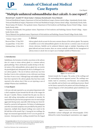
inspection, multiple dome-shaped swellings were noted on the
left side floor of the mouth (Fig 1). Among the two swellings, the
larger one measured approximately 0.5×0.5 cm at its greatest di-
mension. Anteroposteriorly, swelling extended from the lingual
Figure 1: Pre-operative view.
frenum towards the 36 region. The surface of the swellings and
the surrounding mucosa appeared normal. The swellings were
tender, hard in consistency, non-fluctuant, non-compressible,
non-reducible, and not fixed to any underlying structures. Based
on the history reported by the patient and clinical findings, pro-
visional diagnosis of left submandibular gland sialolithiasis was
made.
Figure 2: Occlusal View of the lesion.
Mandibular occlusal radiograph and ultrasonography (USG)
followed by routine hematological examinations were suggest-
3. References
1. Louis Mandel, Salivary Gland Disorders, Dent Clin N Am 55 (2011)
121–140.
2. Krishnappa BD. Multiple submandibular duct (Wharton’s duct) cal-
culi of unusual size and shape. Indian J Otolaryngol Head Neck Surg.
2008 Sep; 60(3):287-8.
3. Harold D. B, Submandibular Salivary Stones, Current Management
Modalities J Oral Maxillofac Surg 62:369-378, 2004.
4. Nahlieli O, Eliav E, Hasson O, et al: Pediatric sialolithiasis. Oral Surg
Oral Med Oral Pathol Oral Radiol Endod 6:709, 2000.
5. Epivatianos A, Harrison JD, Dimtiou T (1987) Ultrastructural and
histochemical observations on micro calculi in chronic submandibular
sialadenitis. J Oral Pathol 16:514–517.
6. Sherman JA, McGurk (2000) Lack of correlation between water hard-
ness and salivary calculi in England. Br J Oral Maxillofac Surg 38:50–53
7. Yoshimura Y, Inoue Y, Odagawa T. Sonographic examination of
sialolithiasis.J Oral Maxillofac Surg 1989; 47: 907-12.
8. Marmary Y. A novel and non-invasive method for the removal of sali-
vary gland stones.Int J Oral Maxillofac Surg 1986;15:585–7.
9. June Sik Park, Jin Ho Sohn, and Jeong Kyu Kim, Factors influencing
intraoral removal of submandibular calculi,Otolaryngology–Head and
Neck Surgery, Vol 135, No 5, November 2006.
Volume 1 Issue 3 -2018 Case Report
3