2. 000
International Dental Journal (2005) Vol. 55/No.1
sensitive teeth. He expressed extreme dissatisfaction
with his appearance, and his father confirmed that the
patient’s social life was affected by this problem. A
detailed medical, dental and social history was
obtained. The patient was examined dentally and
medically; photographs and dental radiographs were
obtained.
The patient had all his teeth except the third molars.
His left mandibular deciduous canine tooth was
persistent and the permanent canine was positioned
lingually. The enamel layer of all teeth was very thin
and yellow-brown in colour and the cuspal structures
were nearly flattened in posterior regions (Figures 1–3).
The molars were severely affected. However, the clini-
cal appearance of cervical and proximal enamel seemed
to be normal. The exposed dentine was brown and
hypersensitive. Periapical and panoramic radiographs
revealed the loss of enamel, especially on the occlusal
surfaces of posterior teeth. The pulp chambers and
root canals were abnormally large and the upper
permanent canines were impacted in the jaw (Figure 4).
The freeway space had increased because of attri-
tion of the molars. In the retruded contact position,
the molars were in Class I relation whereas the anterior
teeth were in an ‘edge to edge’ position. Oral hygiene
was not satisfactory, with evidence of gingivitis. More-
over, because of the poor appearance of the teeth and
their sensitivity, the young patient was reluctant to
brush properly. There were gingival growths on the
distal part of all the second molars due to the short
crown lengths.
The patient was questioned further about the pres-
ence of similar abnormalities in his family including
parents and grandparents. He stated that his father and
father’s mother had similar appearances to their teeth.
Examining the father did not help in diagnosis as he
had full mouth fixed restorations and the grandmother
was not alive. Thus, it was concluded that the patient
probably suffered from a type of X-linked recessive
hypomaturation type of amelogenesis imperfecta.
Treatment
The patient was informed of the diagnosis, and all the
treatment modalities were explained to him and his
parents. The patient refused to undergo any form of
orthodontic treatment for his mandibular canine tooth
because of economic and social factors and was insist-
ent on full mouth rehabilitation. Moreover, he could
not afford to have his anterior teeth restored with
porcelain laminate veneers or bonded crowns.
The patient was placed on an intensive oral hygiene
programme that included scaling and root planing.
After three weeks, he managed to maintain acceptable
oral hygiene and there was a marked improvement in
the soft tissues. Periodontal surgery with crown length-
ening procedures was then carried out in all second
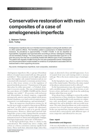
Figure 1. Frontal view of the patient before treatment.
Figure 2. View of the patient’s maxillary arch at diagnosis.
Figure 3. View of the patient’s mandibular arch at diagnosis.
molar areas to create a new, more apically positioned
biologic zone. Impressions were taken and casts were
obtained. The treatment plan was performed with
diagnostic self-curing resin composite restorations made
on the casts with a semi adjustable articulator in order
3. 000
Sebnem Türkün: Restoration of a case of amelogenesis imperfecta
Figure 4. Panoramic radiograph of the patient at diagnosis.
to show a model of the final result to the patient.
The cervical enamel of the six anterior maxillary
teeth was left and was largely bevelled; the discoloured
dentine was removed carefully. The preparation was
not extended to the palatal surfaces of the teeth.
All posterior teeth were prepared for onlay direct
composite restorations. The teeth were isolated with
cotton rolls and high-volume suction and retraction
cords were placed around the teeth. All prepared teeth
were acid-etched for 30s with 34% phosphoric acid
(3M/ESPE St.Paul, MN, USA), thoroughly washed
and dried with cotton pellets. Single Bond adhesive
system (3M/ESPE) was applied for 20s on the enamel
and dentine and light-cured for 10s.
The anterior teeth were restored and contoured by
hand with Filtek A110 (A3 shade, 3M/ESPE) microfill
resin composite. The last layer was incisal colour to
give a natural translucent appearance. Premolar and
molar regions, where strength is more important, were
restored with a hybrid resin composite Filtek Z250
(A3 shade, 3M/ESPE). These teeth were prepared as
for veneer crowns and were restored according to the
anterior occlusion. The vertical dimension was not
increased. Proximal contacts were built-up with a
sectional matrix system and a BiTine ring (Palodent
System, Dentsply De Trey, Konstanz, Germany), while
the premolars were restored similarly to the molars.
The finishing and polishing sequence consisted of the
use of a fine diamond bur for gross contouring under
water spray at high speed followed by the use of a
PoGo micro diamond polisher (Dentsply De Trey,
Konstanz, Germany) for anterior teeth and aluminium
oxide coated brush, Sof-Brush, for posterior teeth
(3M/ESPE).
After the restorative procedures, the patient’s
dental hypersensitivity disappeared completely, and
functional chewing was established. He was recalled
every month for six months and then every two months
for one year. The psychology of the patient was better
after the first recall. He refused to have his left
mandibular deciduous canine tooth extracted and asked
for a restoration at this appointment. We explained to
Figure 5. Frontal view of the patient one year after treatment.
Figure 7. Successfully restored mandibular arch after one year.
Figure 6. Successfully restored maxillary arch after one year.
4. 000
International Dental Journal (2005) Vol. 55/No.1
him the difficulties of maintaining optimal hygiene in
that crosswise region but he insisted and the perma-
nent canine was restored at the first recall.
At the 6-month recall, the upper left lateral incisor’s
restoration was partially fractured. At the one-year
recall, the mandibular deciduous canine had a mesial
proximal carious cavity. This was probably due to the
cleaning difficulties encountered with both canines
present. The local gingiva was slightly inflamed because
of insufficient brushing and some calculus was present
lingually. The patient was motivated again and the teeth
were re-cleaned. There was no other deterioration in
the restored teeth (Figures 5–7). Radiographic examina-
tion revealed no evidence of disease associated with
the teeth or their supporting structures after one year.
Discussion
The treatment plan for cases of amelogenesis imper-
fecta is related to many factors: age and the socio-
economic status of the patient, the type and the sever-
ity of the disorder, and the intraoral situation at the
time the treatment was planned8
.
Historically, some patients with AI have been treated
with multiple extractions followed by the construction
of complete dentures. These options are psychologi-
cally harsh, especially when the patient is in adolescence.
Several studies have illustrated the use of resin compos-
ites, sealants and other bonded resins, polycarbonate
crowns, stainless steel crowns, and space maintainers
to restore a mutilated dentition9. Because of the
advances in aesthetic dentistry, especially in bonding to
dentine; today it is possible to restore function and
aesthetics to an acceptable level and for a long time.
Moreover, with the use of new polishing systems the
aesthetics, colour stability and longevity of the restora-
tions can be achieved successfully.
The dental rehabilitation of a young patient must be
done with regard to the growing potential of the jaws
and the periodontal health. In this case, the patient’s
financial resources were limited and complete coverage
or porcelain laminate veneers could not be a treatment
option. The discoloured dentine and enamel was
removed and the remaining dental tissues were acid
etched with phosphoric acid. The one-step adhesive
system applied did dissolve and penetrate the dentinal
smear layer when polymerised. This mechanism mechani-
cally interlocks into the etched enamel prisms to
strengthen the bonding effect. The resin composites
used for restoring the teeth had excellent colour stabil-
ity and no marginal discolorations were observed at
the end of the one-year.
Venezie et al.10
reported that difficulty in bonding to
hypomineralised enamel can significantly limit the
restorative and orthodontic treatment options for
patients with AI. However, in this case the treatment
was performed over several appointments with only
minor problems. As our goal was to create good
aesthetics at low cost, further orthodontic or surgical
treatment was not planned for the impacted canines.
According to the USPHS criteria11
for clinical evalu-
ation of resin composite restorations, a follow up of
18 months and a level of 95% of acceptable restora-
tions was considered enough for classifying a material
as successful. Although the restorations were performing
well at one year, we still believe that the performance
of the resin composites will be limited in this special
case. However, we could consider the treatment as
successful if 95% of the restorations were free of
major problems after two years.
Conclusion
This case report describes the functional and aesthetic
rehabilitation of a hypomaturated type of amelogen-
esis imperfecta restored with two different resin
composites in a young patient. After one year, all the
restorations were in place and the patient was satisfied
with the result.
Acknowledgment
The author would like to express her sincere gratitude
to Dr. Aycan Kazanç for his contribution to the peri-
odontal and surgical treatment of the patient.
References
1. Shafer WG, Hine MK, Levy BM et al. A textbook of oral
pathology. 4th ed., chapter: developmental disturbances of oral
and paraoral structures. pp 51–58. Tokyo, Japan, 1983.
2. Wright TJ, Robinson C, Shore R et al. Characterization of the
enamel ultrastructure and mineral content in hypoplastic
amelogenesis imperfecta. Oral Surg Oral Med Oral Pathol 1991
72: 594–601.
3. Witkop CS, Kuhlmann W, Sauk J. Autosomal recessive
pigmented hypomaturation amelogenesis imperfecta. Oral Surg
Oral Med Oral Pathol 1973 36: 367–382.
4. Peters E, Cohen M, Altini M. Rough hypoplastic amelogenesis
imperfecta with follicular hyperplasia. Oral Surg Oral Med Oral
Pathol 1992 74: 87–92.
5. Collins MA, Mauriello SM, Tyndall DA et al. Dental anomalies
associated with amelogenesis imperfecta. A radiographic
assessment. Oral Surg Oral Med Oral Pathol 1999 88: 358–364.
6. Alfred MJ, Crawford PJM. Variable expression in amelogen-
esis imperfecta with taurodontism. J Oral Pathol 1989 17: 327–
333.
7. Nel JC, Pretorius JA, Weber A et al. Restoring function and
esthetics in a patient with amelogenesis imperfecta. Int J
Periodontics Restorative Dent 1997 17: 479–483.
8. Sengün A, Özer F. Restoring function and esthetics in a
patient with amelogenesis imperfecta: a case report. Quintes-
sence Int 2002 33: 199–204.
9. Bouvier D, Duprez JP, Bois D. Rehabilitation of young
patients with amelogenesis imperfecta: a report of two cases.
ASDC J Dent Child 1996 63: 443–447.
10. Venezie RD, Vadiakas G, Christense JR et al. Enamel pre-
5. 000
Sebnem Türkün: Restoration of a case of amelogenesis imperfecta
treatment with sodium hypochlorite to enhance bonding in
hypocalcified amelogenesis imperfecta: case report and SEM
analysis. Pediatr Dent 1994 16: 433–436.
11. Ryge G, Snyder M. Evaluating the clinical quality of restora-
tions. JADA 1973 87: 369–70.
Correspondence to: Dr. L. Sebnem Türkün, Ege University School
of Dentistry, Department of Restorative Dentistry and Endodon-
tics, 35100 Izmir, Turkey. E-mail: sebnemturkun@hotmail.com