1. THE JOURNAL O F EXPERIMENTAL ZOOLOGY 217:325-339 (1981)
A Comparison of Cell Proliferation Patterns in the
Digestive Tract of Ascidians
THOMAS H. ERMAK
Scnpps Institution o f Oceanography, La Jolla, California 92037
ABSTRACT Cell proliferation patterns in the postbranchial digestive tract of
a variety of ascidian species were investigated with autoradiography and tritiated
thymidine. Based upon the distribution of radioactive nuclei, gut cell populations
were classified as either static, expanding, or renewing. In most ascidians, renew.
ing populations occur in the esophagus and stomach, but expanding populations
sometimes occur in all or part of the intestine. From species to species, the number
of cell renewal units (pairs of germinal and mature zones) in each portion of the di-
gestive tract increases with increasing organ size, and this multiplication of re-
newal units follows the folding patterns in the gut. Cell turnover is fastest and the
size of a cell renewal unit smallest a t the anterior end of the gut. The smallest CD.
lonial ascidians have a single renewal unit per cell population in the esophagus and
stomach and an expanding population in the intestine. Epithelial folding and mul-
tiplication of cell renewal units occurs in solitary species of increasing body size.
One cell population occurs on the stomach folds of more primitive solitary asci-
dians, two of those of advanced species, The digestive diverticulum, which only
occurs in two families, is renewed much the same as the stomach, and probably
evolved from that organ. In the intestine of primitive solitary ascidians, renewing
populations only occur a t the anterior end; with evolutionary advancement, r e
newing populations line the entire intestine.
In S t y e h chva, a solitary stolidobranch asci- This paper considers cell proliferation pat-
dim, most of the gut epithelia are renewing cell terns in a variety of ascidian species in order to
populations (Ermak, '75a, c, '76a). Cell prolifer- determine those features of gut cell renewal
ation occurs in restricted germinal zones of which are characteristic of the class Ascidiacea
pseudostratified cells. With time, germinal cells as a whole. Examples of each ascidian family
migrate into mature zones of ciliated, secre- (Berrill, '50) from California waters were injec-
tory, or absorptive cells. Aging mature cells ted with tritiated thymidine. In the esophagus,
are presumably extruded into the gut lumen. stomach, and intestine, sites of cell prolifera-
In other ascidian species, the postbranchial tion were detected with autoradiography in
digestive tract (esophagus,stomach, and intes- order to characterize each cell population. In
tine) exhibits a great deal of variability in posi- some cases, individuals were sacrificed a t in-
tion, shape, and structure (Berrill, '50); it may creasing time intervals after injection in order
be posterior or next to the branchial basket; on to determine the fate of the DNA synthesizing
the right or left side of the body; U-shaped, cells. Cell proliferation patterns in different
S-shaped, or twisted into a variety of shapes. A species were compared to each other and to
particularly salient feature is the elaboration those in S t y e h (Ermak, '75c).
of the epithelial lining. This may be unfolded, MATERIALS AND METHODS
folded, or modified into a system of canals and All ascidians were collected in California by
tubules. Ascidians with small bodies (most picking them off the underside of docks, in the
colonial ascidians) have unfolded or smooth intertidal zone a t low tide, or by diving with
gut linings, whereas ascidians with large
T.H. Ermak's present address is Department of Physiology,
bodies (a few colonial species and all solitary University of California School of Medicine, San Francisco. CA
species) have highly folded epithelial linings. 94143.
ALAN R. LISS. INC.
0022-104X/81/2173-0325$04.5001981
2. 326 T.H. ERMAK
SCUBA (see Ermak, '75a, for localities).Those RESULTS
species included in this investigation are listed
in Table 1(fordescriptions and additional illus- General features
trations, see Ritter, '17, and van Name, '45). All Ascidians exhibit a large range in individual
families are represented except the family Dia- body and organ size. Colonial species repro.
zonidae, of which there are no Pacific coast re- duce rapidly through asexual reproduction,
present atives. increasing the number of individuals in a
On the day of collection, specimens were ex- colony, and are several millimeters in length,
posed to tritiated thymidine for one hour (Er- at most a few centimeters. Solitary species re-
mak, '752). In some cases, the solution of tritia- produce only sexually and spend much of their
ted thymidine in sea water was again diluted lives (usually only a year or two) increasing in
with sea water. Solitary ascidians were injec- size and complexity. They may reach 10 cm or
ted with 5 pCi of tritiated thymidine per gm more in length.
animal weight. At least three animals were in- The postbranchial digestive tract, like in
jected for each time interval to be investigated. Styela (Ermak, '75c),is composed of anesopha-
Two or three injections 24 hours apart were gus, stomach, and intestine (Fig. 1).The eso-
given to Molgula and Pyura in order to in- phagus is a short tube in which the food cord is
crease their low labeling index. Colonial asci- formed and is folded in certain solitary species.
dians would usually not take up the radioac- The stomach, the largest organ in which en-
tive label if placed in a solution of tritiated thy- zymes are secreted, has several folding pat-
midine in sea water and were, therefore, per- terns (listed in Table 1).In most colonial asci-
fused with the solution of tritiated thymidine dians, the stomach has a smooth wall (Fig. 1A)
by injecting approximately 15 pCi into the and a raphe of mucous cells along one side. In
common tunic. Each sample of colonial asci- some colonial species, e.g., Distaplia, the epi-
dians injected contained numerous individ- thelium forms small longitudinal ridges (corru-
uals. Polyclinum and Ascidia were sacrificed gated) which apparently do not represent
after one hour, 5 , 10, and 15 days. Ciona was multiple zones of proliferation, as in larger
sacrificed at one hour, 15, and 20 days. Pyura ascidians. The colonial species Euherdmania
was sacrificed at one hour, 5, 10, 20, and 30 has a folded stomach (Fig. 1B). No colonial
days. All other species were sacrificed after one species examined had a folded esophageal or
hour only. Autoradiograms were prepared as intestinal lining.
previously described (Ermak, '75c) and exposed In solitary species, the stomach is always
for two weeks, one month, or two months. folded or has a digestive diverticulum off one
L
1mm
H
1Cm
0.2mm H
H 0.3mm
H 3mm
H
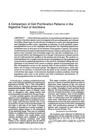
Fig. I . Types of ascidian postbranchid digestive tracts. A) Colonial ascidian, smooth stomach. B) Colonial ascidian.
folded stomach. C) Chelyosoma, pitted (areolatedor waffle patterned) stomach. D) Ascidia, longitudinally folded stomach.
E)Pyura, with digestive diverticulum.
3. CELL PROLIFERATION IN ASCIDIAN GUT 327
TABLE 1. List of ascidian species used in this investigation giving the foldingpattern and distribution of renewing (R)
and expanding (E) cell populations in each region of the postbranchial digestive tract
SPECIES ESO STOM m r
Aplousobranchia
Polyclinum planum (c) smooth (R) smooth (R) smooth (El
Archidistoma ritteri (c) smooth (R) smooth (R) smooth (E)
Didemnum carnulentum (c) smooth (R) smooth (R) smooth (El
Distaplia occidentalis (c) smooth (R) corrugated (R) smooth (E)
Euherdmania claviformis (c) smooth (R) folded (R) smooth (E)
Phlebobranchiata
Perophora annectens (c) smooth (R) smooth (R) smooth (E)
Ciona intestinalis (s) folded (RI folded (R) folded (R-E)
Chelyosoma productum (s) smooth (R) pitted (R) smooth (R-E)
Ascidia ceratodes (s) smooth (R) folded (R) smooth (R-E)
Stolidobranchiata
Styela clava (s) folded (R) folded (R) folded (R)
Botrylloides diegense (c) smooth (ST?) folded (ST?) smooth (ST?)
Molgula verrucifera (s) folded (R) diverticulum (R) smooth (R-E)
Pyuia haustor (s) folded (R) diverticulum (R) smooth (R)
Names from Ahbott ('75). For comparison, Styela clava (from Ermak. '75c) is included in its appropriate place. 'R-E'indicates that renewing and
expanding populations occur in different regions of that organ. c, colonial species; s. solitary species; ST,static population.
side. The folds may be pits (Fig. 1C) as in 250 pm wide) and intestine are approximately
Chelysoma, or longitudinal folds, as in Ascidia circular (Fig. 2A). A one-hour exposure to triti-
(Fig. 1D).The digestive diverticulum may con- ated thymidine labeled germinal cells in the
sist of folds or tubules (Fig. 1E). The molgulid esophagus and stomach. Two germinal zones
diverticulum is actually a specialized region of of mucous cells run opposite one another along
the stomach, whereas the pyurid diverticulum the length of the esophagus (Fig. 3);in the sto-
(Fig. 1E) is a separate organ connected to the mach, germinal zones occurred on each side of
stomach by canals (Fouque, '59). the raphe and around the esophageal and intes-
The intestine is a long tube in which nutri- tinal openings. Both chief and mucous popula-
ents are absorbed and faeces are compacted. In tions were labeled (Fig. 4); the chief germinal
most ascidians, it is smooth walled in the lar- cells, however, were more heavily labeled than
gest species examined, however, a typhlosole mucous germinal cells. Some mature cells were
may pass along one side. also labeled along the circumference of the sto-
The digestive tracts of most ascidian species mach wall (Fig. 51, but most labeled cells were
share several histological features. Most or all along the raphe. In the intestine, labeled cells
of the esophageal wall is lined by mucous cells; were scattered throughout the epithelium.
in advanced species, a narrow strip of band At 5 and 10 days after injection, cells had
cells (Ermak, '75c),whose function is as yet un- migrated away from the germinal zones in the
known, runs from the branchial basket into the esophagus and stomach. Cells appeared to
esophagus. The stomach has a basic pattern re- migrate away from the raphe and centrifugally
gardless of the degree of folding. A narrow away from the esophageal and intestinal open-
raphe of mucous cells like those in the esopha- ings (Fig. 2B). By 10 days, chief cells had
gus passes along one side from the esophagus migrated a third to all the way around the sto-
to the intestine. The rest of the stomach wall is mach wall. In the intestine, little change was
covered by absorptive and zymogen (enzyme observed.
secreting) cells (germinal regions contain un- In Archidistoma ritteri, Didemnum camu-
differentiated cells). This mixed population lentum, and Perophora annectens, other colon-
will here be referred to as the chief cell popula- ial ascidians of approximately the same size as
tion. The intestine has a variable population of Polyclinum, labeling patterns after one hour
absorptive, zymogen, and mucous cells. Sever- were similar. However, in Archidistoma and
al other types of cells, including endocrine Didemnum, only one germinal zone was obser-
cells, also occur in the ascidian gut (Burighel ved in the esophagus.
and Milanesi, '75; Fritsch, '76; Fritsch and
Sprang, '77; Brevis and Thorndyke, '78; Thorn- Distaplia occidentalis
dyke and Brevis, '78). In Distaplia, the esophagus and stomach
Colonial ascidians, smooth stomach were smooth walled, but the stomach some-
In Polyclinum planum, the esophagus is el- times had several low, longitudinal ridges on
liptical in cross section; the stomach (about its internal surface giving the lining a corruga-
4. 328 T.H. ERMAK
C
Fig. 2. Cross sections and cell migration (B)in digestive gula esophagus: J, Molguh stomach (st)and digestive diver-
tracts of aplousobranch (A-D), phlebobranch (E-H), and ticulum: K, Pyuru esophagus: L, Pyuru stomach (st) and
stolidobranch (I-L) ascidians. A, Polyclinum stomach B. digestive diverticulum. Stomach raphe is stipled. bp, Band
Migration in Polyclinum stomach C, Distaplia stomach D, population; ig, intestinal groove; mp. mucous population:
Euherdmania stomach E, Ciona stomach; F, Ciona forein- mj, major fold mn, minor fold t, tubule of digestivediverti-
testine; G , Cionu hind-intestine; H. Ascidia stomach I, Mob culum: ty, typhlosole.
ted appearance (Fig. 2C). The esophagus also Euherdmania claviformis
contained a band cell population.
In the esophagus, germinal zones occurred This is one of the few colonial species with a
on each side of the band population. In the sto- folded stomach wall (Figs. l B , 2D). About six
mach, cells were labeled along the raphe. As in longitudinal folds each measuring about
Polyclinum, several mature cells were labeled, 200-225 pm high occurred on a l sides except
l
but they were not distributed in any pattern the side next to the intestine. At one hour after
related to folding on the stomach wall. As in injection, localized regions of proliferation
other aplousobranchs, labeled cells were scat- occurred in the esophagus and stomach, but
tered in the intestine. not the intestine. Germinal cells were labeled
5. CELL PROLIFERATION IN ASCIDIAN GUT 329
at the axial ends of an esophageal cross section per cross section. The walls of the pits were
and the base of each stomach fold (Fig. 6).Such about 200-250 pm high. An intestinal groove
labeled cells also occurred along the stomach ran a short distance past the stomach before
raphe. terminating.
At one hour after injection, germinal zones
Ciona intestinalis were labeled on each side of the esophageal
T i relatively large solitary species had
hs band cells. In the stomach, proliferative zones
four folds in the esophagus, 30 to 40 alterna- were labeled at the base of each pit and on each
tive major and minor folds in the stomach (Fig. side of the stomach raphe. In the fore-intestine,
2E), and a typhlosole in the intestine (Fig. 2F, cells were laLCled in the intestinal groove; in
G). Each stomach fold measured from 375-500 the hind-intestine, they were scattered
pm in the animals examined. In the intestine, a throughout the epithelium.
deep groove ran opposite the typhlosole for a
short distance past the stomach (Fig. 2F) and
then disappeared, while the typhlosole contin- Ascidia ceratodes
ued throughout the course of the intestine (Fig. Both the esophagus, which had no band cell
2G). Pseudostratified regions of basophilic population, and intestine of Ascidia were
cells occurred at the base of each fold in the smooth walled and oval in cross section. The
esophagus and stomach and in the intestinal stomach, however, had 9-12 longitudinal folds
groove, but not in the intestine after termina- on each side of a single raphe (Figs. ID, 2H);
tion of the groove. each fold was about 500-750 pm high.
One hour after injection, localized regions of At one hour, germinal zones opposite each
cell proliferation occurred in the esophagus, other were labeled in the esophagus and intes-
stomach, and fore-intestine. In the esophagus, tine; in the stomach, germinal zones occurred
the pseudostratified cells but not the mucous a t the base of each fold and on each side of the
or band cells were labeled. At 10 or 20 days raphe (Fig. 7). In the hind-intestine, labeled
after injection, mucous cells on the tops of nuclei were scattered throughout the epithe
folds but no band cells were labeled. lium. At five days labeled cells had migrated
In the stomach, cells a t the base of each towards each other in the esophagus and intes-
major and minor fold and on each side of the tine. In the stomach, cells had migrated up the
raphe were labeled after one hour. At increas- sides of the folds. By ten days, most or all the
ing time intervals, mucous cells on the raphe stomach folds were labeled (Fig. 8), and by 15
and mature cells on the folds became labeled. days most of the esophagus and intestine. No
Three types of proliferative behavior occur- change was observed in the hind intestine.
red in the intestine. In the fore-intestine, pseu-
dostratified cells were labeled in the intestinal
groove. With time, cells migrated onto the side Botrylloides diegense
walls. In the mid-intestine, many columnar As in Styelu cluva (same family as Botry-
cells around the circumference of the intestine b i d e s ) , the stomach wall was folded. However,
were labeled. Labeled cells were not evenly dis- the esophagus and intestine were smooth.
tributed throughout sections but were group- After one hour, no gut cells were labeled, al-
ed into bands of high and low labeling frequen- though many blood cells, which are common to
cies. In the hind-intestine, a small number of all zooids in the colony, were labeled. I t is pos-
labeled cells was scattered throughout the sec- sible that the radiochemical failed to reach the
tions. No cell migration was detected in the DNA synthesizing cells of the gut. The possibi-
mid- or hind-intestine by 20 days after injec- lity that the gut cells cannot incorporate thy-
tion. However, the high percentage of labeled midine into the DNA is remote. Since electron
cells in the mid-intestine suggested that the micrographs of the gut do not reveal any
epithelium might be transitional between the unequivocal mitotic figures or germinal zones
renewing population in the fore-intestine and containing undifferentiated cells either in the
expanding population in the hind-intestine. esophagus or stomach (Burighel and Milanesi,
'73;Ermak, unpublished results), it is conceiv-
Chelyosoma productum able that the gut populations in this species
The esophagus and intestine of Chelyosoma constitute non-dividing, static populations,
were smooth walled and oval in cross section. and that proliferation only occurs during
The stomach (Fig. lC), on the other hand, was development, whether through sexual or
pitted (wafflepatterned) with about 10-15 pits asexual reproduction.
7. CELL PROLIFERATION IN ASCIDIAN GUT 331
Molgula verrucifera radioactive label. Three daily injections, how-
ever, greatly increased the number of labeled
In Molgula, the esophagus had three folds cells and clearly demarcated the zones of cell
with band cells lining one entire groove (Fig. proliferation. Migration rates in Pyura haustor
21) instead of just half a groove as in previous were much slower than in the other species
species (see also Ermak, '75c).The stomach and examined (Polyclinum, Ciona, Ascidia, and
intestine were smooth walled, but a diverticu- Styelu). Even after 30 days, cells had migrated
lum composed of numerous epithelial folds ex- only a short distance. Only in a few tubules of
tended from the stomach maintaining an open the digestive diverticulum did the zones of
connection with no collecting canals (Fig. 25). labeled cells meet each other. The types of cell
At one hour, localized zones of proliferation proliferation were as follows. In the esophagus,
occurred in the esophagus, stomach, diverticu- cells were labeled at the fold bases (Fig. 11)and
lum, and most of the intestine. In the esopha- above the groove lined by band cells (Fig. 12).
gus, germinal regions occurred at the base of Mucous cells migrated toward the crests of
each fold and above the band cells. In the sto- folds; band cells migrated toward the base of
mach, several germinal regions occurred along their groove. In the stomach, labeled cells were
the circumference. In the diverticulum, labeled localized in three longitudinal grooves and at
cells occurred a t the bases of the folds (Fig. 9). the entrance to the diverticulum. Thus, the
Two germinal zones occurred in the fore-intes- stomach is roughly divided into quarters by
tine; one germinal zone occurred in the mid-in- germinal zones. In the canals of the diverticu-
testine (Fig. 10);and no germinal zone was pre- lum, the number of germinal zones depended
sent in the hind-intestine. upon the size of the canals, with several
germinal zones in the large canals, and two
Pyura haustor opposite germinal zones in the small ones. The
tubules of the diverticulum had two germinal
Pyura had an esophagus with four folds, zones, one at each axial end of a cross section
a smooth stomach with a diverticulum, and a (Fig. 13). Cell migration occurred along the
smooth intestine. In the esophagus, band cells sides of the tubule cross section. In the intes-
lined the entire base of one groove as in Mol- tine, there were two germinal zones in the mid-
gula (Fig. 2K). Three shallow grooves usually and fore-intestine and one germinal zone in the
ran the length of the stomach. In the mid- hind-intestine (Fig. 14).
intestine, however, the grooves were reduced
t o two, and in the hind-intestine, only one DISCUSSION
groove remained. Each groove simply termin- The cell populations lining the ascidian post-
ated while the others continued further along branchial digestive tract share several kinetic
the length of the digestive tract. features (Table 1). Except in Botrylloides,
The digestive diverticulum (Fig. 2L) extend- which may well have nondividing populations
ed from the distal end of the stomach and con- throughout the adult digestive tract (Ermak,
sisted of numerous tubules and branching '75a; Burighel and Milanesi, '77; see also
canals. The tubules were oval in cross section, below), renewing populations consistently
about 100-150 pm in the long axis. Several occurred in the esophagus and stomach of all
tubules usually joined together a t an entrance ascidian species surveyed. Such renewing pop-
to a canal. In living material, the tubules were ulations are characterized by a high rate of cell
bright orange with clear bands of cells running proliferation, migration of cells from germinal
along each side and joining at the tips. These zones into mature zones, and loss of aging ma-
bands corresponded to small zones of basophi- ture cells at secalled extrusion zones (Ermak,
lic cells in histological sections. '7513. The populations turn over rapidly, and,
After a single injection of tritiated thymi- at the steady state, the rate of cell production
dine, only a small number of nuclei took up the is carefully balanced by the rate of cell loss.
Figs. 3-8. Autoradiograms of digestive tracts from Fig. 5. Stomach of Polyclinum, 5 days after injection.
aplousobranch (Figs. 3-6) and phlebobranch (Figs. 7-8) Labeled mature cells (arrows) occur outside the region of la-
ascidians. beled germinal cells. X 315.
Fig. 3. Esophagus of Polyclinum, one hour after Fig. 6. Stomach of Euherdmania one hour after injec-
injection of tritiated thymidine. showing two germinal tion, showing germinal cells at the base of each fold. X 150.
zones (arrows).X 315. Fig. 7 Stomach fold of Ascidiu, one hour after injection.
.
Fig. 4. Stomach of Polyclinum, one hour after injection, Only germinalcells at the base of each fold are labeled. X 100.
showing labeled germinal cells (arrows)in chief population Fig. 8. Stomach fold of Ascidiu, 10 days after injection.
(cp)and mucous population (mp). X 400. Maturecellsalong entire height of fold arenow labeled. X 150.
9. CELL PROLIFERATION IN ASCIDIAN GUT 333
Germinal cells have basophilic cytoplasm, are ther colonial species, has four folds which have
smaller in size than mature cells, and in many mitotic figures at their bases (Ermak, '75a),
species form a pseudostratified epithelium. indicating that cell renewal also occurs in this
Mitotic figures frequently occur along the lu- ascidian. The small folds of Distaplia forming a
menal edge of the epithelium. The germinal corrugated appearance do not appear to
cells are relatively undifferentiated in compar- represent multiple regions of cell renewal,
ison to the ciliated, secretory, or absorptive since only a single pair of germinal zones was
cells of mature zones (Thomas, '70; Ermak, observed for the chief population. A similar
'75a; Thorndyke, '77). situation might also occur on the corrugated
Both renewing and expanding populations stomach of other colonial ascidians (Ermak,
occurred in the ascidian intestine. Expanding '75a),but this possibility needs further testing.
populations have a slower rate of cell prolifera- In solitary ascidians, the stomach has nu-
tion, and cell division is not confined to a speci-merous folds. Ciona, one of the largest ascidian
fic region or group of cells (germinal cells). In species, has 40 or more stomach folds. Surface
this case, mature cells divide and maintain the area in the stomach is increased by longitudi-
cell population. Expanding populations line nal folding in Ciona, Ascidia, and Styela Both
the entire intestine of all colonial ascidians and longitudinal and latitudinal infolding create
a t least the posterior regions of most solitary the pits of Chelyosoma whereas folding in
ascidians. Only Styela (Ermak, '75c) and three dimensions produces the digestive diver-
Pyura had renewing populations throughout ticulum of Pyura, with hundreds of tubules. A
the entire digestive tract. pit differs from a tubule in its method of renew-
al. That is, the germinal zone for a pit only
Cell renewal units occurs a t the base and not along the side walls
The renewing populations of the ascidian gut of the epithelium. The germinal zone for a tu-
are adapted to different degrees of body size, bule, however, is a strip which goes down one
organization, and evolutionary advancement. side, across the base, and up the opposite side.
The basic unit of cell renewal on a fold, pit, or The number of folds and cell renewal units in
tubule is a pair of germinal and mature zones, the stomach does not necessarily correspond
here defined as a cell renewal unit. With an in- to absolute animal size. For example, Molgula
crease in body size, the digestive organs under- and Pyura have the greatest number of cell re-
go extensive folding and the number of cell re- newal units but are not the largest species.
newal units increases, usually in proportion to However, they belong to the most evolutiona-
the increase in folds. Only species with large- rily advanced ascidian families (Berrill, '50).
bodied individuals exhibit folding of the gut In the intestine, folding of the gut lining may
lining. Colonial ascidians, because of their occur independently of cell renewal; thus, a
small body size limitations, usually have large fold, the typhlosole, runs the entire intes-
smooth digestive tracts, whereas solitary spe- tine of Ciona However, the germinal zone ter-
cies (phlebobranch and stolidobranch asci- minates after the foreintestine and an expan-
dians) have the greatest degree of folding. ding population continues through the rest of
In the esophagus, folding usually only oc- the intestine.
curs in solitary species, where they may reach The size of a cell renewal unit is characteris-
at most three or four folds. The stomach is the tic for each region of the gut, the largest ones
most folded and largest of the postbranchial occuring in the intestine. The smallest ascidian
organs, and the number of folds usually increa- gut cell renewal units examined occurred in a
ses with increasing body size. Large colonial tubule of the pyurid digestive diverticulum.
ascidians have a few folds and cell renewal Each region apparently has a size limitation
units. Euherdmania, a relatively large colonial for each population, for with an increase in
ascidian, has about six folds. Clavelina, ano- organ size, the number of germinal and mature
Figs. 9-14. Autoradiogramsof digestive tracts from sto- Fig. 12. Esophageal groove of Pyuru, 30 days after three
lidobranch ascidians. daily injections. Mucous cells (mc) above the groove are
Fig. 9. Digestive diverticulum of Molgula, one day after heavily labeled, but most band cells (bc)within the groove
two consecutive daily injections of tritiated thymidine. Ger- are still unlabeled. X 125.
minal cells at the base of each fold are labeled. X 150. Fig. 13. Tubules of digestive diverticulum of Pyura, 30
Fig. 10. Intestine of Molgula, one day after two daily in- days after three injections, showing two opposite germinal
jections, showing a single germinal zone. X 150. regions for each tubule. X 220.
Fig. 11. Mucous cells on an esophageal fold of Pyura. 30 Fig. 14. Intestine of Pyuru, 30 days after three injec-
days after three consecutive daily injections. Cells are still tions, showing one of two germinal regions extending along
only labeled at the base of the fold. X 150. opposite w l s X 150.
al.
10. 334 T.H. ERMAK
compartments increased. Both the number creas of the crayfish (Davis and Burnett, '64)
and size of cell renewal units increased with on- and, among lower vertebrates, in larval lam-
togenic growth of a single species (Ermak, preys (Hansen and Youson, '78), fish Wickers,
'76a). In Stye& most folds are apparently '62; Hyodo-Taguchi, '70; Garcia and Johnson,
added between metamorphosis and sexual ma- '72; Gas and Noaillac-Depeyre, '74), amphi-
turity, reaching 20-30 in large individuals. bians (O'Steen and Walker, '60; Patten, '60;
Those animals 1-30 gm in weight had folds Martin, '71; McAvoy and Dixon, '77), and rep-
ranging in height from about 0.60 to over 1.5 tiles (Wurth and Mussachia, '64). The method
mm. Stomach folds in other solitary species of cell renewal in the lamprey is similar to that
averaged about 500 pm in height. In Ciona, the of ascidians in that it occurs on simple folded
size of the cell renewal units alternated be- epithelia. Renewal of gut epithelia in the am-
tween major and minor folds. The correspon- phibian and reptile, however, involves nests of
ding length of a cell renewal unit in the smooth germinal cells in stratified epithelia.
stomach of a colonial ascidian was about The similarity of renewing populations in the
250-300 pm. digestive tract of ascidians and mammals has
been particularly noted (Ermak, '75c). Mucous
Transit time cells in the ascidian esophagus and stomach
In all the ascidian species surveyed, cell are renewed much like similar mucous cells of
turnover was faster in the esophagus and the surface epithelium in the mammalian sto-
stomach than in the intestine, suggesting that mach (Messier, '60; Hunt and Hunt, '62; Mac-
the stresses placed upon the esophageal and Donald et al, '64). and renewal of absorptive
stomach epithelia are greater than those upon and zymogen cells of the chief population in as-
the intestine (see Ermak, "75c).This is possibly cidian stomach resembles renewal of absorp-
related to the fact that digestive enzymes are tive and goblet cells on villi of the mammalian
secreted in the anterior portion of the gut, es- intestine (Leblond and Messier, '58; Messier
pecially the stomach and digestive diverticu- and Leblond, '60; Cheng and Leblond, '74). As-
lum. The decrease in turnover in the intestine cidian band cells were reminiscent of mamma-
was accomplished by a decreased rate of cell lian Paneth cells (Cheng et al., '69; Cheng, '74)
renewal or by the transition to an expanding in that they are both renewed slowly and ori-
population. ginate from the same germinal cells which give
Transit times for the renewing populations in rise to the rapidly renewed cells of the diges-
Polyclinum, Ciona, and Ascidia appeared to be tive tract. Unlike in most ascidians, however,
on the same order as in Styela, about 2.5 weeks renewal in mammals occurs in pits or on villi.
in the esophagus and stomach and 2-5 weeks in Several of the renewing populations in asci-
the intestine. In Pyura, however, transit times dians, notably the digestive folds and tubules,
were much longer, on the order of one or more the stigmata (Ermak, "75c), and the dorsal
months. The factors responsible for this differ- tubercle (Ermak, '75c) undergo extensive mor-
ence are as yet unknown. Transit times in asci- phological alterations during ontogeny. The
dians are longer at lower temperatures (Er- precise role of the germinal and mature com-
mak, '76a).They are also significantly longer in partments during development or during the
poikilotherms (weeks to months) than in home- budding of colonial species has yet to be
otherms (days)(Gas and Noaillac-Depeyre, '74; determined.
Garcia and Johnson, '72; Hyodo-Taguchi, '70; Evolutionary patterns
Hansen and Youson, '78; Messier and Leblond, The evolution of ascidians has proceeded
'60; O'Steen and Walker, '60). mainly in two directions: 1)Toward the elabor-
Phylogeny and ontogeny ation of structures for the maintenance of large
solitary animals, and 2) toward the elaboration
In addition to their amazing powers of regen- of structures advantageous to the colonial ha-
eration and budding, the ascidians have a high bitat. In general, solitary forms have complex
number of renewing cell populations, compar- adult structures and simple or reduced larvae
able to mammals. Renewal of gonads (Ermak, whereas colonial forms have simple adult
'76b) and blood cells (Ermak, '75b; '77) as it oc- structures and complex tadpoles. Ciona is gen-
curs in ascidians also occurs in many other erally considered to be one of the most primi-
phyla; renewal of the digestive tract, however, tive ascidians, and the cionid juvenile is consi-
has been reported to occur mainly in the verte- dered to most closely resemble the postulated
brates. Among invertebrates, renewing gut ancestral ascidian (Berrill, '36; Millar, '66).
epithelia have been found in the hepatopan- Aplousobranch colonial ascidians apparently
11. CELL PROLIFERATION IN ASCIDIAN GUT 335
evolved from these primitive ancestors where The simplest condition for the renewal of a
as the stolidobranch ascidians (Styela, Mol- cell population is by a single germinal zone, as
gula, and Pyura) probably arose from advan- occurred in the esophagus of several colonial
ced phlebobranch ancestors (Berrill, '36). ascidians and in the intestine of many solitary
In colonial ascidians, adult structures usu- ascidians (Fig. 15A). Duplication of germinal
ally become reduced and larval structures zones (Fig. 15B) was common in these organs
more complex; their zooids become fully func- and also defined the method of renewal on the
tional units soon after metamorphosis. Berrill stigmata of the branchial basket (Ermak, '75c).
('36)warns against using elaboration of an in- In the esophagus of more complex ascidians,
ternal organ to explain evolutionary patterns further multiplication produced a folded epi-
in ascidians. Since structure is dependent upon thelium with a germinal zone at the base of
size, the process of budding, which induces each fold (Fig. 15C).
dwarfing, is likely to induce simplification of In the esophagus of advanced stolidobranch
parts. The question arises whether a simple ascidians, there was a duplication of the 'half
structure is primitive or the result of size r e cell renewal unit for the band population (Fig.
duction. The primitive nature of the branchial 15D) as occurred in Styela (Ermak, '75c). In
basket in colonial ascidians has been question- Molgula and Pyura, a pair of germinal and ma-
ed by Berrill ('36) and such questioning may ture zones or a 'whole' cell renewal unit occu-
also be applied to the rest of the digestive pied the entire groove (Fig. 15E).Both germin-
tract. In colonial forms present today, a al zones produced band cells from one side and
smooth gut lining might have resulted from mucous cells from the other side.
the loss of epithelial folds. For example, Pero- Multiplication of cell renewal units, an in-
phora, which has a smooth gut lining, is gener- crease in the number of cell populations per
ally believed to have evolved from groups fold, and the formation of the digestive diverti-
which today have highly folded gut epithelia. culum have all been part of stomach evolution.
The colonial habitat might also result in the The most primitive condition in the stomach
loss of cell renewal from the gut. In the Botryl- was most likely similar to the smooth stomach of
lidae, renewing populations on styelid type colonial ascidians. A single mucous cell re-
folds might have been lost from the gut in con- newal unit occurred on the raphe and a single
nection with the short lives of individual zoo- chief cell renewal unit occurred on the stomach
ids in the colony. The botryllids represent an wall (Fig. 16A). In response to epithelial fold-
independent line of evolution stemming from ing and stomach growth, the chief cell popula-
rather advanced styelid stock. They have un- tion first underwent multiplication of renewal
dergone size reduction and have lost complex units (Fig. 16B) as in some aplousobranch and
structures such as folds in the branchial basket most phlebobranch ascidians. This was later
but have developed complex larvae and spe- followed in styelid ascidians by multiplication
cialized budding patterns. Botryllus zooids of the mucous cell population, apparently de-
live only about a week before they are absorbed rived from the raphe, on the crests of each fold
into the colony to make room for the next gen- (Fig. 16C).
eration of zooids (Burighel and Milanesi, '73). In the most advanced ascidian species, the
In such a case, a renewing population would be folds of the stomach underwent infolding to
obsolete. With a life span of only one week, form numerous epithelial sacs. The folded part
none of the cells in the digestive tract need be of the stomach wall formed a new organ, the di-
replaced (see also Burighel and Milanesi, '77). gestive diverticulum. In its simpler form (Fig.
With an increase in body size, solitary asci- 16D),the diverticulum maintained an intimate
dians developed greater demands for food, connection with the stomach, thereby forming
oxygen, and waste removal. These animals, part of the stomach wall (see also Fouque, ' 9 .
5)
thus, have greater feeding and respiratory sur- In this case, each sac of the diverticulum was
faces in the branchial basket, which can be lined only by a chief population and not a mu-
very elaborate in advanced species. Likewise, cous population. The presence of only one cell
these forms exhibit a greater degree of post- population in this type of diverticulum sug-
branchial gut folding. Large bodied ascidians gests that the Molgulidae evolved from asci-
exhibited several evolutionary trends: 1) Mul- dians with only one cell population on each
tiplication of cell renewal units; 2) an increase fold. Possibly, the molgulids evolved from a
in the number of cell populations per fold 3) stolidobranch ancestor which had not yet
formation of the digestive diverticulum; and 4) evolved a styelid stomach. The pyurid diverti-
an increase in the renewal of the intestine. culum (Fig. l6E) probably evolved from the
12. 336 T.H. ERMAK
molgulid condition by further separation of the renewal unit and, most likely, an expanding
folds from the stomach to form tubules and the population toward the posterior region. Grad-
formation of canals by mucous cells. The ar- ally, the entire intestine became renewed.
rangement of germinal zones up and down However, even in this case, the number of cell
each side of the pyurid tubules can be derived renewal units decreased posteriorly.
from the molgulid folds by folding the molgu- The distribution of expanding and renewing
lid diverticulum grooves upon themselves to cell populations in ascidians provides a clue to
form pyurid tubules. Thus, the folds and tu- the possible origin and evolution of cell renew-
bules of the digestive diverticulum are appar- al in epithelial populations, i.e., that renewing
ently homologous to the stomach folds and not populations might have evolved from expand-
the pyloric caecum, as once suggested by ing populations by increased proliferation and
Berrill (’50). cell loss and by the separation of proliferative
The intestine exhibits increasing degrees of and mature compartments. In response to a
renewal with evolutionary advancement. In greater need for turnover or epithelial growth,
the primitive condition, the entire intestinal the number of cell renewal units might have
epithelium was most likely an expanding popu- multiplied. In light of the possibility that asci-
lation. Renewal was developed first in the an- dians gave rise to the vertebrates (Berrill, ’55),
terior portion of the intestine with a single cell it is possible that renewing populations in ver-
Fig. 15. Multiplication of cell renewal units. Black r e minal zone. C) Further multiplication in esophagus. D) Band
gions represent germinal zones, small arrows directions of population ‘half d renewal unit in esophagus. E) Band popu-
cell migration, medium arrows sites of cell extrusion, and lation ‘whole’cell renewal unit in esophagus of molgulid and
large arrows presumed evolutionary pathways. A) Single pyurid ascidians.
germinal zone in esophagus or intestine. B) Duplication of ger-
Fig. 16. Presumed evolutionary pathways (large arrows) newal units on epithelial folds, as in larger colonial ascidians
of cell renewal in the stomach (and digestive diverticulum)of and phlebobranch solidary ascidians. C) Multiplication of
ascidians. Black regions represent germinal zones. Chief cell mucous cell renewal units on top of epithelial folds. as in
populations in white; mucous cell populations stipled. Small styelids. D) Formation of the molgulid digestive diverticu.
arrows represent directions of cell migration. Medium arrows lum lined by chief cell population. E) Formation of pyurid di-
represent main sites of cell extrusion. A) Smooth stomach gestive diverticulum with mucous cell population in canals
w a l l with single chief and mucous cell population. as in most and chief cell population in tubules.
small colonial ascidians. B) Multiplication of chief cell re-
14. 338 T.H. ERMAK
tebrates evolved through similar stages of cell Ermak. T.H. (1977)The hematogenic tissue of Tunicates. In:
proliferation patterns. The Phylogeny of Thymus and Bone Marrow-Bursa Cells.
R.K. Wright and E.L. Cooper, eds.. ElsevidNorth
ACKNOWLEDGMENTS Holland, Amsterdam, pp. 45-56.
Fouque. G. (1959) Observations sur la “foie”de quelques as-
I am indebted to Dr. Nicholas D. Holland for cidies stolidobranches. Rec. Trav. Sta. Mar., Endoume,
his support, guidance, and critical evaluation 29; 18 1- 19 1.
Fritsch, H.A.R. (1976) The occurrence of argyrophilic and
during the course of this study. I thank Dr. argentaffin cells in the gut of Ciona intestinalis L. Cell
Donald P. Abbott for valuable discussion and Tiss. Res., 175:131-135.
comments. Fritsch, H.A.R. and R. Sprang (1977) On the ultrastructure
of polypeptide hormone-producing cells in the gut of the
LITERATURE CITED ascidian. Ciona intestinalis L. and the bivalve, Mytilus
edulis L. Cell Tiss. Res., 177:407-413.
Abbott, D.P. (1975) Phylum cbordata: Introduction and Garcia, N.N. and H.A Johnson (1972)Cell proliferation kine-
Urochordata. In: Light’s Manual. R.I. Smith and J.T. tics in goldfish acclimated to various temperatures. Cell
Carlton. eds., University of California Press, Berkeley, pp. Tiss. Kinet., 5:331-339.
638-655. Gas, N. and J. Noaillac-Depeyre (1974) Renouvellement de
Berrill, N.J. (1936)Studies in tunicate development. V. Evo- I’epithelium intestinal de la Carpe (Cyprinus carpio L.).
lution and Classification. Phil. Trans. Roy. SOC. Lond., Influence de la saison. C.R. Acad. Sci. Paris,
226:43-70. 279~1085- 1088.
Berrill, N.J. (1950) The Tunicata. Ray Society, London. Hansen, S.J. and J.H. Youson (1978)Cell renewal in the epi-
Berrill, N.J. (1955) The Origin of Vertebrates. Oxford Uni- thelium of the alimentary tract of the larval lamprey,
versity Press, London. Petromyron marinus L. J. Morphol., 155219-236.
Brevis, P.J.R. and M.C. Thorndyke (1978)Endocrine cells in Hunt, T.E., and E.A. Hunt (1962) Radioautographic study
the oesophagus of the ascidian Styela C ~ Q U Q ,a cytochemi- of proliferation in the stomach of the rat using
cal and immunofluorescence study. Cell Tiss. Res.. 187: thymidine-H’ and compound 48/80. Anat. Rec.,
153-158. 142:505-517.
Burighel. P. and C. Milanesi (1973)Fine structure of the gas- Hyodo-Taguchi, Y. (1970) Effect of X-irradiation on DNA
tric epithelium of the ascidian Botryllus schlosseri. synthesis and cell proliferation in the intestinal epithelial
Vacuolated and zymogen cells. Z . Zellforsch., cells of goldfish a t different temperatures with special re-
145:541-555. ference to recovery process. Radiation Res., 41t568-578.
Burighel, P. and C. Milanesi (1975)Fine structure of the gas- Leblond, C.P. and B. Messier (1958) Renewal of chief and
tric epithelium of the ascidian Botryllus schlosseri. Mu- goblet cells in the small intestine as shown by radioauto-
cous, endocrine and plicated cells. Cell Tiss. Res., graphy after injection of thymidine-H’ into mice. Anat.
I58:481-496. Rec., 132:247-259.
Burighel, P. and C. Milanesi (1977) Fine structure of the in- MacDonald. W.C., J.S. Trier, and N.B. Everett (1964) Cell
testinal epithelium of the colonial ascidian Botryllus sch- proliferation and migration in the stomach, duodenum,
losseri. Cell Tiss. Res., 182357-369. and rectum of man: radioautographic studies. Gastroen-
Cheng. H. (1974) Origin, differentiation and renewal of the terology. 46:405-4 17.
four main epithelial cell types in the mouse small Martin, R. (1971) Etude autoradiographique de renouvelle-
intestine. IV. Paneth cells. Am. J. Anat., 141:521-536. ment de l’bpith8liurn intestinal de I’Axolotl (Amphibien
Cheng, H., J. Merzel, and C.P. Leblond (1969) Renewal of Urodble). C.R. Acad. Sci. Paris, 2722816-2819.
Paneth cells in the small intestine of the mouse. Am. J. McAvoy, J.W. and K.E. Dixon (1977)Cell proliferation and r e
Anat.. 126:507-524. newal in the small intestinal epithelium of metamorphos-
Cheng, H. and C.P. Leblond (1974) Origin, differentiation ing and adult Xenopus laeuis. J. Exp. Zool.. 202;129-138.
and renewal of the four main epithelial cell types in the Messier, B. (1960)Radioautographic evidence for the renew-
mouse small intestine. I. Columnar cell. Am. J. Anat., al of the mucous cells in the gastric mucosa of the rat.
141:461-480. Anat. Rec., 136:242.
Davis, L.E. and A.L. Burnett (1964) A study of growth and Messier, B. and C.P. Lehlond (1960) Cell proliferation and
cell differentiation in the hepatopancreas of the crayfish. migration a s revealed by radioautography after injection
Dev. Biol. lQ122-153. of thymidine-H’ into male rats and mice. Am. J. Anat.,
Ermak, T.H. (1975a) Cell Proliferation in the Ascidian 106:247-265.
Styela CLQUQ~ An Autoradiographic and Electron Micro- Millar, R.H. (1966) Evolution in ascidians. In: Some Con-
scopic Investigation Emphasizing Cell Renewal in the Di- temporary Studies in Marine Science. H. Barnes, ed.,
gestive Tract of This and Fourteen Other Species of Asci- Allen and Unwin, Ltd.. London, pp. 519-534.
dians. Ph.D. dissertation, University of California, San O’Steen, W.K. and B.E. Walker (1960) Radioautographic
Diego. studies of regeneration. Anat. Rec., 137501-509.
Ermak. T.H. (1975b)An autoradiographic demonstration of Patten, Jr. S.F. (1960) Renewal of the intestinal epithelium
blood cell renewal in Styela C ~ Q U Q(Urochordata: Ascidia- of the urodele. Exp. Cell Res., 20:638-641.
cea). Experientia, 312337-838. Ritter. W.E. and R.A. Forsyth (1917) Ascidians of the Lit-
Ermak, T.H. ( 1 9 7 5 ~ ) proliferation in the digestive tract
Cell toral Zone of Southern California. University of California
of Styela claua (Urochordata: Ascidiacea) a s revealed by Publ. 2001..16t439-512.
autoradiography with tritiated thymidine. J. Exp. Zool., Thomas, N.W. (1970)Morphology of cell types from the gas-
194:449-466. tric epithelium of Ciona intestinalis. J. Mar. Biol. Ass.
Ermak. T.H. (1976a) Cell migration kinetics in the stomach U.K. 50:737-746.
of Styela C ~ Q U Q(Urochordata: Ascidiacea). J. Exp. Zool., Thorndyke, M.C. (1977) Observations of the gastric epithe-
197:339-346. lium of ascidians with special reference to Styela claua.
Ermak. T.H. (1976b) Renewal of the gonads in Styela cluua Cell Tiss. Res.. 184.539-550.
(Urochordata: Ascidiacea) a s revealed by Thorndyke, M.C. and P.J.R. Brevis (1978)Endocrine cells in
autoradiography with tritiated thymidine. Tiss. Cell, the gut of the ascidian Styela C ~ Q U Q . Cell Tiss. Res.,
8:471-478. 187:159-165.
15. CELL PROLIFERATION IN ASCIDIAN GUT 339
Van Name, W.G. (1945) North and South American As-
The salts of cobalt and manganese. Quart. J. Microscop. Sci.,
cidians. Bull. Am. Mus. Nat. Hist., 84:l-476. 103:93-110.
A
Vickers, T. (1962) study of the intestinal epithelium of the Wurth, M.A. and X.J.Mussachia (1964) Renewal of intes-
goldfish Carassius auratus: I t s normal structure, the dy- tinal epithelium in the freshwater turtle, Chrysemys
namics of cell replacement, and the changes induced by &a. Anat. Rec., 148:427-439.