
Recommended
Recommended
More Related Content
What's hot
What's hot (20)
Similar to Protesis echelon revision st
Similar to Protesis echelon revision st (20)
Recently uploaded
Recently uploaded (20)
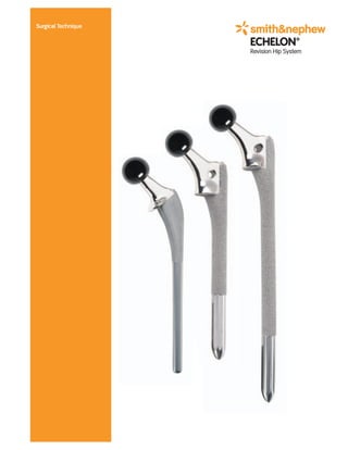
Protesis echelon revision st
- 3. Nota Bene The technique description herein is made available to the healthcare professional to illustrate the author's suggested treatment for the uncomplicated procedure. In the final analysis, the preferred treatment is that which addresses the needs of the specific patient. ECHELON™ Revision Hip System Surgical Technique Contents ECHELON Reamer Chart ......................................................................2 Porous Implants....................................................................................3 Cemented Implants ..............................................................................4 Porous-Coated Implant Specifications ................................................5 Porous-Coated Implant Surgical Technique ........................................7 Cemented Implant Specifications ........................................................20 Cemented Implant Surgical Technique ................................................22 Catalog Information ..............................................................................34 Important Medical Information..............................................................46
- 4. 2 Cemented Stem Standardcollar300mm Standardcollar315mm Cemented 300mm stem length 190mm Straight 190mm 260mm 175mm 160mm 260mm 245mm 230mm 245mm 230mm 175mm 260mm Bowed 175mm 225mm 190mm Porous Stem ECHELON™ Reamer Chart 300mm
- 5. 3 Porous Implants 190mm Straight * * 260mm Bowed 1 Effect of Cementless Femoral Stem Design on Bone Strains and Torsional Stability, Yoshihiro Suzuki, MD; Glen Renowitzky, B.S.; Jeff Lotz, Ph.D.; Robert L. Barrack, MD; Robert B. Bourne, MD; Cecil H. Rorabeck, MD; Michael D. Ries, MD Porous Coating – RoughCoat porous coating increases the friction between the implant and bone, improving implant stability and providing a porous surface for bone ingrowth. Driving platform – The ECHELON™ implants feature a threaded driving platform with an elliptical slot for rotational and axial implant control during insertion. Neck geometry – Circulotrapezoidal neck provides increased range of motion compared to a circular neck of the same strength. Collar options – A standard collar and two calcar platforms are available to match the implant to the proximal defect. Distal Slot – The distal slot eases stem insertion, reduces the risk of fracture,1 and reduces distal stem stiffness. * Hydroxyapatite – A 50 micron layer of hydroxyapatite is applied to the fully porous coated stem. Shoulder Relief – The lateral shoulder is rounded to minimize the risk of fracturing the greater trochanter during stem insertion. Lateral proximal flare – The ECHELON system has a 3° proximal anterior/posterior flare to improve proximal fill, without preventing implant seating. Distal Flutes – The ECHELON system offers distal flutes to increase rotational stability. Distal bullet tip – The bullet tip reduces the stress between the distal implant tip and the bone to minimize thigh pain. Size Range – The ECHELON system porous stems are offered in 1mm increments to minimize bone removal and provide optimum canal fill.
- 6. 4 Cemented Implants Double Taper Proximal Geometry – Limits shear stresses and promotes compressive stress transfer between the cement and implant. Proximal A/P Groove – Increases rotational stability without increasing cement stresses. Trapezoidal Distal Cross Section – Improves resistance to rotation. Neck geometry – Circulotrapezoidal neck provides increased range of motion compared to a circular neck of the same strength. Collar options – A standard collar and two calcar platforms are available to match the implant to the proximal defect. 225mm 300mm ECHELON™ Implants are made from Cobalt Chromium material. An optimized 12/14 taper is used to lock the modular head to the stem. 175mm
- 7. 5 Size –3 +0 +4 +8 +12 +16 11-12 38 40 43 46 49 52 13-17 43 45 48 51 54 57 18-22 48 50 53 56 59 62 Size –3 +0 +4 +8 +12 +16 11-12 34 37 41 45 49 53 13-17 36 39 43 47 51 55 18-22 38 41 45 49 53 57 Neck Length (mm) Neck Offset (mm) Neck Height (mm) Porous-Coated Implant Specifications General Specifications: Cobalt Chromium Material Neck Shaft Angle 131° Porous Straight Stem Length 190mm* Extensively Coated Straight Stem Porous Coating Length 140mm** Porous Bowed Stem Length 260mm* Bowed Stem Porous Coating Length 210mm** The porous stem trial is 1.25mm smaller than the implant. The broach is .5mm smaller than the implant. * Stem length is measured from the standard collar area to the distal tip. ** Porous coating length is measured from the shoulder to the distal end of the coating. Size –3 +0 +4 +8 +12 +16 Standard Collar 11-20 33 35 38 40 43 46 +15mm Calcar 11-20 48 50 53 55 58 61 +30mm Calcar 11-22 63 65 68 70 73 76
- 8. 6 Porous Straight Stem-190mm* Straight Stem Porous Coating Length-140mm Porous Bowed Stem-260 mm Bowed Stem Porous Coating Length-210 mm Neck Length Neck Offset Neck Height Neck Length Neck Offset Neck Height Calcar Stem Stem Size Stem Size Available in standard collar and +15mm calcar options. Available in standard collar, +15mm calcar, and +30mm calcar options. *Porous coating length of ECHELON™ Porous Plus HA is 175mm. Porous-Coated Implant Specifications
- 9. 7 Porous-Coated Implant Surgical Technique Optional Surgical Approach Surgical exposure can be improved by an extended trochanteric osteotomy. The greater trochanter, in continuity with a strip of the lateral proximal femoral cortex, is osteotomized to permit intramedullary access to remove the femoral component. The revision stem should bypass the distal extent of the osteotomy by two to three canal diameters. Place a cerclage cable slightly distal to the osteotomy before reaming, broaching, and inserting the stem to minimize the risk of propagating a crack or fracture. Once the final components are implanted, the osteotomy is reduced and secured with cables. In order to reduce the osteotomized bone fragment in its anatomic position, it may be necessary to shape the endosteal surface of the bone fragment with a curette or burr to fit against the lateral portion of the femoral component. Because of the intramedullary exposure gained by extended trochanteric osteotomy, over-reaming is less likely to be necessary to insert a bowed femoral stem than if the surgery is performed without an extended trochanteric osteotomy.
- 10. 8 Femoral Neck Osteotomy An osteotomy guide is available for proximal bone resection. The angled slot is for a standard collar stem, the proximal horizontal slot is for a +15mm calcar stem, and the distal horizontal slot is for a +30mm calcar stem. The osteotomy guide has a vertical scale in 5mm increments to help gauge neck height. Resect the proximal bone by cutting through the osteotomy slot that corresponds to the implant collar type. When a calcar-type stem is selected, ensure that sufficient bone has been removed to allow the collar to fully seat.
- 11. 9 Reaming Depth from Collar Options Medial Resection Level Standard Collar 190mm +15mm Calcar 175mm Standard Collar (190mm) +15mm Calcar (175mm) Femoral Reaming (Straight Stems) Rigid femoral reamers in 0.5mm increments are available for the porous straight stems. The stem size is measured at the maximum diameter of the distal porous coating. The maximum diameter of the flutes is equal to the diameter of the porous coating. Start reaming with a reamer 4 to 6mm smaller than the templated size or a reamer that has little or no resistance in the femoral canal. For a line-to-line fit, ream the canal in 0.5mm increments until the last reamer matches the selected implant size. The canal can also be reamed 0.5mm smaller than the size for a tighter distal fit. The final reamer size should be based on bone quality, anatomy, and surgeon preference. The stem length is measured from the standard collar area to the distal tip of the implant. Reaming depth is measured from the selected collar level to the distal tip of the implant. Use the straight implant reaming chart to determine the reaming depth for the porous straight implants. Seat the reamer to the appropriate depth mark on each reamer. Porous Straight Stem Standard Collar- 190mm Porous Straight Stem +15mm Calcar- 175mm Stem Size Porous Straight 190mm Implant Reaming Chart
- 12. 10 Femoral Reaming (Bowed Stems) Thin shaft reamers are available for the porous bowed stems. Ream the canal in 0.5mm increments until the reamer size is .5mm larger than the selected implant. The canal can be reamed larger if required to seat the implant. The final reamer size should be based on bone quality, anatomy, and surgeon preference. The stem length is measured from the standard collar area to the distal tip of the implant. Reaming depth is measured from the selected collar level to the distal tip of the implant. Use the bowed implant reaming chart to determine the reaming depth for the bowed porous implants. I often ream the bowed stems line-to-line. If the fit is too tight as determined during implant insertion, the stem is removed and the canal reamed 0.5mm larger. – Douglas Becker, MD Reaming Depth from Collar Options Medial Resection Level Standard Collar 260mm +15mm Calcar 245mm +30mm Calcar 230mm Porous Bowed 260mm Implant Reaming Chart Stem Size Porous Bowed Stem Standard Collar– 260mm Porous Bowed Stem +15mm Calcar- 245mm Porous Bowed Stem +30mm Calcar- 230mm
- 13. 11 4b. 4a. +15mm Calcar +30mm Calcar Begin broaching two sizes smaller than the size of the last femoral reamer. The final broach should match the size of the selected implant. The femoral broaches are 0.5mm smaller than the porous coating level of the implant. Standard Collar Implant Broaching 4a. For a standard collar implant, the proximal medial aspect of the broach should be flush with the osteotomy level. Calcar Implant Broaching 4b.The femoral broach has two black lines on the proximal medial section. The proximal line indicates the level of the +15mm calcar. The distal line indicates the level of the +30mm calcar. When implanting a calcar-style implant, seat the broach so that the appropriate black line meets the medial resection level. If I have used an extended trochanteric osteotomy for removal of cement and/or the femoral components to be revised, I like to close the osteotomy prior to canal preparation. I do this with bone clamps during preparation of the canal and then trial with the bone clamps in place. If I am satisfied with what I learned from the trial, I then apply my definitive cable fixation to the osteotomy prior to inserting the final stem. I find this greatly enhances the fixation that I obtain proximally as opposed to the more usual technique which is to insert the stem and then re-attach the osteotomized trochanter with cables. – James Waddell, MD, FRCS(C) Femoral Broaching
- 14. 12 Standard Collar Calcar Preparation A calcar reamer is available for standard collar implants. With the final broach fully seated, remove the broach handle and ream the calcar bone with the calcar reamer.
- 15. 13 6a. Trial Head Color 22mm 26mm 28mm 32mm 36mm Green — — –3 –3 –3 Yellow +0 +0 +0 +0 +0 Red +4 +4 +4 +4 +4 White +8 +8 +8 +8 +8 Blue +12* +12* +12* +12* +12 Black — — +16* +16* — *Skirted Femoral Head Femoral Neck Length Options Trialing with the Broach 6a. Seat the final broach to the appropriate level for the selected implant (fully seated for standard collar, proximal black line for +15mm calcar, and distal black line for +30mm calcar). Remove the broach handle and place the matching trial neck onto the broach post. Place the desired trial femoral head on the trial neck and reduce the hip to assess stability and range of motion. Note: The broach length is shorter than the implant. Also the broach is .5mm smaller than the implant. Trialing
- 16. 14 6b. Standard Collar Trialing Calcar Trialing Trialing with the Trial Stem 6b.When trialing with the trial stem, select the trial size that corresponds to the last broach used. The trial stem is 1.25mm smaller than the implant (diameter only) to prevent the trial from locking in the canal. A modular collar is available to convert the standard collar trial to a +15mm or +30mm calcar trial. Insert the trial collar into the proximal hole for a +15mm calcar implant and the distal hole for a +30mm calcar implant. Place the desired trial femoral head on the trial stem and reduce the hip to assess stability and range of motion. To get a better feel for the distal fit of the stem, I like to use the next larger trial size from the last reamer diameter. On a straight stem, for example, if the last reamer is a 14.5mm, it's probably useful to insert the 16mm trial. This gives me a good idea how tight the actual implant will be. – Cecil Rorabeck, MD, FRCS(C) Trialing (Cont.)
- 17. 15 Trialing with the Trial Stem Assemble the threaded stem inserter by inserting the stem inserter pommel through the stem inserter frame. Engage the tip of the stem inserter frame into the stem driver slot on the selected implant and turn the pommel to thread the inserter onto the implant. Fully tighten the pommel before impaction. Straight Stem Insertion 7a. Insert the implant into the canal with hand pressure and verify proper implant version. Use firm mallet blows to seat the implant to the desired level. Note: Once the implant flutes have engaged the bone, the implant version cannot be changed without removing the implant. The implant can be removed by striking the underside of the threaded stem driver with a mallet. When using a cylindrical stem design, I believe it is important to know the exact dimensions of the reamers and implants used. Therefore, especially with bowed stems, I will use a ring gauge to measure the reamer and implant to 0.5mm. Based on these measurements, I can decide whether to ream line-to-line or otherwise. – Robert Barrack, MD In cases of considerable bone ectasia proximally, I find it helpful to use prophylactic cables or wires about the proximal femur. The bone is very fragile and propagation cracks can occur easily. – James Waddell, MD, FRCS(C) Implant Insertion 7a.
- 18. 16 Bowed Stem insertion 7b. Insert the bowed stem with the same technique as the straight stem. Implant version must be correct before the implant encounters resistance in the canal. If the bowed stem stops progressing during insertion, remove the implant and enlarge the canal with a larger size thin shaft reamer. Note: A fully seated bowed stem will be extremely difficult to remove from the femur and may require an osteotomy to remove. If the implant does not advance visibly with forceful blows, the distance from the collar to the calcar should be measured in millimeters and re-measured after a series of blows to insure that it’s advancing steadily. If there's no advancement with several hard blows, the pitch has changed, and the stem is still proud, the stem should be extracted and the canal over-reamed. – Robert Barrack, MD For a bowed stem, I find it occasionally necessary to introduce the stem initially for the first few centimeters with the bow turned 90º to its ultimate orientation and, once the tip of the stem has passed the initial proximal bow (anteversion), rotate the stem back to its desired position and gradually impact the stem. – James Waddell, MD, FRCS(C) Standard Collar Calcar Preparation
- 19. 17 8. Once the implant is fully seated, perform a final trial reduction to determine appropriate neck length. Place the desired trial femoral head on the implant and reduce the hip to assess stability and range of motion. Implant Trialing
- 20. 18 Femoral Head Assembly Clean and dry the taper with a sterile cloth, place the prosthetic femoral head on the neck taper and firmly impact several times with a femoral head impactor and a mallet.
- 21. 19
- 22. 20 Neck Offset (mm) Neck Length (mm) Stem Specifications Neck Height (mm) Stem Stem Length* Final Broach Final Reamer Distal Stem Cement Size (mm) Size Size Size Mantle 12 175 / 225 / 300 12 12mm 9mm 1.5mm 14 175 / 225 / 300 14 14mm 11mm 1.5mm 16 175 / 225 16 16mm 13mm 1.5mm General Specifications: Cobalt Chromium Material Neck Shaft Angle 131° *Stem length is measured from the standard collar area to the distal tip. Size –3 +0 +4 +8 +12 +16 12 34 37 41 45 49 53 14 36 39 43 47 51 55 16 36 39 43 47 51 55 Size –3 +0 +4 +8 +12 +16 12 38 40 43 46 49 52 14 43 45 48 51 54 57 16 43 45 48 51 54 57 Size –3 +0 +4 +8 +12 +16 Standard Collar 12, 14, 16 33 35 38 40 43 46 +15mm Calcar 12, 14, 16 48 50 53 55 58 61 +30mm Calcar 12, 14, 16 63 65 68 70 73 76 Cemented Implant Specifications
- 23. 21 Cemented Implant Specifications Cemented Stem 175mm Cemented Stem 225mm* Neck Offset Neck Height Neck Offset Neck Height Calcar Stem Neck Length Neck Length *300mm length also available
- 24. 22 Cemented Implant Surgical Technique Femoral Neck Osteotomy An osteotomy guide is available for proximal bone resection. The angled slot is for a standard collar stem, the proximal horizontal slot is for a +15mm calcar stem, and the distal horizontal slot is for a +30mm calcar stem. The osteotomy guide has a vertical scale in 5mm increments to help gauge neck height. Resect the proximal bone by cutting through the osteotomy slot that corresponds to the implant collar type. When a calcar-type stem is selected, ensure that sufficient bone has been removed to allow the collar to fully seat.
- 25. Stem Length Measured Reaming Depth from Collar Options from Collar Medial Resection Level* Standard Collar 175mm 190mm +15mm Calcar 160mm 175mm +30mm Calcar 145mm 160mm Standard Collar (190mm) +15mm Calcar (175mm) +30mm Calcar (160mm) Cemented 175mm Implant Reaming Chart *Reaming depth includes 1.5cm for the distal cement plug. Femoral Reaming (175mm Stems) Rigid femoral reamers are available for the 175mm cemented stems. Cemented implants are offered in three sizes: 12, 14, and 16. It is important to note that the size corresponds to the size of the recommended final reamer and broach, not the actual implant size. Set the appropriate depth mark at the medial resection level. The stem length is measured from the standard collar area to the distal tip for all cemented implants. Ream to the size of the implant for a 1.5mm cement mantle per side. Use the following chart to determine the reaming depth for the 175mm cemented implants. Reaming depth is measured from the medial resection to the distal cement plug. 23 Stem Size Standard Collar– 175mm +15mm Calcar- 160mm +30mm Calcar- 145mm Cemented 175mm Stem Height
- 26. 24 Stem Length Measured Reaming Depth from Collar Options from Collar Medial Resection Level* Standard Collar 225mm / 300mm 240mm / 315mm +15mm Calcar 210mm 225mm +30mm Calcar 195mm 210mm *Reaming depth includes 1.5cm for the distal cement plug. Cemented 225mm and 300mm Implant Reaming Chart Femoral Reaming (225mm and 300mm Stems) Use thin shaft reamers for the 225mm and 300mm cemented stems. Ream the canal in 0.5mm increments until the reamer size matches the selected implant. This reaming method will provide a minimum 1.5mm cement mantle on each side of the implant. It is unusual that axial reaming is necessary in this revision surgery. Preservation of cancellous bone is critical for allowing intrusion of cement into the cancellous bed thus enhancing fixation. Prior to broaching it is necessary to remove the neocortex that has formed around the previous implant. Failure to do this prevents the cement-bone bond. The stem length is measured from the standard collar area to the distal tip of the implant. Reaming depth is measured from the selected collar level to the distal cement plug. Stem Size Standard Collar– 225mm +15mm Calcar- 210mm +30mm Calcar- 195mm Cemented 225mm Stem Length
- 27. 25 4a. 4b. +15mm Calcar +30mm Calcar Begin broaching two sizes smaller than the size of the last femoral reamer. The final broach should match the size of the selected implant and the last reamer used. The femoral broach provides a 1.5mm cement mantle per side. Standard Collar Implant Broaching 4a. For a standard collar implant, the proximal medial aspect of the broach should be flush with the osteotomy level. Calcar Implant Broaching 4b.The femoral broach has two black lines on the proximal medial section. The proximal line indicates the level of the +15mm calcar. The distal line indicates the level of the +30mm calcar. When implanting a calcar-style implant, seat the broach so that the appropriate black line meets the medial resection level. Femoral Broaching 25
- 28. 26 Standard Collar Calcar Preparation A calcar reamer is available for standard collar implants. With the final broach fully seated, remove the broach handle and ream the calcar bone with the calcar reamer.
- 29. 27 6a. 6b. Standard Collar Trialing Calcar Trialing Trial Head Color 22mm 26mm 28mm 32mm 36mm Green — — –3* –3* –3 Yellow +0* +0* +0* +0* +0 Red +4* +4* +4* +4* +4 White +8* +8* +8* +8* +8 Blue +12* +12* +12* +12* +12 Black — — +16* +16* — *Skirted Femoral Head Femoral Neck Length Options Trialing can be performed using either the broach or trial stem. Trialing with the Broach 6a. Seat the final broach to the appropriate level for the selected implant (fully seated for standard collar, proximal black line for +15mm calcar, and distal black line for +30mm calcar). Remove the broach handle and place the matching trial neck onto the broach post. Place the desired trial femoral head on the trial neck and reduce the hip to assess stability and range of motion. Note: The broach length is shorter than the implant. Trialing with the Trial Stem 6b.When trialing with the trial stem, select the trial size that corresponds to the last broach used. The trial stem is the same size as the implant. If increased stability is needed when trialing, use the femoral broach as a trial. A modular collar is available to convert the standard collar trial to a +15mm or +30mm calcar trial. Insert the trial collar into the proximal hole for a +15mm calcar implant and the distal hole for a +30mm calcar implant. Place the desired trial femoral head on the trial stem and reduce the hip to assess stability and range of motion. Trialing
- 30. 28 Preparing the Femoral Canal Use a curette to remove any grossly loose cancellous bone. Irrigate the canal with saline solution and pulsatile lavage to remove all debris. Continue preparing the femur with the femoral canal brush to remove any remaining weak cancellous bone, blood clots, and marrow fats. Repeat lavaging as necessary to remove all remaining debris. While awaiting the appropriate cement texture, I find it helpful to remind the anesthesiologist to keep the patient’s blood pressure stable and relatively low. Epinepherine-soaked rags are placed in the canal at this time as well, preventing additional bleeding. – Kevin Garvin, MD
- 31. 29 Placing The BUCK™ Cement Restrictor The proximal flange of the cement restrictor should always be larger than the distal canal diameter. Screw the cement restrictor onto the inserter using a clockwise motion. Insert the device to the level of the medullary canal that has been predetermined. Once this level is reached, disengage the restrictor from the inserter using a counterclockwise twisting motion. Remove the inserter from the medullary canal. If it is necessary to remove the restrictor prior to cement insertion, it can be reattached to the inserter rod and pulled out of the canal. The surgeon may adjust the restrictor as many times as required. I find it helpful to place a small 10cc volume of cement distal to the plug if the canal is large and the plug does not remain stable. – Kevin Garvin, MD Drying The Femoral Canal Connect OR suction to the femoral suction absorber handle. Insert the femoral absorber into the femoral canal to dry the canal while mixing the cement.
- 32. 30 Injecting Cement After removing the femoral canal suction absorber, immediately insert the nozzle of the cement gun deep into the femoral canal. Beginning at the distal end of the femoral canal, inject cement into the canal in retrograde fashion. Continue injecting cement until the canal is completely full and the distal tip of the nozzle is clear of the canal.
- 33. 31 Pressurizing Cement Break off the long nozzle and place the femoral pressurizer over the short nozzle. Place the pressurizer against the proximal femur. This will occlude the canal and pressurize the cement. Maintain firm pressure for 30–60 seconds, depending on cement viscosity, to allow good cement interdigitation into trabecular bone. Withdraw the pressurizer from the canal and remove any extruded cement around the periphery of the pressurizer.
- 34. 32 Implant Insertion Engage the tip of the stem inserter into the stem driver slot on the selected implant. Insert the implant into the canal with hand pressure while verifying proper implant alignment. Implant Trialing Once the implant is fully seated and the cement has cured, perform a final trial reduction to determine appropriate neck length. Place the desired trial femoral head on the implant and reduce the hip to assess stability and range of motion.
- 35. 33 Femoral Head Assembly Clean and dry the taper with a sterile cloth, place the prosthetic femoral head on the neck taper and firmly impact several times with a femoral head impactor and a mallet.
- 36. 34 Standard +15mm Extensively Coated Porous Straight Implants (190mm) Size. Standard Collar +15mm Calcar 11 7134-0111 7134-0211 12 7134-0112 7134-0212 13 7134-0113 7134-0213 14 7134-0114 7134-0214 15 7134-0115 7134-0215 16 7134-0116 7134-0216 17 7134-0117 7134-0217 18 7134-0118 7134-0218 19 7134-0119 7134-0219 20 7134-0120 7134-0220 Catalog Information Porous Plus HA (190mm) Size. Standard Collar +15mm Calcar 11 7134-2011 7134-3011 12 7134-2012 7134-3012 13 7134-2013 7134-3013 14 7134-2014 7134-3014 15 7134-2015 7134-3015 16 7134-2016 7134-3016 17 7134-2017 7134-3017 18 7134-2018 7134-3018 19 7134-2019 7134-3019 20 7134-2020 7134-3020 Standard +15mm Porous Bowed Implants (260mm) Size. Standard Collar +15mm Calcar +30mm Calcar 12L 7134-0412 7134-0612 7134-0812 13L 7134-0413 7134-0613 7134-0813 14L 7134-0414 7134-0614 7134-0814 15L 7134-0415 7134-0615 7134-0815 16L 7134-0416 7134-0616 7134-0816 17L 7134-0417 7134-0617 7134-0817 18L 7134-0418 7134-0618 7134-0818 19L 7134-0419 7134-0619 7134-0819 20L 7134-0420 7134-0620 7134-0820 12R 7134-0512 7134-0712 7134-0912 13R 7134-0513 7134-0713 7134-0913 14R 7134-0514 7134-0714 7134-0914 15R 7134-0515 7134-0715 7134-0915 16R 7134-0516 7134-0716 7134-0916 17R 7134-0517 7134-0717 7134-0917 18R 7134-0518 7134-0718 7134-0918 19R 7134-0519 7134-0719 7134-0919 20R 7134-0520 7134-0720 7134-0920 21L 7134-0421 7134-0621 7134-0821 22L 7134-0422 7134-0622 7134-0822 21R 7134-0521 7134-0721 7134-0921 22R 7134-0522 7134-0722 7134-0922 Standard +15mm +30mm
- 37. 35 +15mm +30mmStandard Cemented Implants Size Length Standard +15mm +30mm (mm) Collar Calcar Calcar 12 175 7131-0112 7131-0312 7131-0612 12 225 7131-0212 7131-0412 7131-0712 14 175 7131-0114 7131-0314 7131-0614 14 225 7131-0214 7131-0414 7131-0714 16 175 7131-0116 7131-0316 7131-0616 16 225 7131-0216 7131-0416 7131-0716 *12L 300 7131-4112 — — *14L 300 7131-4114 — — *12R 300 7131-5112 — — *14R 300 7131-5114 — — *Note: These sizes available by special request. OXINIUM™ 12/14 Taper Femoral Heads Neck Length 22mm 26mm 28mm 32mm 36mm –3 — — 7134-2803 7134-3203 7134-3603 +0 7134-2200 7134-2600 7134-2800 7134-3200 7134-3600 +4 7134-2204 7134-2604 7134-2804 7134-3204 7134-3604 +8 7134-2208 7134-2608 7134-2808 7134-3208 7134-3608 +12 7134-2212 7134-2612 7134-2812 7134-3212 7134-3612 +16 — — 7134-2816 7134-3216 7134-3616 Alumina 12/14 Taper Femoral Heads Neck Length 22mm 26mm 28mm 32mm 36mm –3 — — — — — +0 — — 7133-2800 7133-3200 7133-3600 +4 — — 7133-2804 7133-3204 7133-3604 +8 — — 7133-2808 7133-3208 7133-3608 +12 — — — — — +16 — — — — — CoCr 12/14 Taper Femoral Heads Cobalt Chromium – ASTM F 799 Neck Length 22mm 26mm 28mm 32mm 36mm –3 — — 7130-2803 7130-3203 7130-3603 +0 7130-2200 7130-2600 7130-2800 7130-3200 7130-3600 +4 7130-2204 7130-2604 7130-2804 7130-3204 7130-3604 +8 7130-2208 7130-2608 7130-2808 7130-3208 7130-3608 +12 7130-2212 7130-2612 7130-2812 7130-3212 7130-3612 +16 — — 7130-2816 7130-3216 — Trial 12/14 Taper Femoral Heads Neck Color Length Code 22mm 26mm 28mm 32mm 36mm –3 Green — — 7135-2803 7135-3203 7135-3603 +0 Yellow 7135-2200 7135-2600 7135-2800 7135-3200 7135-3600 +4 Red 7135-2204 7135-2604 7135-2804 7135-3204 7135-3604 +8 White 7135-2208 7135-2608 7135-2808 7135-3208 7135-3608 +12 Blue 7135-2212 7135-2612 7135-2812 7135-3212 7135-3612 +16 Black — — 7135-2816 7135-3216 —
- 38. 36 ECHELON™ Starter Tray Cat. No. 7136-6001 Osteotomy Guide Cat. No. 7136-4100 Box Osteotome Cat. No. 7136-4002 Femoral Canal Finder Cat. No. 7136-4001 T-Handle Cat. No. 7136-4006 Anteversion Handle (2 per set) Cat. No. 7136-4012 ECHELON Reamer Tray Cat. No. 7136-6002
- 39. 37 Broach Handle (2 per set) Cat. No. 7136-4007 Proximal Reamer Cat. No. 7136-4015 Rigid Reamer Cat. No. Size 7135-0090 9mm 7135-0095 9.5mm 7135-0100 10mm 7135-0105 10.5mm 7135-0110 11mm 7135-0115 11.5mm 7135-0120 12mm 7135-0125 12.5mm 7135-0130 13mm 7135-0135 13.5mm 7135-0140 14mm 7135-0145 14.5mm 7135-0150 15mm 7135-0155 15.5mm 7135-0160 16mm 7135-0165 16.5mm 7135-0170 17mm 7135-0175 17.5mm 7135-0180 18mm 7135-0185 18.5mm 7135-0190 19mm 7135-0195 19.5mm 7135-0200 20mm ECHELON™ Broach Tray Cat. No. 7136-6003 ECHELON Straight Stem Trial Tray Porous and Cemented Cat. No. 7136-6004
- 40. 38 Broach Cat. No. Size 7136-7010 10 7136-7011 11 7136-7012 12 7136-7013 13 7136-7014 14 7136-7015 15 7136-7016 16 7136-7017 17 7136-7018 18 7136-7019 19 7136-7020 20 Trial Neck Cat. No. Size 7136-7101 11-12 7136-7102 13-17 7136-7103 18-20 Calcar Reamer Cat. No. 7136-4004 Stem Inserter Frame Cat. No. 7136-4008 Stem Inserter Pommel Cat. No. 7136-4011 Femoral Head Impactor Cat. No. 7136-4009 Trial Collar (2 per set) Cat. No. 7136-7034
- 41. 39 Porous Straight Stem Trial Cat. No. Size 7136-8011 11 7136-8012 12 7136-8013 13 7136-8014 14 7136-8015 15 7136-8016 16 7136-8017 17 7136-8018 18 7136-8019 19 7136-8020 20 Cemented Stem Trial Cat. No. Size Length 7136-8512 12 175mm 7136-8514 14 175mm 7136-8516 16 175mm 7136-8612 12 225mm 7136-8614 14 225mm 7136-8616 16 225mm 7136-8712 12 left 300mm 7136-8714 14 right 300mm 7136-8812 12 left 300mm 7136-8814 14 right 300 mm Cemented Stem Inserter Cat. No. 7136-4014 ECHELON™ Bowed Stem Trial Tray Cat. No. 7136-6015
- 42. 40 ECHELON™ Thin Shaft Reamer Tray Cat. No. 7136-6006 ECHELON 300mm Bowed Cemented Stem Trial Tray Cat. No. 7136-6008 ECHELON 21-22mm Bowed Stem Trial Thin Shaft Reamer Tray Cat. No. 7136-6007
- 43. 41 Thin Shaft Reamer Cat. No. Size 7135-1900 9mm 7135-1100 10mm 7135-1110 11mm 7135-1115 11.5mm 7135-1120 12mm 7135-1125 12.5mm 7135-1130 13mm 7135-1135 13.5mm 7135-1140 14mm 7135-1145 14.5mm 7135-1150 15mm 7135-1155 15.5mm 7135-1160 16mm 7135-1165 16.5mm 7135-1170 17mm 7135-1175 17.5mm 7135-1180 18mm 7135-1185 18.5mm 7135-1190 19mm 7135-1195 19.5mm 7135-1200 20mm 7135-1205 20.5mm 7135-1210 21mm 7135-1215 21.5mm 7135-1220 22mm 7135-1225 22.5mm 7135-1230 23mm 7135-1235 23.5mm 7135-1240 24mm Porous Bowed Stem Trial Cat. No. Size 7136-8312 12 Left 7136-8313 13 Left 7136-8314 14 Left 7136-8315 15 Left 7136-8316 16 Left 7136-8317 17 Left 7136-8318 18 Left 7136-8319 19 Left 7136-8320 20 Left 7136-8321 21 Left 7136-8322 22 Left 7136-8412 12 Right 7136-8413 13 Right 7136-8414 14 Right 7136-8415 15 Right 7136-8416 16 Right 7136-8417 17 Right 7136-8418 18 Right 7136-8419 19 Right 7136-8420 20 Right 7136-8421 21 Right 7136-8422 22 Right
- 44. 42 VORTEX™ Vacuum Mixer Cat. No. 7127-0070 MIXOR Hose Only Cat. No. 7127-0041 MIXOR™ Vacuum Mixing System with Syringe Cat. No. 7127-0020 VORTEX Nozzles Cat. No. Description 7127-0080 Standard Breakaway 7127-0081 Long Tapered 7127-0082 Angled Nozzle 7127-0084 Revision 7127-0085 Umbrella 7127-0071 Re-use Kit (not shown) 7127-0072 Adaptor Catalog Information - Cement Accessories Mixer Components MIXOR Pump Only Cat. No. 7127-0042 Stryker Foot Pump Adapter Cat. No. 7127-0060 VORTEX Gun Cat. No. 7127-2001
- 45. 43 Catalog Information - Cement Accessories MIXOR™ Pump Connectors Connector, Schraeder Cat. No. 7127-0050 Connector, Drager Cat. No. 7127-0051 Connector, D.I.S.S. Cat. No. 7127-0052
- 46. 44 Catalog Information - Cement Accessories Cement and Accessories VERSABOND™ Cat. No. 7127-1340 VERSABOND™ AB Cat. No. 7127-1440 PREP-IM™ Total Hip Preparation Kit Cat. No. 12-1010 Includes the following: 2 Buck Cement Restrictors 1 Femoral Canal Brush 1 Buck Disposable Inserter 1 Femoral Canal Suction Absorber 2 Concise Cement Sculps 1 Medium Femoral Pressurizer Jet Vacs Cat. No. Description 89-0105 Universal Tip (shown) 89-0106 Hip Tip 89-0107 Intermediate Tip BUCK™ Cement Restrictors Cat. No. Description 91-4535 13mm 12-9418 18.5mm 12-9419 25mm 7127-9420 30mm 7127-9421 35mm Small Femoral Pressurizer Cat. No. 7127-0026 Medium Femoral Pressurizer Cat. No. 7127-0027 Large Femoral Pressurizer Cat. No. 7127-0028
- 47. 45 Handpiece with Synthes Connection Cat. No. 7127-7006 Catalog Information - Cement Accessories POWERPULSE™ Lavage System Handpiece with Zimmer Coupling Cat. No. 7127-7000 Powerhose with Zimmer Coupling Cat. No. 7127-7001 ECHELON™ Hip and Knee with Suction Cat. No. 7127-7004 ECHELON Hip and Knee without Suction Cat. No. 7127-7005
- 48. 46 Important Note Total hip replacement (THR) arthroplasty has become a successful procedure for relieving pain and restoring motion in patients who are disabled from hip arthropathy. The goals of total hip replacement are to decrease pain, increase function, and increase mobility. Materials Femoral components are cobalt chromium alloy, titanium 6Al-4V alloy or stainless steel. Femoral heads are cobalt chromium alloy, zirconia ceramic, alumina ceramic, OXINIUM™ oxidized zirconium or stainless steel. Acetabular liners are ultra-high molecular weight polyethylene or alumina ceramic. All poly acetabular components are ultra-high molecular weight polyethylene. Acetabular shells are titanium 6Al-4V alloy. The component material is provided on the outside carton label. Some of the alloys needed to produce orthopedic implants contain some metallic components that may be carcinogenic in tissue cultures or intact organism under very unique circumstances. Questions have been raised in the scientific literature as to whether or not these alloys may be carcinogenic in implant recipients. Studies conducted to evaluate this issue have not identified conclusive evidence of such phenomenon, in spite of the millions of implants in use. Description of System The Total Hip System consists of femoral components, proximal sleeves, taper sleeves, acetabular components, fixation screws and pegs, hole covers, centralizers, and femoral heads. Components may be grit blasted, porous coated, hydroxylapatite (HA) coated, or HA porous coated. All implantable devices are designed for single use only. Femoral Components Femoral components are available in a variety of sizes. Porous coated components are coated for biological ingrowth. Modular femoral components accept proximal sleeves. Non-porous femoral components can feature PMMA centralizers that help produce a uniform thickness of cement. Femoral components are available with a Small (10/12), Large (14/16), or 12/14 global taper. Small taper femoral components mate and lock directly with a 22 mm metal, oxidized zirconium or ceramic head. The Small taper also mates with a taper sleeve which, in turn, mates with either metal or ceramic heads (26, 28, or 32 mm), bipolar or unipolar components. Large taper femoral components mate and lock with either metal heads (26, 28, or 32 mm), ceramic heads (28 or 32 mm), oxidized zirconium (28, 32, or 36mm), bipolars or unipolar components. Femoral components with a 12/14 taper mate and lock with either metal heads, oxidized zirconium heads, ceramic heads, bipolar or unipolar components. Small, Large, and 12/14 taper femoral component tapers are machined to mate and lock with ceramic heads, thus preventing rotation of the ceramic head on the stem, which would cause wear of the stem taper. Taper Sleeves A taper sleeve is required to be impacted on the Small taper femoral component prior to impacting a Large (14/16) taper femoral head size 26, 28, or 32 mm. A taper sleeve is required to attach a unipolar head. Unipolar taper sleeves are available in Small, Large, and 12/14 tapers. Never place more than one taper sleeve on a femoral component. Femoral Heads Cobalt chromium, stainless steel, oxidized zirconium, and ceramic heads are available in multiple neck lengths for proper anatomic and musculature fit. Heads are highly polished for reduced friction and wear. The following zirconia ceramic heads are available for use only with Small and Large taper femoral components: Zirconia Head Neck Ceramic Diameter Length 42-7815 32mm Standard 0mm 42-7816 32mm Long + 4mm 42-7817 32mm X-Long + 8mm 42-7818 28mm Standard 0mm 42-7819 28mm Long + 4mm 42-7820 28mm X-Long + 8mm Note: 32 mm heads with a -3 mm neck length are not available for use with the Small taper stems. In addition to the components listed above, the following components are available for use only with Small taper femoral components: Zirconia Head Neck Ceramic Diameter Length 7132-0002 22mm Long + 4mm 7132-0006 22mm X-Long + 8mm Note: 22mm Zirconia Ceramic Heads used with Small taper femoral components are not available in the USA The following zirconia ceramic heads are available for use only with 12/14 taper femoral components: Zirconia Head Neck Ceramic Diameter Length 7132-0028 28mm Standard 0mm 7132-0428 28mm Long + 4mm 7132-0828 28mm X-Long + 8mm 7132-0026 26mm Standard 0mm 7132-0426 26mm Long + 4mm 7132-0826 26mm X-Long + 8mm 7132-0422 22mm Long + 4mm 7132-0822 22mm X-Long + 8mm The following alumina ceramic heads are available for use only with 12/14 taper femoral components: Alumina Head Neck Ceramic Diameter Length 7133-2800 28mm Standard 0mm 7133-2804 28mm Long + 4mm 7133-2808 28mm X-Long + 8mm 7133-3200 32mm Standard 0mm 7133-3204 32mm Long + 4mm 7133-3208 32mm X-Long + 8mm Acetabular Components Acetabular components can be one piece all polyethylene, two-piece component consisting of a titanium shell and a polyethylene liner or a titanium shell and an alumina ceramic liner. Please see Warnings and Precautions for specific information on screws, pegs and hole covers use. Acetabular reinforcement and reconstruction rings are used with an all polyethylene acetabular component. The BIRMINGHAM HIP™ Resurfacing (BHR) prosthesis is a metal-on-metal bearing component consisting of a stemmed femoral head resurfacing component designed for cemented insertion and a hemispherical acetabular cup designed for cementless interference fit into the acetabulum. The acetabular cup has hydroxylapatite coating applied to the external surface and porous coating. Cement should not be used with this type of implant. Note: The BHR prosthesis is not available in the USA Note: 10 Mrad cross-linked polyethylene (UHMWPE) REFLECTION™ acetabular liners may be used with metal (CoCr), oxidized zirconium, alumina ceramic or zirconia ceramic femoral heads. Femoral components and femoral heads are designed for use with any Smith & Nephew polyethylene acetabular component or polyethylene-lined, metal-backed acetabular component having an appropriately-sized inside diameter. Indications, Contraindications, and Adverse Effects Hip components are indicated for individuals undergoing primary and revision surgery where other treatments or devices have failed in rehabilitating hips damaged as a result of trauma or noninflammatory degenerative joint disease (NIDJD) or any of its composite diagnoses of osteoarthritis, avascular necrosis, traumatic arthritis, slipped capital epiphysis, fused hip, fracture of the pelvis, and diastrophic variant. Hip components are also indicated for inflammatory degenerative joint disease including rheumatoid arthritis, arthritis secondary to a variety of diseases and anomalies, and congenital dysplasia; old, remote osteomyelitis with an extended drainage free period, in which case, the patient should be warned of an above normal danger of infection postoperatively; treatments of nonunion, femoral neck fracture and trochanteric fractures of the proximal femur with head involvement that are unmanageable using other techniques; endoprosthesis, femoral osteotomy, or Girdlestone resection; fracture-dislocation of the hip; and correction of deformity. The BIRMINGHAM HIP Resurfacing (BHR) arthroplasty system is indicated for use for reduction or relief of pain and/or improved hip function in patients who are candidates for a total hip replacement but who have evidence of good femoral bone stock. These patients should also be skeletally mature with the following conditions: noninflammatory degenerative joint disease such as osteoarthritis, avascular necrosis, ankylosis, protrusio acetabuli, and painful hip dysplasia; inflammatory degenerative joint disease such as rheumatoid arthritis; correction of functional deformity; and are of an age such that total hip revision is likely at some future point. Acetabular reinforcement and reconstruction rings are intended to be used in primary and revision surgeries where the acetabulum has the deficiencies of the acetabular roof, anterior or posterior pillar, medial wall deficiency, and / or protrusion as a result of the indications listed previously. Some of the diagnoses listed above and below may also increase the chance of complications and reduce the chance of a satisfactory result. Contraindications 1. Conditions that would eliminate or tend to eliminate adequate implant support or prevent the use of an appropriately- sized implant, e.g.: a. blood supply limitations; b. insufficient quantity or quality of bone support, e.g., osteoporosis, or metabolic disorders which may impair bone formation, and osteomalacia; and c. infections or other conditions which lead to increased bone resorption. 2. Mental or neurological conditions which tend to impair the patient's ability or willingness to restrict activities. 3. Physical conditions or activities which tend to place extreme loads on implants, e.g., Charcot joints, muscle deficiencies, multiple joint disabilities, etc. 4. Skeletal immaturity. 5. The zirconia ceramic head is contraindicated for use with any other product than an UHMW polyethylene cup or a metal backed UHMW polyethylene cup. 6. The alumina ceramic liner is contraindicated for use with any product other than the metal shell with the correlating inner taper geometry and the appropriate sized alumina ceramic head. The alumina ceramic liner should only be used with the alumina ceramic head. Contraindications may be relative or absolute and must be carefully weighed against the patient's entire evaluation and the prognosis for possible alternative procedures such as non-operative treatment, arthrodesis, femoral osteotomy, pelvic osteotomy, resection arthroplasty, hemiarthroplasty and others. Conditions presenting increased risk of failure include: osteoporosis, metabolic disorders which may impair bone formation, and osteomalacia. Possible Adverse Effects 1. Wear of the polyethylene and ceramic articulating surfaces of acetabular components may occur. Higher rates of wear may be initiated by the presence of particles of cement, metal, or other debris which can develop during or as a result of the surgical procedure and cause abrasion of the articulating surfaces. Higher rates of wear may shorten the useful life of the prosthesis and lead to early revision surgery to replace the worn prosthetic components. 2. With all joint replacements, asymptomatic, localized, progressive bone resorption (osteolysis) may occur around the prosthetic components as a consequence of foreign-body reaction to particulate wear debris. Particles are generated by interaction between components, as well as between the components and bone, primarily through wear mechanisms of adhesion, abrasion, and fatigue. Secondarily, particles may also be generated by third-body particles lodged in the polyethylene or ceramic articular surfaces. Osteolysis can lead to future complications necessitating the removal or replacement of prosthetic components. 3. Loosening, bending, cracking, or fracture of implant components may result from failure to observe the Warnings and Precautions below. Fracture of the implant can occur as a result of trauma, strenuous activity, improper alignment, or duration of service. 4. Dislocations, subluxation, decreased range of motion, or lengthening or shortening of the femur caused by improper neck selection, positioning, looseness of acetabular or femoral components, extraneous bone, penetration of the femoral prosthesis through the shaft of the femur, fracture of the acetabulum, intrapelvic protrusion of acetabular component, femoral impingement, periarticular calcification, and/or excessive reaming. Important Medical Information Warnings and Precautions – Total Hip System
- 49. 47 5. Fracture of the pelvis or femur: post-operative pelvic fractures are usually stress fractures. Femoral fractures are often caused by defects in the femoral cortex due to misdirected reaming, etc. Intraoperative fractures are usually associated with old congenital deformity, improper stem selection, improper broaching, and/or severe osteoporosis. 6. Infection, both acute post-operative wound infection and late deep wound sepsis. 7. Neuropathies; femoral, sciatic, peroneal nerve, and lateral femoral cutaneous neuropathies have been reported. Temporary or permanent nerve damage resulting in pain or numbness of the affected limb. 8. Wound hematoma, thromboembolic disease including venous thrombosis, pulmonary embolus, or myocardial infarction. 9. Myositis ossificans, especially in males with hypertrophic arthritis, limited pre-operative range of motion and/or previous myositis. Also, periarticular calcification with or without impediment to joint mobility can cause decreased range of motion. 10. Trochanteric nonunion usually associated with early weight bearing and/or improper fixation of the trochanter, when a transtrochanteric surgical approach is used. 11.Although rare, metal sensitivity reactions and/or allergic reactions to foreign materials have been reported in patients following joint replacement. 12. Damage to blood vessels. 13. Traumatic arthrosis of the knee from intraoperative positioning of the extremity. 14. Delayed wound healing. 15. Aggravated problems of the affected limb or contralateral extremity caused by leg length discrepancy, excess femoral medialization, or muscle deficiency. 16. Failure of the porous coating/ substrate interface or hydroxylapatite coating/ porous coating bonding may result in bead separation delamination. 17. Stem migration or subsidence has occurred in conjunction with compaction grafting procedures usually resulting from insufficient graft material or improper cement techniques. Varus stem alignment may also be responsible. Warnings and Precautions The patient should be warned of surgical risks, and made aware of possible adverse effects. The patient should be warned that the device does not replace normal healthy bone, that the implant can break or become damaged as a result of strenuous activity or trauma, and that it has a finite expected service life and may need to be replaced in the future. Do not mix components from different manufacturers. Additional Warnings and Precautions may be included in component literature. Preoperative 1. Use extreme care in handling and storage of implant components. Cutting, bending, or scratching the surface of components can significantly reduce the strength, fatigue resistance, and/or wear characteristics of the implant system. These, in turn, may induce internal stresses that are not obvious to the eye and may lead to fracture of the component. Implants and instruments should be protected from corrosive environments such as salt air during storage. Do not allow the porous surfaces to come in contact with cloth or other fiber- releasing materials. 2. Allergies and other reactions to device materials, although infrequent, should be considered, tested for (if appropriate), and ruled out preoperatively. 3. Fixation and expected longevity of components expected to be left in place at revision surgery should be thoroughly assessed. 4. Surgical technique information is available upon request. The surgeon should be familiar with the technique. Refer to medical or manufacturer literature for specific product information. 5. Intraoperative fracture or breaking of instruments can occur. Instruments which have experienced extensive use or excessive force are susceptible to fracture. Instruments should be examined for wear, or damage, prior to surgery. 6. Do not cold water quench ceramic components and never sterilize ceramic heads while fixed on the stem taper. (See sterilization section, below.) 7. Select components such that the zirconia ceramic and oxidized zirconium heads always articulate with a UHMW polyethylene cup or a metal backed UHMW polyethylene cup and alumina heads articulate with UHMW polyethylene or alumina liners. Zirconia ceramic, oxidized zirconium, and alumina heads should never articulate against metal because severe wear of the metal will occur. 8. Select only Smith & Nephew femoral components that indicate their use with ceramic heads. This is important because the taper on the stem is machined to tightly mate and lock with the ceramic head thus preventing rotation of the ceramic head on the stem. Also, an improperly dimensioned taper could result in fracture of the ceramic head. 9. The zirconia ceramic head is composed of a new ceramic material with limited clinical history. Although mechanical testing demonstrates that when used with a polyethylene acetabular component the yttria stabilized zirconia ball produces a relatively low amount of particulates, the total amount of particulate remains undetermined. Because of the limited clinical and preclinical experience, the biological effect of these particulates can not be predicted. 10.Alumina ceramic should never articulate against metal because severe wear could occur. 11.If a computer assisted surgery system is used, consult the applicable software and hardware reference manuals provided by the manufacturer to ensure proper operation of this equipment. Intraoperative 1. The general principles of patient selection and sound surgical judgment apply. The correct selection of the implant is extremely important. The appropriate type and size should be selected for patients with consideration of anatomical and biomechanical factors such as patient age and activity levels, weight, bone and muscle conditions, any prior surgery and anticipated future surgeries, etc. Generally, the largest cross-section component which will allow adequate bone support to be maintained is preferred. Failure to use the optimum- sized component may result in loosening, bending, cracking, or fracture of the component and/or bone. 2. Correct selection of the neck length and cup, and stem positioning, are important. Muscle looseness and/or malpositioning of components may result in loosening, subluxation, dislocation, and/or fracture of components. Increased neck length and varus positioning will increase stresses which must be borne by the stem. The component should be firmly seated with the component insertion instruments. 3. Care should be taken not to scratch, bend (with the exception of the Reconstruction Rings) or cut implant components during surgery for the reasons stated in Number One of the "Pre-Operative" section of "Warnings and Precautions." 4. A +12 mm or +16 mm femoral head should not be used with any Small taper stems. 5. MATRIX™ Small taper stem sizes 8S - 10L must have a minimum neck length of +8 mm when used with a bipolar component; and Small taper stem sizes 12S - 16L must have a minimum neck length of +4 mm when used with a bipolar component. 6. Modular heads and femoral components should be from the same manufacturer to prevent mismatch of tapers. 7. Stainless steel heads and stainless steel stems should only be used together. Neither should be used with other metal components. 8. Use only REFLECTION Liners with REFLECTION Shells. 9. Clean and dry stem taper prior to impacting the femoral head or taper sleeve. The modular femoral head component must be firmly seated on the femoral component to prevent disassociation. 10.Take care, when positioning and drilling screw and peg holes, to avoid penetration of the inner cortex of the pelvis, penetration of the sciatic notch, or damage to vital neurovascular structures. Perforation of the pelvis with screws that are too long can rupture blood vessels causing the patient to hemorrhage. Do not place a screw in the center hole of the acetabular prosthesis. Placement of drills and screws in the anterior or medial portions of the prosthesis is associated with a high risk of potentially fatal vascular injury. Bone screws must be completely seated in the holes of the shell to allow proper locking for the acetabular component liner. If the tapered pegs need to be removed from the shell after impaction of the pegs, do not reuse the pegs or the peg shell holes. Use new pegs and different shell holes, or a new shell if necessary. 11.USE ONLY REFLECTION TITANIUM SPHERICAL HEAD BONE SCREWS, UNIVERSAL CANCELLOUS BONE SCREWS, TAPERED PEGS, AND HOLE COVERS with the REFLECTION Acetabular Components. REFLECTION SP3, FSO and INTERFIT™ Shells accept both REFLECTION spherical head screws and Universal cancellous bone screws. REFLECTION FSO and INTERFIT Shells accept the Modified REFLECTION screw hole covers. The REFLECTION V Shell only accepts Universal Cancellous, REFLECTION screws, and tapered screw-hole covers, not pegs. REFLECTION Peripheral Hole Screws should only be used with REFLECTION Peripheral Hole Shells. Locking Head Pegs and REFLECTION SP Screw Hole Covers are only for use with SP3 Shells. Tapered pegs can only be used with REFLECTION V Shells. The threaded center hole in REFLECTION Shells only accepts the threaded hole cover, not screws or pegs. The INTERFIT threaded hole cover is only for use with REFLECTION INTERFIT, SP3, Spiked and No Hole Shells. The REFLECTION threaded hole cover can be used with all REFLECTION shells. Refer to product literature for proper adjunctive fixation and hole cover usage. 12.Prior to seating modular components, surgical debris including tissue must be cleaned from the surfaces. Debris, including bone cement, may inhibit the component locking mechanism. If the shell is to be cemented in place, remove extraneous cement with a plastic sculps tool to ensure proper locking of the liner. During liner insertion, make sure soft tissue does not interfere with the shell/liner interface. Chilling the liner reduces the impaction force required to seat the liner. Modular components must be assembled securely to prevent disassociation. Debris inhibits the proper fit and locking of modular components which may lead to early failure of the procedure. Failure to properly seat the acetabular liner into the shell can lead to disassociation of the liner from the shell. 13.Avoid repeated assembly and disassembly of the modular components which could compromise the critical locking action of the locking mechanism. 14.Care is to be taken to assure complete support of all parts of the device embedded in bone cement to prevent stress concentration which may lead to failure of the procedure. During curing of the cement, care should be taken to prevent movement of the implant components. 15.If the head is removed from a femoral component that will be left in place at revision surgery, it is recommended that a metal head be used. A ceramic head may fracture from irregularities on the femoral component taper. 16.If components are to be left in place at revision surgery, they should first be thoroughly checked for signs of looseness, etc. and replaced if necessary. The head/neck component should be changed only when clinically necessary. 17.Once removed from the patient, implants previously implanted should never be reused, since internal stresses which are not visible may lead to early bending or fracture of these components. 18.With the congenitally dislocated hip, special care should be taken to prevent sciatic nerve palsy. Also, note that the femoral canal is often very small and straight and may require an extra-small straight femoral prosthesis; however, a regular-sized prosthesis should be used when possible. Note that the true acetabulum is rudimentary and shallow. A false acetabulum should not ordinarily be utilized as a cup placement site for anatomical and biomechanical reasons. 19. With rheumatoid arthritis, especially for those patients on steroids, bone may be extremely osteoporotic. Care should be taken to prevent excessive penetration of the acetabular floor or fracture of the medial acetabular wall, femur, or greater trochanter. 20. Revision procedures for previous arthroplasty, Girdlestone, etc., are technically demanding and difficult to exercise. Common errors include misplacement of the incision, inadequate exposure or mobilization of the femur, inadequate removal of ectopic bone, or improper positioning of components. Postoperative instability as well as excessive blood loss can result. In summary, increased operative time, blood loss, increased incidence of pulmonary embolus and wound hematoma can be expected with revision procedures. 21. Prior to closure, the surgical site should be thoroughly cleaned of cement, bone chips, ectopic bone, etc. Ectopic bone and/or bone spurs may lead to dislocation or painful or restricted motion. Range of motion should be thoroughly checked for early contact or instability. 22. Proper positioning of the components is important to minimize impingement which could lead to early failure, premature wear, and/or dislocation. 23. In order to minimize the risks of dislocation and loosening of the shell-acetabular bone or shell-bone cement interface that may occur when using a metallic shell intended for biological fixation or cemented use only, surgeons should consider providing immediate resistance to tensile forces between the metallic shell and the acetabular bone or bone cement interface through the use of orthopedic bone fixation devices such as bone screws, spikes, screw threads, pegs, fins, or other bone fixation devices. 24. Physicians should consider component malposition, component placement, and the effect on range of motion when using modular heads (with sleeves or skirts) and extended liners. 25. For computer assisted surgery systems, it is extremely important to correctly select input parameters (e.g. bony landmarks). Operators of this equipment should be familiar with the anatomy relevant to the procedure. Failure to provide proper input could cause problems such as violation of critical anatomical structures and malpositioned implants. Postoperative 1. Postoperative directions and warnings to patients by physicians, and patient care, are extremely important. Gradual weight bearing is begun after surgery in ordinary total hip arthroplasty. However, with trochanter
- 50. 48 osteotomy or certain complex cases, weight-bearing status should be individualized with the non or partial weight-bearing period extended. 2. Patients should be warned against unassisted activity, particularly use of toilet facilities and other activities requiring excessive motion of the hip. 3. Use extreme care in patient handling. Support should be provided to the operative leg when moving the patient. While placing the patient on bedpans, changing dressings, and clothing, and similar activities, precautions should be taken to avoid placing excessive load on the operative part of the body. 4. Postoperative therapy should be structured to regain muscle strength around the hip and a gradual increase of activities. 5. Periodic x-rays are recommended for close comparison with immediate postoperative conditions to detect long-term evidence of changes in position, loosening, bending and/or cracking of components or bone loss. With evidence of these conditions, patients should be closely observed, the possibilities of further deterioration evaluated, and the benefits of early revision considered. 6. Prophylactic antibiotics should be recommended to the patient similar to those suggested by the American Heart Association for conditions or situations that may result in bacteremia. Packaging and Labeling Components should only be accepted if received by the hospital or surgeon with the factory packaging and labeling intact. If the sterile barrier has been broken, return the component to Smith & Nephew, Inc. Sterilization/Resterilization Most implants are supplied sterile and have been packaged in protective trays. The method of sterilization is noted on the package label. All radiation sterilized components have been exposed to a minimum of 25 kiloGrays of gamma radiation. If not specifically labeled sterile, the implants and instruments are supplied non-sterile and must be sterilized prior to use. Inspect packages for punctures or other damage prior to surgery. Metal Components Nonporous or non-HA coated metal components and oxidized zirconium heads may be initially sterilized or resterilized, if necessary, by steam autoclaving in appropriate protective wrapping, after removal of all original packaging and labeling. Protect the devices, particularly mating surfaces, from contact with metal or other hard objects which could damage the product. The following process parameters are recommended for these devices: • Prevacuum Cycle: 4 pulses (Maximum = 26.0 psig (2.8 bars) & Minimum = 10.0 inHg (339 millibars)) with a minimum dwell time of 4 minutes at 270°F to 275°F (132°C to 135°C), followed by a 1 minute purge and at least 15 minutes of vacuum drying time. • For the United Kingdom, sterilization should be carried out in accordance with HTM 2010. The recommended prevacuum sterilization cycle is: Evacuation to 100 mbar for 2-3 minutes, Negative Pressure pulsing (5): 800 mbar-100 mbar, Positive Pressure pulsing (5): 2.2 bar - 1.1 bar, Sterilization exposure: 3 minutes at 134°-137°C, Drying vacuum 40 mbar for 5-10 minutes. Note: mbar absolute. • World Health Organization Steam Cycle: 4 pulses (Maximum - 26.0 psi, Minimum - 10.0 inHg (339 mbars)) with a minimum exposure time of 18 minutes at 134°C, followed by a 1 minute purge and at least 15 minutes of vacuum drying time. • Gravity Cycle: 270°F to 275°F (132°C to 135°C) with a minimum dwell time at temperature of 15 minutes, followed by a 1 minute purge and at least 25 minutes of vacuum drying time. Smith & Nephew does not recommend the use of low temperature gravity cycles or flash sterilization on implants. Do not resterilize femoral prostheses with ceramic heads seated on the stem. Do not steam autoclave femoral prostheses with proximal or distal centralizers attached. If resterilization is required for femoral prostheses with proximal or distal centralizers attached, use ethylene oxide gas. If porous coated or HA coated implants are inadvertently contaminated, return the unsoiled prosthesis to Smith & Nephew for resterilization. DO NOT RESTERILIZE porous coated or HA coated implants. The porous coating requires special cleaning procedures. Plastic Components Plastic components may be resterilized by ethylene oxide gas. The following parameters are recommended as starting points for cycle validation by the health care facility: Sterilant Temperature Humidity 100% EtO 131°F (55°C) 40-80% (70% Target) Maximum Concentration Exposure Pressure Time 10 PSIA 725 mg/l 60-180 (689 millibar) minutes Suggested initial starting point for aeration validation is 12 hours at 120°F (49°C) with power aeration. Consult aerator manufacturer for more specific instructions. Ceramic Components Do not resterilize ceramic femoral heads or liners. Information For further information, please contact Customer Service at 1-800-238-7538 for calls within the continental USA and 1-901-396-2121 for all international calls. Manufacturing facilities and EC representative: Smith & Nephew Inc., Orthopaedic Division 1450 Brooks Road Memphis, TN 38116 USA Tel.: 1-901-396-2121 Smith & Nephew Orthopaedics GmbH Alemannenstrasse 14 78532 Tuttlingen, Germany Tel.: 07462/208-0 Fax: 07462/208-135 Caution: Federal Law (USA) restricts this device to sale by or on the order of a physician. H2O2 - hydrogen peroxide sterilization
- 52. 45770102 7138-0826 03/06 Orthopaedics Smith & Nephew, Inc. 1450 Brooks Road Memphis, TN 38116 USA Telephone: 1-901-396-2121 Information: 1-800-821-5700 Orders/inquiries: 1-800-238-7538 www.smith-nephew.com ™Trademark of Smith & Nephew. Certain Marks Reg. US Pat. & TM. Off.