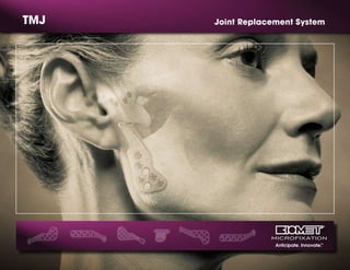
2. An Innovative Implant for Total Joint Replacement
Anticipation and innovation. These two qualities have made Biomet Microfixation
an industry leader. Founded by Walter Lorenz more than thirty years ago, Biomet
Microfixation offers instrumentation, plating systems and related products for a
wide range of surgical procedures.
The Total TMJ Replacement System is a temporomandibular joint
prosthesis. Biomet Microfixation incorporates Biomet’s 30 years of
Orthopedic total joint experience into the design and materials
utilized in the Total TMJ Replacement System.
The Total TMJ replacement system has been manufactured
and clinically used since July 1995 under an approved
investigational device exemption (IDE) from the FDA. Since
1995, over 400 patients have been implanted with the
prosthesis. The clinical study yielded a 96% patient success
rate.* For complete information, we invite you to contact us
or visit www.biometmicrofixation.com today.
*Success rate derived from clinical study conducted for system approval.
3. Mandibular Components
The mandibular (condylar) prosthesis is designed to replace the
articular surface of the mandibular condyle. The mandibular
prosthesis is offered in 3 sizes (45mm, 50mm, and 55mm)
designated left and right. The mandibular prosthesis is offered
in 3 styles (standard, offset, and narrow) to fit a diverse range
of mandibular sizes and shapes. The mandibular prosthesis
is made of Cobalt Chromium Alloy. The undersurface of the
prosthesis is coated with titanium plasma spray for increased
bony integration to the mandibular prosthesis.
Fossa Prosthesis
The fossa prosthesis is designed to replace the articulating
surface of the temporomandibular joint comprised of the
glenoid fossa and the articular eminence of the temporal
bone. The fossa prosthesis is made of Arcom®
Ultra High
Molecular Weight Polyethylene (UHMWPE). UHMWPE is
the same material used effectively in total hip and knee
surgery for over 30 years. The fossa prosthesis is offered in 3
sizes (small, medium, and large) in which all sizes are freely
interchangeable with every size and style of the mandibular
prosthesis. The spherical head of the mandibular prosthesis is
designed similar to the spherical radius of the fossa prosthesis
allowing for excellent articulation of the joint.
Fossa and Mandibular Screws
The system screws are made of 6AL/4V titanium. The screws are
self-retaining and self-tapping to facilitate ease of insertion.
The fossa screws (2.0mm) are specially designed to fit the fossa
prosthesis. The fossa screws have a flat screw head to ensure a
proper fit onto the fossa prosthesis and to create an extremely
low-profile implant. The mandibular prosthesis screws (2.7mm)
have a spherical radius on the head of the screw to mate with
the counter sink in the screw holes of the mandibular prosthesis
resulting in a low-profile implant.
System Components
Standard Mandibular Implant
(Underside view on right)
Fossa
Prosthesis
Narrow Mandibular Implant
(Available in 45mm and 50mm)
Mandibular
and Fossa
Trials
2.0mm Fossa Screws
and 2.7mm Mandibular
Screws
4. Surgical Technique
Pre-Operative
After satisfactory induction of general nasotracheal anesthesia, the patient is prepped
and draped in the usual manner. Twenty-six and twenty-four gauge stainless steel wires are
used to place Ivy loop fixation wires in both the maxilla and the mandible, so that during
the critical time of condylar prosthesis screw placement, the patient can be placed in
inter-maxillary fixation.
1. Incisions
A standard endaural incision is made with dissection along the tragal cartilage down to
the joint space itself, avoiding any damage to the upper trunk of the facial nerve. The
root of the zygoma is identified and periosteum is stripped off to expose the zygoma. The
dissection is then carried down to expose the capsule of the joint. A modified posterior
mandibular incision is then made approximately two finger breadths below, and posterior
to, the angle of the mandible. This is truly a retro-mandibular incision, which allows better
visualization of the entire ramus and also permits rapid access to the terminal branch of the
external carotid if any troublesome hemorrhage is encountered from the internal maxillary
artery during the condylectomy. Dissection is carried through to the subcutaneous tissue
until the marginal mandibular branch of the facial nerve is identified and retracted
superiorly. Dissection is then carried in a plane between the anterior border of the sterno-
cleidomastoid muscle and the submandibular gland. The facial artery and vein can be
ligated at this time and then the aponeurosis between the masseter and pterygoid muscle
is identified and incised. After stripping off the masseter muscle, the surgeon should be
able to have full visualization of the lateral ramus up to the neck of the condyle.
2. Exposure of the Joint
Attention is then directed back to the joint itself where an incision is made along the
posterior root of the zygoma and a full-thickness mucoperiosteal flap is elevated to expose
the entire lateral surface of the glenoid fossa and the capsule of the joint itself. The neck of
the condyle is isolated with condylar retractors and a two step condylectomy is performed.
3. Performing the Osteotomy
A 1mm fissure burr is first used to make a traditional condylectomy cut at the level of
the sigmoid notch. After the condyle is removed, the angle of the mandible is grasped
securely with bone holding forceps and the ramus is pushed superiorly so that more of
the superior ramal stump is visualized in the endaural exposure. This allows the second
osteotomy to be performed, which is approximately 5mm below the sigmoid notch and
can include the coronoid to be removed in a single section osteotomy. This is necessary
to accommodate the thickness of the glenoid fossa implant.
2nd
1st
1
2
3
5. 4. Preparing for the Fossa Prosthesis
A specially designed, large diamond burr or reciprocating diamond burr can be used to
flatten the articular eminence. This removes the majority of the variability in the glenoid
fossa. The end of the burr has a radial shape, which matches the medial radial surface of
the fossa component.
5. Occlusion Placement
Surgical wounds are now packed with antibiotic-soaked sponges. (Irrigant for the entire
case is antibiotic infused solution). The surgeon now re-enters the oral cavity and places
the patient in the optimum desired occlusion with secure twenty-six and twenty-four gauge
inter-maxillary wires. IMF screws can also be used as an alternative.
6. Sizing and Implantation of the Fossa Prosthesis
The surgeon changes his/her gown and gloves to place the fossa implant. The fossa trial
(sizer) can now be used to assess the initial fit of the prosthesis, with the goal to have
a tripod stable fit of the fossa with minimal dead space. At this point, the surgeon can
determine which of the three sizes of the fossa prosthesis would best allow a minimum of
four 2.0mm zygomatic screws to be placed. The difference between sizes is strictly in the
flange area for screw placement options. The articular surface is identical between sizes.
It is extremely important to reduce the bone on the medial surface of the glenoid fossa
adequately enough to allow proper seating of the medial edge of the fossa prosthesis.
This ensures that the fossa is positioned so that it is approximately parallel to the Frankfurt-
Horizontal line, and it avoids an anterior-posterior or medio-lateral tilt to the implant. Once
the proper fit is achieved, two 2.0mm screws are used for the initial fixation, followed by a
minimum of two additional 2.0mm screws.
7. Fitting of the Mandibular Component
The mandibular trial (sizer) is used to assess whether a 45mm, 50mm, or 55mm prosthesis will be
used. It is also used to determine whether an optional narrow component or offset component
could be appropriate. There is unlimited size interchangeability between the mandibular
components and the fossa components. The trial also allows the surgeon to determine whether
the large diamond burr or rasp needs to be utilized to contour the lateral surface of the ramus
so there is a flush fit of the mandibular component against the
ramus. The specially designed diamond rasp can be also used
to radius the edge of the resected ramus. The mandibular
implant may not be bent in any way.
4-5
6
7
6. Surgical Technique
8. Range of Motion
Once the head of the permanent mandibular prosthesis is positioned in the mid
portion of the glenoid fossa, two 2.7mm screws are placed temporarily to secure
the prosthesis. When drilling the holes for the ramus prosthesis, it is important to
approximate the position of the inferior alveolar nerve to avoid any damage while
placing the screws. If desired, a drill guide is available to assist in drilling. Once the
prosthesis is placed temporarily, and the wounds are covered with sterile drapes,
the surgeon then goes back into the oral cavity and removes the intermaxillary
fixation. The mandible should be put through a reasonable range of motion with
an interincisal opening of 30 to 35mm to assess the mechanical functioning of
the joint, and to look for any subluxation, dislocation, or mechanical obstruction.
If there is any question that the patient has increased muscle tone under a light
anesthetic, it may be necessary to request that the anesthesiologist administer a
short acting muscle relaxant to make sure that there is proper mechanical joint
function with a reasonable range of motion. If, under full muscle relaxation, there
appears to be significant impairment to range of motion, it may be important
to assess whether a coronoidectomy is necessary or whether further stripping of
the soft tissue attachments would be appropriate. If the range of motion and the
occlusion are satisfactory, the maxillary and mandibular fixation wires could be
removed at that time and an occlusive dressing is placed over the oral cavity.
9. Final Screw Placement
The remaining screws are then placed with an average of 4-6 screws
recommended for securing the mandibular component. The implant may not
be bent and great care should be taken to avoid scratching or damaging the
articular surface of the mandibular component.
Closure
The surgical wounds are then irrigated with antibiotic solution; the deep layers of
the wounds are closed with 3-0 chromic, the subcutaneous layer is closed with
4-0 chromic, and the skin incisions are closed with 5-0 nylon. The pressure
dressing is applied. At the end of the procedure, all of the fixation wires of the
maxilla and mandible are removed, and the patient is extubated.
Rehabilitation and Follow Up
The day following surgery, a regimen of jaw exercises may be recommended and
should be continued until maximum opening is achieved, or for at least six weeks. The
post-operative evaluation form must be filled out at the prescribed follow-up intervals.
8
9
7. Ordering Information
Fossa Trials*
Fossa Left Right Left Right
Small 24-6563 24-6562 24-6563TR 24-6562TR
Medium 24-6561 24-6560 24-6561TR 24-6560TR
Large 24-6565 24-6564 24-6565TR 24-6564TR
Mandibular Trials*
45mm 24-6546 24-6545 24-6546TR 24-6545TR
50mm 24-6551 24-6550 24-6551TR 24-6550TR
55mm 24-6556 24-6555 24-6556TR 24-6555TR
Mandibular (Narrow) Mandibular (Offset)
45mm 01-6546 01-6545 24-6646 24-6645
50mm 01-6551 01-6550 24-6651 24-6650
Implants
Part # Description
01-6505 TMJ Implant Container
01-6507 TMJ Instrument Case
01-7600 2.0/2.4mm Screwdriver Handle
01-7390 Green 1.5/2.0 Screwdriver Handle
24-1194 2.4/2.7mm X-Lock Blade, Short
24-1196 2.4/2.7mm X-Lock STD Blade
01-7694 2.0mm X-Lock Blade, Short
01-7696 2.0mm X-Lock STD Blade
24-6520 TMJ Double Ended Drill Guide
24-6610 1.5 x 50mm, 11mm Stop Twist Drill
24-6612 1.5 x 105mm, 11mm Stop Twist Drill
24-6614 2.0 x 70mm, 12mm Stop Twist Drill
24-6616 2.0 x 115mm, 12mm Stop Twist Drill
24-6618 2.2 x 70mm, 12mm Stop Twist Drill
24-6620 2.2 x 115mm, 12mm Stop Twist Drill
24-6526 TMJ Diamond Rasp
24-6530 TMJ Diamond Burr, Coarse (Gold)
24-6532 TMJ Diamond Burr, Medium
24-6534 TMJ Diamond Short Burr, Coarse*
24-6536 TMJ Diamond Short Burr, Medium*
Additional Instrumentation
24-6510 PDQ Zygoma Retractor
24-6512 PDQ Condylar Retractor
24-6514 PDQ Coronoid Retractor
01-0200 Condylar Stripper
01-0194 Posterior Condylar Retractor
27-0066 Dingman Bone Holding Forceps
01-0431 Dunn-Dautrey Spatula Osteotome
24-1110 Plate Holding Forceps
15-1150 Tray Rack System*
01-9095 Bone Plate Holding Forceps*
Instruments
* Optional Instrumentation
Fossa Screws Mandibular Screws
X-Drive HT X-Drive
Part # Description Part # Description
99-6577 2.0 x 7mm 91-2708 2.7 x 8mm
99-6579 2.0 x 9mm 91-2710 2.7 x 10mm
99-6581 2.0 x 11mm 91-2712 2.7 x 12mm
X-Drive Emergency HT X-Drive Emergency
99-6587 2.3 x 7mm 99-9948 3.2 x 8mm
99-6589 2.3 x 9mm 99-9950 3.2 x 10mm
Screws
99-Singles, 01-5/pk 91-Singles, 95-5/pk
Add -int for International Parts
Add ti for Titanium Implants
Titanium Implants available for patients with nickel allergies
* Trials are not implantable devices
8. Caution
Federal Law (USA) restricts this device to sale, distribution, or use, by or on the
order of a physician.
Description
The Total Temporomandibular Joint (TMJ) Replacement System is implanted
in the jaw to functionally reconstruct a diseased and/or damaged
temporomandibular joint.
The Total TMJ Replacement System is a two-component system comprised of
mandibular condyle and glenoid fossa components.
Both components are available in multiple sizes as right and left side specific
designs and are attached to the bone by screws. Included in the system are
trials, instruments and instrument cases.
Materials
Mandibular Component—Cobalt-Chromium-Molybdenum (Co-Cr-Mo)
alloy with titanium alloy coating or Titanium (Ti-6Al-4V) alloy with titanium
alloy coating
Fossa Component—ultra high molecular weight polyethylene (UHMWPE)
Screws—titanium alloy
Trials: Mandibular—aluminum, Fossa—Radel®
plastic
Instruments: TMJ flat diamond rasp, TMJ diamond burrs, TMJ double-ended
drill guide, retractors—stainless steel
Instrument Case—stainless steel, silicone, Radel®
plastic
Indications
The Total Temporomandibular Joint Replacement System is indicated for
reconstruction of the temporomandibular joint. The reconstruction is necessary
due to one of the following diagnoses:
Arthritic conditions: osteoarthritis, traumatic arthritis, rheumatoid arthritis
Ankylosis including but not limited to recurrent ankylosis with excessive
heterotopic bone formation
Revision procedures where other treatments have failed (e.g. alloplastic
reconstruction, autogenous grafts)
Avascular necrosis
•
•
•
•
•
•
•
•
•
•
Multiply operated joints
Fracture
Functional deformity
Benign neoplasms
Malignancy (e.g. post-tumor excision)
Degenerated or resorbed joints with severe anatomic discrepancies
Developmental abnormality
Contraindications
Active or chronic infection.
Patient conditions where there is insufficient quantity or quality of bone to
support the components.
Systemic disease with increased susceptibility to infection.
Patients with extensive perforations in the mandibular fossa and/or bony
deficiencies in the articular eminence or zygomatic arch that would
severely compromise support for the artificial fossa component.
Partial TMJ joint reconstruction.
Known allergic reaction to any materials used in the components. NOTE:
Patients with known or suspected nickel sensitivity should not have Co-Cr-
Mo devices implanted since this material contains nickel.
Patients with mental or neurological conditions who are unwilling or unable
to follow postoperative care instructions.
Skeletally immature patients.
Patients with severe hyper-functional habits (e.g. clenching, grinding etc.)
Patients with a foreign body reaction due to previous implants.
Warnings
Mandibular and fossa components are provided STERILE. DO NOT RESTERILIZE.
Screws, trials, instruments and instrument cases are provided NON-STERILE.
CLEAN AND STERILIZE BEFORE USE.
DO NOT USE if there is a loss of sterility of the devices.
DO NOT USE damaged implants and only use implants that are packaged
in unopened or undamaged containers.
•
•
•
•
•
•
•
•
•
•
•
•
•
•
•
•
•
•
•
•
•
Warnings and Precautions for Use of the Total TMJ Replacement
9. DO NOT USE the individual components of this total system (e.g. mandibular
components, fossa components, or screws) for partial joint reconstruction.
Bone cement or other grouting agents should not be used when
implanting these devices. Safety and efficacy have not been established
for the use of bone cement or other grouting agents with these implants.
DO NOT USE IN CHILDREN. The Total TMJ Replacement was designed for
skeletally mature patients.
Precautions
The device is limited to surgeons who are adequately trained in the use of the
device through hands-on and educational course work. In all cases sound
medical practice is to be followed and the surgeon must select the type of
device appropriate for treatment.
The patient is to be warned that the system does not replace normal healthy
bone in their TMJ and they may continue to have chronic pain and limited
range of motion. The system can break or loosen as a result of stress, activity, or
trauma. Patients with severe hyper-functional habits may have an undesirable
outcome. The presence of existing mandibular and/or zygomatic arch screws
or screw holes may compromise fixation. Note that placement of the implant
in one joint only may result in harmful effects to the joint on the opposite side.
Placement of the implant may produce an improper relationship between
teeth surfaces that should contact during biting. The patient is to be made
aware of surgical risks and possible adverse effects prior to surgery and
warned that failure to follow postoperative care instructions can cause failure
of the implant and the treatment.
Specialized instruments/trials are designed for use with the Total TMJ
Replacement System to aid in the accurate implantation of the components.
DO NOT USE trials/instruments or cases that are disfigured, cracked, corroded,
or otherwise damaged. Instruments/trials are subject to wear with normal
usage and are susceptible to fracture when exposed to extensive use or
excessive force. All trials/instruments and cases should be regularly inspected
for wear or disfigurement. These should be disposed of appropriately.
Adverse Events
Adverse events that may occur following placement of the Total TMJ
Replacement System are listed below. See Tables 7 and 8 located in the insert #
01-50-1000 for more detailed information on adverse events from the clinical trial.
•
•
•
• Removal of components(s) including, but not limited to the following:
- implant changes caused by loading and/or wear
- degenerative changes within the joint surfaces from disease
or previous implants
- implant materials producing particles or corroding
• Loosening or displacement with or without removal of the implant
• Infection (systemic or superficial)
• Foreign body or allergic reaction to implant components
• Fossa wear through
• Facial swelling and/or pain
• Facial nerve dysfunction
• Excision of tissue
• Heterotopic bone formation
• Neuroma formation
• Ear problems
• Dislocation
Patient Counseling Information
Discussion of the following points is recommended prior to surgery.
The importance of prompt medical attention if they experience unusual
swelling in the area of the implant.
The risks associated with a Total TMJ System (see Warnings and Adverse Events).
Post-operative pain relief and return of function varies from patient to patient.
Additional treatment may be required including but not limited to extended
physical therapy, bite splint, dental braces, and/or orthognathic and
reconstructive surgery.
Sterility
The Total Temporomandibular Joint Replacement System mandibular
and fossa components are sterilized by exposure to a minimum of 25 kGy
of gamma irradiation. DO NOT RESTERILIZE. Screws, trials, and the TMJ
Instrument Case containing instruments are supplied non-sterile and should
be wrapped with an FDA cleared sterilization wrap prior to steam sterilization
in order to maintain sterility.
•
•
•
•
•
10. For more information on TMJ, please contact us at:
GLOBAL HEADQUARTERS
1520 Tradeport Drive • Jacksonville, FL 32218-2481
Tel (904) 741-4400 • Toll-Free (800) 874-7711• Fax (904) 741-4500 • Order Fax (904) 741-3059
www.biometmicrofixation.com
EUROPE
Toermalijnring 600 • 3316 LC Dordrecht • The Netherlands
Tel +31 78 629 29 10 • Fax 31 78 629 29 12
e-mail: europe@biomet.com
What fascinates you about the body is also what drives us. That’s why we’re always
pushing the boundaries of engineering to make products that help you keep the
human form as glorious as it was intended. To learn more about our breadth of
products, call 800-874-7711 or visit us online at biometmicrofixation.com.
We’d love to join you in a conversation about the future.
This brochure is presented to demonstrate the technique and post-operative care regimen of Peter Quinn, MD. As the manufacturer of this device, Biomet Microfixation does not practice medicine and does not
recommend this product or surgical technique for use on a specific patient. The surgeon who performs any implant procedure must determine the appropriate device and surgical procedure for each individual patient.
Devices shown in this brochure may not be cleared or licensed for use or sale in your individual country. Please contact your local distributor for information regarding availability of this product. Information contained
in this brochure is intended for surgeon or distributor information only and is not intended for patient distribution. All surgeries carry risks. For additional information, please see package insert, or visit our web site at
www.biometmicrofixation.com or call 1-800-874-7711.
r05k0705 BMF00-2166
REVISED: 9/25/07