
Recommended
More Related Content
What's hot
What's hot (20)
Viewers also liked
Similar to Lethal Cardiac Rhythms - Manual Defibrillation
Similar to Lethal Cardiac Rhythms - Manual Defibrillation (20)
More from ryanhall911
More from ryanhall911 (11)
Recently uploaded
Recently uploaded (20)
Lethal Cardiac Rhythms - Manual Defibrillation
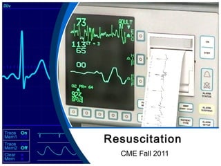
- 1. Resuscitation CME Fall 2011
- 3. Agenda • Morning – Welcome & Introduction – Housekeeping – CPR Recert – New Base Hospital Arrest Protocols • Lunch & Flu shots
- 4. Agenda • Afternoon – Autopulse rounds presentation (Base Hospital) – Dissection of Arrest ECGs – Lethal Rhythms – Manual Defibrillation – Autopulse Plus (shock / synch) – Skill Stations – Test • Go Home
- 5. New Training Tools • Audience Response Systems – ‘Clickers’ – Will be used for games, challenges, tests • SimMan 3G – State of the art patient simulator – Allows us to practice in a safe environment – Might seem spooky at first but great learning tool
- 6. Background • Recent Changes in Resuscitation – 2010 AHA ECC Guidelines • Reduce interruptions to compressions – Base Hospital Arrest Protocols • Medical TORs – Autopulse Plus (shock / synch) • Minimizes pauses in CPR
- 7. Housekeeping • New Woodstock General Hospital – Emerg Patients enter through garage • Garage holds 2 trucks • 1st truck in, clear out quickly for next vehicle • Caution leaving garage – blind corner to left – Give report to RN at desk across from Trauma Rm • Do not go behind desk – patient confidentiality – Non-Emerg / Transfers • Do Not Enter through Emerg / Garage • Use side entrance, park trucks outside
- 9. TDMH • When patching in give: – Family MD (allows them time to contact doc) – MRSA / VRE status if known (from MARS sheet) • When arriving: – Give health card to clerk with reason for visit • Allows them to start registration • Can help expedite tests, labs, x-rays, etc • May not always be possible / practical (Code 4s)
- 10. All Hospitals • When Patching give FRI status (+ve or –ve) – Any new or worsening cough – Shortness of Breath – Fever over 38 deg C. – Allows staff to prepare isolation precautions
- 11. IVs • Please refrain from pre-spiking IV bags – New drip set piece is sharp – Causes bags to leak if pre-spiked – Will most likely be switching to Baxter drip sets • IV Locks – Will probably start stocking locks – Good for use when transporting to TDMH
- 12. ACRs • Doing a great job uploading ECGs • Procedures performed by 1 medic – Unless its lifting, stairchair, extricate, etc • Oxford policy – ACRs are completed for any call where you arrive scene (even if no pt) • Please don’t use ‘Z’ procedure codes (ie Z301) • Will be placing OmniDrives in each truck soon • Working on having ability to upload calls from hospital or on the road
- 14. 2012 Base Hospital New Arrest Protocols
- 15. Medical Arrests • Introduction of Medical TOR Protocol – ≥ 18 years – Unwitnessed Arrest – No ROSC – No Shocks Delivered > BHP Patch for TOR
- 16. Medical Arrests • Introduction of EPI where Anaphylaxis is suspected as the cause of arrest – Give 0.01 mg/ kg to a max of 0.5 mg EPI 1:1000 IM
- 17. Traumatic Arrests • Merging of Blunt and Penetrating Trauma protocols • > 30 days old • VF/VT – 1 shock ER • Trauma TOR > 16 yrs • Asystole – Patch for TOR • PEA & Transport >30 mins – Patch for TOR
- 19. Autopulse Rounds Dr. Sameer Mal - SWORBHP
- 27. Lethal Rhythms & Manual Mode • Review of the 4 lethal rhythm types • Nothing new, reviewed annually during recerts • Work on rapid recognition (5 seconds)
- 28. Lethal Rhythm
- 29. Ventricular Tachycardia • 3 or more consecutive ventricular complexes occurring at a rate of more than 100 bpm • Could have an associated pulse or be pulseless
- 30. Ventricular Tachycardia • Causes – Usually starts suddenly, triggered by a PVC – Usually a result of myocardial ischemia or significant cardiac disease
- 31. Ventricular Tachycardia • Other Causes – Electrolytic imbalance (Acid/Base, Na+, K+…) – CHF – Stimulants (ETOH, tobacco, C8H10N4O2) – Drug Toxicity (digitalis, trycylics, antidepressants) – Sympathomimetics (cocaine, meth) – Prolonged QT
- 32. Ventricular Tachycardia • Interpretation – QRS is WIDE – ≥ 0.12 seconds (same as LBB interpretation) – May appear distorted or bizarre – P waves may or may not be present – if present usually dissociated from QRS – Rate > 100 bpm
- 33. Ventricular Tachycardia • Types – Monomorphic - one form, derives from one focus - every wave appears the same
- 34. Ventricular Tachycardia • Types – Polymorphic - generated by multiple foci - waveform appearance variable
- 35. Ventricular Tachycardia • Types ALS Warning: Do NOT use antidysrhythmic drugs on Torsades – Torsades de Pointes - ‘twisting of the points’ - conduction rotates, form of polymorphic
- 36. Ventricular Tachycardia • Action – No Pulse? – Fast? – Wide? SHOCK
- 37. Lethal Rhythm 2
- 38. Ventricular Fibrillation • Chaotic ventricular rhythm results in ventricular ‘quivering’ and pulselessness • Always pulseless • Most common initial rhythm in sudden cardiac arrest
- 39. Ventricular Fibrillation • Causes – Myocardial ischemia – AMI – 30 AV block with a slow ventricular escape rhythm – Cardiomyopathy – Digitalis Toxicity – Acidosis – Electrolyte Imbalance – Electrical Injury – Drug Overdose (cocaine, tricyclics)
- 40. Ventricular Fibrillation • Interpretation – Chaotic – No discernible P waves or QRS complexes
- 41. Ventricular Fibrillation • Types – Coarse VF • Amplitude of > 3mm – Fine VF • Amplitude < 3mm • May be very difficult to differentiate from asystole
- 42. Ventricular Fibrillation • Action SHOCK
- 43. Lethal Rhythm 3
- 44. Pulseless Electrical Activity • Used to be called ‘Electromechanical Dissociation’ • Electrical activity is present but there are no resultant contractions
- 45. Pulseless Electrical Activity • Causes – The 6 H’s and the 6 T’s – Hypothermia – Hypoxia – Hydrogen ions (Acidosis) – Hyper/Hypokalemia – Hypoglycemia – Hypothermia
- 46. Pulseless Electrical Activity • Causes – The 6 H’s and the 6 T’s – Tablets / Toxins (Drug overdose) – Cardiac Tamponade – Tension pneumothorax – Thrombosis (MI) – Thrombosis (PE) – Trauma (Hypovolemia)
- 47. Pulseless Electrical Activity • Interpretation – Patient is pulseless, apneic – Rhythm appears organized (anything from an escape rhythm to normal sinus) – Slow & Wide -> PEA – Fast & Wide -> V Tach
- 48. Pulseless Electrical Activity • Action – Ensure Pulselessness – Continue CPR
- 49. Lethal Rhythm IV
- 50. Asystole • Flatline, absence of any electrical activity • Causes – 6H’s, 6 T’s, prolonged VF / VT / PEA
- 51. Asystole • Interpretation Continue CPR – Flat line – Slow, wide, thin wave – May be fine V-Fib – Look at possible causes of death to help differentiate from VF
- 53. Agonal Rhythms
- 54. Paced Rhythms
- 55. And now you know… And Knowing is half the battle
- 56. Manual Mode Do not be afraid
- 57. Using the E Series in Manual Mode • Turn on Defib as you normally would – Press ‘Manual Mode’ soft key
- 58. Using the E Series in Manual Mode • Turn on Defib as you normally would – Then press ‘Confirm’ soft key
- 59. Using the E Series in Manual Mode • Ensure ‘Pads’ are selected (not Ld I,II or III…)
- 60. Using the E Series in Manual Mode • 120 Joules will be the default energy • After shock is delivered, energy will increase – 150 J, 200 J
- 61. Using the E Series in Manual Mode • To evaluate a rhythm – Stop CPR – Check Pulse – NOT MORE THAN 5 SECONDS – Press ‘Recorder’ button and print off strip (also marks event on summary)
- 62. Using the E Series in Manual Mode • Resume CPR immediately then make your defibrillation decision • (Shock / No Shock) – You can use the rhythm strip you printed to make the decision after the pause
- 63. Using the E Series in Manual Mode • If choosing to shock, press ‘Charge’ – (no need to press ‘Analyze’) – Confirm you have selected the proper energy setting
- 64. Using the E Series in Manual Mode • Continue CPR until ready to shock then once all rescuers are clear, press ‘Shock’ then resume CPR immediately. – There should be a only very brief pause in compressions
- 65. Using the E Series in Manual Mode To dump a shock, just hit the ‘Energy Select’ button (either arrow) If really unsure whether to shock or not, the ‘Analyze’ button is always an option.
- 66. Using the E Series in Manual Mode For Paeds Keep Defib in Semi Automatic and use pediatric attenuator pads
- 67. Autopulse Plus AKA ‘Shock/synch’
- 69. Using Autopulse Plus • The Autopulse now has the ability to coordinate defibrillation with the contraction cycle • Allows for minimal interruption to compressions • Can be hooked up initially or at any point in the call
- 71. Using Autopulse Plus • Connecting the Defib to the Autopulse – Connector site is located at the top of the Autopulse next to the battery bay
- 72. Using Autopulse Plus • Connecting the electrodes to the Autopulse – Connect the defib pad electrodes by plugging them into the connector site (1) – Ensure connector is firmly seated in the connector site
- 73. Using Autopulse Plus • Connecting Defibrillator to Autopulse – Connect defibrillator cable into connector site (marked ‘2’) – Ensure cable is firmly seated
- 75. Using Autopulse Plus –When ready to interpret cardiac rhythm, pause compressions briefly for interpretation and pulse check if applicable –Resume compressions immediately
- 76. Using Autopulse Plus • Ensure appropriate energy and charge defibrillator if applicable • Press ‘Shock’ – Shock may be delayed as long as 800 ms to coordinate with the upstroke of compressions from the Autopulse.
Editor's Notes
- Demonstrate lock preparation
- This is the last slide before CPR recert
- This will lead to death notification training
- After this shows, play Autopulse Inservice Video – EMS deployment