
Recommended
More Related Content
What's hot
What's hot (20)
Similar to Cervical Fractures(clay shovelers,hangmans,odontoid)
Similar to Cervical Fractures(clay shovelers,hangmans,odontoid) (20)
Recently uploaded
Recently uploaded (20)
Cervical Fractures(clay shovelers,hangmans,odontoid)
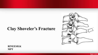
- 2. Clay Shoveler’s Fracture • Clay shoveler’s fracture derives its name from a common occurrence in clay miners in Australia during the 1930s • Stable avulsion fracture through the spinous process of a vertebra • Occurring at any of the lower cervical or upper thoracic vertebrae • Classically at C6 or C7
- 3. Mechanism of Injury • It occurs with abrupt flexion of the head such as found with motor vehicle accidents, diving, or wrestling injuries. • Occurs with repeated stress caused by the pulling of the trapezius and rhomboid muscles. • Direct blows or trauma to the base of the neck.
- 4. Symptoms –Sudden onset of pain between the shoulder blades or base of neck –Reduced head/neck ROM –Tenderness
- 5. Diagnosis • Radiographs Cervical & Thoracic x-rays that should always be obtained on evaluation • CT SCAN Indications Routine CT imaging in high-energy trauma patients Clinical criteria – altered consciousness – midline spinal pain or tenderness – impaired CCJ motion – lower cranial nerve paresis – motor paresis
- 6. Treatment Nonoperative – NSAIDS, rest, immobilization in hard collar for comfort • indications – most common treatment for pain control • modalities – short term treatment with hard collar • outcomes – usually high union rates and excellent clincal outcomes Operative – surgical excision indications – persistent pain or non-union – failed conservative treatment
- 8. Hangman’s Fracture • The second most common fracture of the second cervical vertebra. • Involves a bilateral arch fracture of the C2 pars interarticularis with variable displacement of C2 on C3
- 9. Mechanism of Injury The injury mainly occurs from falls, usually in older adults, and motor accidents mainly due to impacts of high force causing Extension of the neck and great axial load onto the C2 vertebra. The mechanism of the injury is forcible hyperextension of the head, usually with distraction of the neck.
- 10. Classification Type I: Non-displaced fractures with no angulation between C2 and C3 and a fracture dislocation of less than 3 mm Type II: significant angulation (>11°) and displacement (>3.5 mm)
- 11. type IIA: minimum displacement and significant angulation (>11°) type III: severe angulation and displacement and concomitant unilateral or bilateral facet dislocation C2–3.
- 12. Symptoms • The most common symptom of hangman’s fracture is neck pain following a fall or motor vehicle accident • The most important concern with hangman’s fracture is injury to the spinal cord. • If the spinal cord is damaged, symptoms can include pain, sensory loss, weakness, paralysis, and/or death.
- 13. Tests and Diagnosis • X-ray - flexion and extension radiographs show subluxation • Computed tomography scan (CT scan) - study of choice to delineate fracture pattern • Magnetic resonance imaging (MRI) - consider if suspicious of a vascular injury to the vertebral artery
- 14. Treatment Nonoperative Rigid cervical collar x 4-6 weeks Indications :Type I fractures (< 3mm horizontal displacement)
- 15. Closed reduction followed by halo immobilization for 8-12 weeks Indications – Type II with 3-5 mm displacement – Type IIA Reduction technique – Type II use axial traction combined + extension – Type IIA use hyperextension (avoid axial traction in Type IIA)
- 16. Operative Reduction with surgical stabilization Indications • Type II with > 5 mm displacement and severe angulation • Type III (facet dislocations) Technique • anterior C2-3 interbody fusion • posterior C1-3 fusion • bilateral C2 pars screw osteosynthesis
- 18. Odontoid Fracture • The most common axis injury is a fracture through the odontoid process. • Translational motion of C1 on C2 is restricted by the transverse atlantal ligaments that center the odontoid process to the anterior arch of C1. • With a fracture of the odontoid process, restriction of translational atlantoaxial movement is lost.
- 19. Classification Type I: oblique fractures through the upper portion of the odontoid process. According to the classification of Anderson and D’Alonzo:
- 20. Type II: across the base of the odontoid process at the junction with the axis body.
- 21. • Type III: through the odontoid that extends into the C2 body.
- 22. Mechanism of Injury Flexion loading is the cause in the majority of patients, and results in anterior displacement of the dens Extension loading (forward fall onto forehead) occurs in a minority of patients, and results in posterior displacement of the dens;
- 23. Treatment • A variety of non-operative and operative treatment alternatives have been proposed for odontoid fractures based on: – fracture type – degree of (initial) dens displacement – extent of angulation – patient’s age
- 24. Type II and Type III odontoid fractures should be considered for surgical fixation in cases of: – dens displacement of 5 mm or more – dens fracture (Type IIA) – inability to achieve fracture reduction – inability to achieve main fracture reduction with external immobilization
- 25. • Anterior trans articular screw fixation: As an augmentation of the anterior dens screw or in cases of a salvage procedure. • Screws can be inserted over Kirschner wires from a medial- anterior-caudal to a lateral-posterior-cranial direction crossing the atlantoaxial joint.