Fallopian Tubes and Ovary Anatomy
•
0 likes•60 views

The fallopian tubes and ovaries are paired reproductive organs in females. The fallopian tubes are hollow muscular tubes located on each side of the uterus that transport eggs from the ovaries to the uterus. Each tube has an opening into the uterus and another opening near the ovary. The ovaries are oval shaped organs located laterally in the pelvis that contain eggs at different stages of development and produce hormones. The ovaries and fallopian tubes work together to allow for fertilization and early embryo development.

Recommended
More Related Content
Similar to Fallopian Tubes and Ovary Anatomy
Similar to Fallopian Tubes and Ovary Anatomy (20)
More from ramveer sharma
More from ramveer sharma (19)
Recently uploaded
Recently uploaded (20)
Fallopian Tubes and Ovary Anatomy
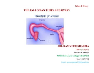
- 1. Tubes & Ovary THE FALLOPIAN TUBES AND OVARY fMEcokfguh ,oa v.Mk”k; DR. RAMVEER SHARMA M.D. Ayu. (Gynae) I.P.G.T.&R. Jamnagar MMM Govt. Ayu. College UDAIPUR Mob. 9414757932 Email. ramveersharma1960@gmail.com
- 2. Fallopian Tubes Definition:- The fallopian tubes are - Tubes & Ovary Definition:- The fallopian tubes are - One on each side, Are hollow and muscular tube like structure. • Meant for ovum transportation, • Ovum fertilization, and • Transportation of fertilized ovum. Dr. Ram Veer Sharma
- 3. Situation:- They are situated • In medial three fourth of the upper free margin of broad ligament. Openings:- Each tube has got Two opening - a. Uterine opening :- one is communicating with the lateral angle of the uterine cavity. Measures:- 1mm. in diameter. Tubes & Ovary angle of the uterine cavity. Measures:- 1mm. in diameter. b. Pelvic opening :- other is one lateral end of the tube , or abdominal ostium close to ovary. Measures:- 2 mm. in diameter. Dr. Ram Veer Sharma
- 4. Size :- Is about - Long =10 cm. • Breath = 1 cm. at thickest part. Course :- • The tube pass laterally and backwards up to the ovary. • Over which it arches, and finally turns downwards to end in relation to its free Tubes & Ovary relation to its free • Posterior border and medial surface. Dr. Ram Veer Sharma
- 5. . Tubes & Ovary Dr. Ram Veer Sharma
- 6. Parts:- 4 Interstitial(Intramural) Isthmus Ampulla Infundibullum Tubes & Ovary Dr. Ram Veer Sharma
- 7. 1) Interstitial (Intramural) :- • It is narrowest part, it lies in only uterine wall. • Size :- 1.25 cm X 1 mm. Tubes & Ovary 2) The Isthmus :- Is narrow and cord like, • Straight , succeeding 1/3 of the tube. • Size :-2.5 cm. X 2.5 mm. Dr. Ram Veer Sharma
- 8. 3) Ampulla :- • Is lateral, widest and tortuous part. • Size :- 5.0 cm. X 4 mm. 4) Infundibullum :- Tubes & Ovary 4) Infundibullum :- • Is funnel shaped and fimbriated opening (abdominal ostium), is surrounded by a number of radiating fimbrias about - 25. • One of these larger is attached to outer pole of the ovary called Ovarian Fimbriae. • Size :- 1.5 Cm. X 6mm. Dr. Ram Veer Sharma
- 9. Structure :- Has 3 layers. a)Serous Layer :- It consist of peritoneum (Broad ligament) and covers of all side except at the floor, where about 1/6 of its circumference is a bare area. • The minimum bare area is on isthmus part. b)Muscular Layer :- Consist of two layers, Tubes & Ovary b)Muscular Layer :- Consist of two layers, i. Outer layer:- is longitudinal, ii. Inner layer :- Are circular. • The fibers are best developed in the isthmus - 0.5mm and thinned out near the Fimbrial end - 0.1 mm. Dr. Ram Veer Sharma
- 10. 3) Mucous layer:- (Endosalpinx) :- • The layer is direct rest on muscular layer, with out sub mucous layer. Have no glands. It consist of - • Surface epithelium :- Consist of tall and • broad ciliated columnar cells = 20 -30% Tubes & Ovary • broad ciliated columnar cells = 20 -30% • With intervening secretory non ciliated cells = 50 – 60 % are show variations during menstrual cycle. • A third type of peg cells lying in between ciliated and secretory cells. Dr. Ram Veer Sharma
- 11. 4) Stroma :- these cells are lying between epithelial and muscular layer, is rich in vessels and lymphatic's. Tubes & Ovary Dr. Ram Veer Sharma
- 12. . Tubes & Ovary Dr. Ram Veer Sharma
- 13. Coats of the Fallopian Tubes. Tubes & Ovary Dr. Ram Veer Sharma
- 14. Coats of the Fallopian Tubes Tubes & Ovary Dr. Ram Veer Sharma
- 15. Coats of the Fallopian Tubes Tubes & Ovary Dr. Ram Veer Sharma
- 16. Mucous layer of the Fallopian Tubes Tubes & Ovary Dr. Ram Veer Sharma
- 17. Coats of the Fallopian Tubes Tubes & Ovary Dr. Ram Veer Sharma
- 18. Coats of the Fallopian Tubes Tubes & Ovary Dr. Ram Veer Sharma
- 19. Peculiarities of the Tube i. The tubal lumen enlarges from uterine to ovarian end. Tubes & Ovary ii. The mucous membrane is thrown in to longitudinal folds and rugaes, three or four rugaes are present in inner end of the tube. iii. The motion of the ciliary current of tube are directed towards uterus. Dr. Ram Veer Sharma
- 20. iv. The peristaltic movements are also directed to wards the same direction. v. The contractile activities of tubal muscles is effected by prostaglandins. vi.The epithelium under goes cyclic changes like the endometrium during menses Tubes & Ovary endometrium during menses vii.10 % Decidual changes are seen in mucous cells during normal pregnancy. Dr. Ram Veer Sharma
- 21. Secretion:- • Is serous and Transudent. • Controlled by ovarian hormones. • During embryo transport to the uterus, the secretion flows towards the uterus. Tubes & Ovary Dr. Ram Veer Sharma
- 22. Blood Supply Arteries:- Middle 2/3 supplied by tubal branch of uterine artery. Outer 1/3 “ “ “of ovarian artery. • Veins :- Pampiiniform plexus drain to ovarian vein. Dr. Ram Veer Sharma
- 23. Lymphatic's :- Drain to ovarian lymphatic's to Para aortic lymph nodes. Nerves:- (Motor and sensory) • Sympathetic:- T11 - T12- Ovarian and uterine nerve. • Prasymphathatic:- Outer ½ - from vagal fibrous. • Inner ½ - from pelvic splanchic nerve. Tubes & Ovary • Inner ½ - from pelvic splanchic nerve. Dr. Ram Veer Sharma
- 24. Functions :- • Transport of ovum, ovary to uterus. • The tubal fimbrias has getup ovum pickup function. • The Ampulla is the seat of fertilization. • The tubal secretion provide nutrition to ovum, sperm and embryo. Tubes & Ovary embryo. • The tubal secretion forms a medium for capacitation of sperm, and maturation of morulla. Dr. Ram Veer Sharma
- 25. THE OVARY • The ovaries are paired, sex glands or gonads in female, • Functioning germ maturation, storage, and release. Produce sex hormones. • Anatomy.:- No - One on each side. Tubes & Ovary • Shape :- Oval shape. • Color:- Pinkish gray. Surface is scared during reproductive life. • Structure :- Solid, flat and ramiform. Dr. Ram Veer Sharma
- 26. Measures :- Length - 1.5" =3.5 cm. Breath - = 2 cm. Thickness - = 2.5 cm. Weight - = 5 to 10 gm. Tubes & Ovary Weight - = 5 to 10 gm. Ends :- Two - Tubal and Uterine end. Border :- Two - Mesovarium and Free posterior. Surface :- Two - Medial and Lateral. Dr. Ram Veer Sharma
- 27. Site :- It is situated - to close to lateral wall of the true pelvic wall in ovarian Fossa. Ligaments :- • Laterally it is attached to the Posteriorly to broad ligament by mesovarium at hylum of the ovary – Infundibulo pelvic ligament. Tubes & Ovary ligament. • And suspended from uterine cornu by - Ovarian ligament. Dr. Ram Veer Sharma
- 28. Relations:- • Lateral :- It is lies in relation to the ovarian Fossa on lateral pelvic wall. Tubes & Ovary • Middle surface :- Covered to a large extent by fallopian tube. • Anterior border :- Attached to mesovarium and directed to the obliterated umbilical artery. • Posterior :- Border is closely related to tubal Ampulla and is a convex free border. Dr. Ram Veer Sharma
- 29. Structure - Is divided two zones. a) Cortex :- Is the outer or Functioning zone. b) Medulla :- It is inner zone. a) Cortex :- • It is covered on its surface by cubical epithelium called germinal epithelium. Tubes & Ovary germinal epithelium. • Surface germinal epithelium :- Is covering of a single layered cuboidal cells - instead of peritoneal covering. • The junction of germinal epithelium with peritoneum is called white line. Dr. Ram Veer Sharma
- 30. • This is functional part of the ovary consist of stromal cells - which are thickened beneath the germinal epithelium to form Tunica blugenica. Tubes & Ovary Dr. Ram Veer Sharma
- 31. Tubes & Ovary Dr. Ram Veer Sharma
- 32. • The tunica albugenica is condensed of fibrous tissue surrounds the whole ovary except hilum. • The rest part of the cortex is composed of. Tubes & Ovary Dr. Ram Veer Sharma
- 33. Primordial follicles :- Called functional unit of the ovary. • The follicles are in various phases of their development - with hormone and ovum producing unit called Graffian follicles. The structures includes - Primordial follicles, Maturing follicles, Graffian follicles, and Corpus luteum (Yellow body). Atresia of the structures results in formation of atratic follicles Tubes & Ovary Atresia of the structures results in formation of atratic follicles (Corpus albucans) Dr. Ram Veer Sharma
- 34. B) The Stroma:- consist of highly cellular, spindle shape cells, very richly supplied, blood vessels, lymphatic's and nerves. C) Medulla :- it consist of loose connective tissue, non striated plain muscles cells, spiral blood vessels, lymphatic's, nerves and islands of stromal cells. Tubes & Ovary and islands of stromal cells. • Some remnants of Wolffian duct are visible at medulla, more over at hilum. • There are small collection of hiatus cells considered homologues to the interstitial cells of testes. Dr. Ram Veer Sharma
- 35. Blood Supply Artery:- i. Ovarian artery– hilum– arterioles are spiral types. ii. Uterine artery. Veins :- From -Tube and Uterine body • Pampiniform plexus To Tubes & Ovary To • Ovarian vein. a. Right ovarian vein, drain to Inferior vana cava b. Left ovarian vein, drain to left renal vein. Dr. Ram Veer Sharma
- 36. Lymphatic's :- • Run with ovarian vessels drain to Para aortic lymph nodes. Nerves :- T10 - Coelic - Renal plexus. Parasympathetic - Pelvic splenic nerve. Tubes & Ovary Parasympathetic - Pelvic splenic nerve. Dr. Ram Veer Sharma
- 37. Functions - Two main functions are - a. Maturation of Graffian follicles in to the ovum. b. Production of Gonadal steroids ( hormones). Tubes & Ovary Dr. Ram Veer Sharma
- 38. Tubes & Ovary Dr. Ram Veer Sharma THANKS