More Related Content
Similar to Mat weight ga fetal frac nipt pnd
Similar to Mat weight ga fetal frac nipt pnd (20)
Mat weight ga fetal frac nipt pnd
- 1. DOI: 10.1002/pd.4119
ORIGINAL ARTICLE
Gestational age and maternal weight effects on fetal cell-free DNA
in maternal plasma
Eric Wang*, Annette Batey, Craig Struble, Thomas Musci, Ken Song and Arnold Oliphant
Ariosa Diagnostics, San Jose, CA, USA
*Correspondence to: Eric Wang. E-mail: ewang@ariosadx.com
ABSTRACT
Objective To determine the effects of gestational age and maternal weight on percent fetal cell-free DNA (cfDNA) in
maternal plasma and the change in fetal cfDNA amounts within the same patient over time.
Methods The cfDNA was extracted from maternal plasma from 22 384 singleton pregnancies of at least 10 weeks
gestation undergoing the HarmonyTM Prenatal Test. The Harmony Prenatal Test determined fetal percentage via
directed analysis of cfDNA.
Results At 10 weeks 0 days to 10 weeks 6 days gestation, the median percent fetal cfDNA was 10.2%. Between 10 and
21 weeks gestation, percent fetal increased 0.1% per week (p<0.0001), and 2% of pregnancies were below 4% fetal
cfDNA. Beyond 21 weeks gestation, fetal cfDNA increased 1% per week (p<0.0001). Fetal cfDNA percentage was
proportional to gestational age and inversely proportional to maternal weight (p = 0.0016). Of 135 samples that were
redrawn because of insufficient fetal cfDNA of the initial sample, 76 (56%) had greater than 4% fetal cfDNA in the
sample from the second draw.
Conclusion Fetal cfDNA increases with gestation, decreases with increasing maternal weight, and generally improves
upon a blood redraw when the first attempt has insufficient fetal cfDNA. © 2013 John Wiley & Sons, Ltd.
Funding Sources: The study was supported by Ariosa Diagnostics.
Conflicts of interest: EW, AB, CS, TM, KS, and AO are paid employees of Ariosa Diagnostics.
INTRODUCTION
Over this past year, non-invasive prenatal testing (NIPT) via
the analysis of cell-free DNA (cfDNA) from maternal plasma
for the detection of trisomy 21, 18, and 13 has been
introduced into clinical practice. NIPT can detect fetal
trisomy with high sensitivity (up to 99%) and at low false
positive rates (less than 0.1%)1–9 making it a promising
alternative to current screening modalities that have
trisomy detection rates of up to 95% and false positive rates
of 5%.10,11 Several different approaches have been reported
for the analysis of cfDNA chromosomal fragments utilizing
next-generation DNA sequencing technology, including
directed or chromosome-selective sequencing of cfDNA,
random analysis of cfDNA fragments by massively parallel
shotgun sequencing, or sequencing of single-nucleotide
polymorphisms (SNPs) in cfDNA.2,6,12
Regardless of the approach, the ability to complete an
analysis and report out a reliable clinical result is related
to the proportion of fetal to maternal cfDNA in maternal
plasma, where the minimum fetal cfDNA needed for
analysis is approximately 4%.2,6 Although others have
shown an increase in fetal cfDNA by using other
technologies,13 previous reports using next-generation DNA
sequencing technology showed no statistical difference
week to week (between 10 and 22weeks gestation) in the
percentage of fetal cfDNA in maternal plasma. The average
fetal cfDNA percent was 11%–13.4%, although a large
variance in percentage exists among patients.2,6 In addition,
the clinical factors of maternal age, prenatal screening
results, and nuchal translucency measurement, commonly
associated with a priori trisomy risk, do not appear to
influence fetal cfDNA percentage.14 Moreover, in high risk
pregnancies between 11 and 13weeks gestation, there is no
correlation between percent fetal cfDNA and fetal karyotype,
crown rump length, or other maternal characteristics. Fetal
percentage in maternal blood cfDNA is associated with an
increase with serum pregnancy-associated plasma protein
A and free beta hCG, and decreases with increasing
maternal weight.15
Although a small percentage of patients will not receive a
NIPT result primarily because of low fetal cfDNA percentage,
a specific threshold relative to maternal weight where results
cannot be obtained has not been established. The objective
of this study was to further define the relationship between
gestational age and maternal weight on the percent fetal in
maternal plasma cfDNA.
Prenatal Diagnosis 2013, 33, 662–666 © 2013 John Wiley & Sons, Ltd.
- 2. Maternal factors affecting fetal cell-free DNA in maternal plasma 663
METHODS
The study set included 22 384 maternal plasma samples drawn
between 20 July 2012 and 31 December 2012 from women with
singleton pregnancies and a stated gestational age on the test
requisition form of at least 10 weeks. Not all gestational ages
were confirmed by ultrasound. Blood samples were collected
into Cell-Free DNA BCT TM tubes (Streck, Omaha, NE) and
underwent testing with the Harmony Prenatal Test (Ariosa
Diagnostics, San Jose, CA) if received within 5 days of being
drawn. Samples were anonymized and used in compliance
with all applicable laws.
Maternal plasma cfDNA was isolated and analyzed by using
the DANSRTM assay (Ariosa Diagnostics, San Jose, CA, USA),
which determines chromosome proportion and fetal cfDNA
percentage simultaneously as previously described.1,3
Measurement of fetal cfDNA percentage was determined against
a set of 192 SNP-containing loci on chromosomes 1–12 to query
each SNP. The HapMap 3 dataset was used to optimize the SNPs
for minor allele frequency (http://hapmap.ncbi.nlm.nih.gov/).
Based on measurements of informative polymorphic loci where
the maternal alleles were different from the fetal alleles, a
maximum likelihood estimate using the binomial distribution
was utilized to determine themost likely fetal cfDNA percentage.
Linear regression was used for continuous variables and chi-squared
test with 10 000 000 simulated replicates was used for
categorical comparisons.
RESULTS
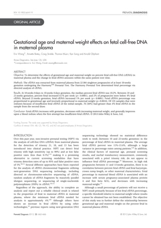
Fetal percentage of cfDNA was measured in 22 384 patients.
The mean reported gestational age in the group was 15.8 weeks
(range: 10–40 weeks), while the mean maternal weight was
73 kg (range: 32–284 kg) (Figure 1). At 10 weeks 0 days to
10 weeks 6 days gestation, the median percent fetal cfDNA
was 10.2%. Between 10 and 21 weeks gestation, percent fetal
increased at 0.10% per week (p<0.0001) (Figure 2), and 2% of
the pregnancies during this period were below 4% fetal cfDNA.
Starting at 21 weeks gestation, the percent fetal cfDNA
increased at a rate of 1% per week (p<0.0001), a tenfold
increase in the amount of percent fetal per week compared
with the weeks between 10 and 21 weeks gestation (Figure 2).
Table 1 shows the fraction of samples having at least 4% fetal
cfDNA in relation to maternal weight. The negative correlation
between fetal cfDNA and maternal weight was statistically
significant (p = 0.0003). Samples were grouped by weight
categories or bins in 10 kg increments. Within the cohort, 95%
of the samples came from women between the weight bin
categories of <50–120 kg. Of women with maternal weight less
than 90 kg, 99.1% had a fetal cfDNA greater than 4%. Both
gestational age and maternal weight significantly influence
the amount of fetal cfDNA, explaining as much as 27% of the
variations seen in percent fetal (p<0.0001).
In our study, approximately 1.9% of our tests resulted in
redraw requests because of low fetal percent. At the close of
this study, we have made 357 requests because of insufficient
fetal cfDNA, of which 135 women underwent a second blood
draw and sent the sample to our laboratory. As compared with
the entire cohort, the patients who underwent a second blood
draw were on average at a younger gestational age (13.9 weeks
Distribution of Gestational Age
Mean: 15.79
SD: 4.55
Median: 13.86
IQR: 6.14
Range: 10-40.43
10 15 20 25 30 35 40
Gestational Age (Weeks)
Frequency
0 500 1000 1500 2000 2500
Distribution of Maternal Weight
50 100 150 200 250 0 500 1000 1500
Maternal Weight (kg)
Frequency
Mean: 73.23
SD: 18.66
Median: 68.95
IQR: 22.23
Range: 31.75-284.41
Figure 1 Distribution of gestational age and maternal weight
vs 15.8 weeks, p<0.0001) and had higher mean maternal
weight (103 kg vs 73 kg, p<0.0001). We observed a mean 1%
fetal cfDNA gain in the second draw as compared with the first
draw with an average interval time between the two draws of
3.6 weeks. Of the 135 samples that had insufficient percent fetal
cfDNA with the first blood draw, 76 (56%) resulted in greater
than 4% fetal percent on the second blood draw. The
proportion of women with sufficient fetal percent on the
second draw decreased with increasing maternal weight
(Table 2). If we accounted for gestational age between blood
draws within the same pregnancy, the difference in percent
fetal cfDNA between first draw and second draw is no longer
significant with respect to maternal weight (p = 0.76),
suggesting that waiting for a later gestational age could
overcome low initial percent fetal cfDNA because of high
maternal weight.
Prenatal Diagnosis 2013, 33, 662–666 © 2013 John Wiley & Sons, Ltd.
- 3. 664 E. Wang et al.
DISCUSSION
This study represents the largest clinical data set on the
percent fetal cfDNA related to both gestational age and
maternal weight. We found that the fetal cfDNA increased
incrementally between 10 and 21 weeks of gestation and that
over this gestational age window, an overall 1% increase in
Table 2 Proportion of women with ≥4% fetal cfDNA on repeat
blood draw
Maternal
weight bin (kg)
# ≥4% fetal on
2nd draw
# of total
patients
% with ≥4% on
2nd draw
<90 30 42 71.4%
≥90<100 14 23 60.9%
≥100<110 13 22 59.1%
≥110<120 10 17 58.8%
≥120<130 2 7 28.6%
≥130<140 5 13 38.5%
≥140 2 11 18.2%
total fetal percent can be anticipated. However, starting at
21 weeks of gestation, a greater weekly increase in fetal percent
can be expected (1%). It is unlikely that a 0.1% average weekly
increase would be clinically relevant and warrant a general
change in blood draw protocol for the general population
greater than 10 weeks gestational age.
For those patients who had insufficient fetal cfDNA on their
first blood draw and underwent a second blood draw, the
maternal weight and gestational age characteristics were both
significantly different from the entire cohort. These patients
had higher maternal weight and were earlier in gestation.
Although 56% of these pregnancies resulted in greater than
4% fetal upon redraw, it is puzzling why the rate of increase
in fetal cfDNA in these samples were threefold higher than
the expected, given their gestational age range (0.28% per week
vs 0.1% per week observed in the general cohort). One simple
explanation is that the rate of cfDNA increase is different when
measured longitudinally in the same individual versus
aggregating results from different individuals. However, when
we looked at the entire cohort of redraws for various reasons,
which includes these 135 pregnancies requested because of
low initial percent fetal cfDNA, the rate of increase with respect
to gestational age is 0.09% per week within the same pregnancy
(data not shown). This rate is not dissimilar from the overall
rate of 0.1% computed from the entire cohort of pregnancies.
Alternatively, compounded with a higher maternal weight, it
is entirely possible that some of these patients with low initial
fetal percent did not have accurate dating of their pregnancies
as not all dating was based on ultrasound. We have observed a
significant decrease in fetal percent before 10 weeks gestation
(data not shown) and the rate of fetal percent increase may
be higher during this period. Therefore, in addition to the
anticipated limitations of maternal weight, accurate
gestational age dating at the time of first blood sample is
critical to the likelihood of receiving a result and in
determining when to schedule a redraw if necessary or desired.
This study confirms the previously reported relationship
between increasing maternal weight and decreasing fetal
percent.15 However, the majority of women, based on this set
of 22 000 commercial samples, will have greater than 4% fetal
DNA, regardless of gestational age or maternal weight, and will
be able to receive a result from NIPT. As the ability to detect
trisomy depends on the precision of the assay and fetal
10 15 20 25 30 35 40
0 10 20 30 40
Gestational Age (Weeks)
Fetal cfDNA (%)
Figure 2 Relationship between percentage of fetal cfDNA and
gestational age. Fetal cfDNA percentage for 22 384 pregnancies is
plotted with respect to gestational age (weeks). At 10 weeks 0 days
to 10 weeks 6 days gestation, the median fetal cfDNA percentage
was 10.2%. Between 10 and 21 weeks gestation (black open circles),
fetal cfDNA increased at 0.10% per week (p<0.0001; blue dash
line within black open circles) and 2% of the pregnancies during this
period were below 4% fetal cfDNA. Starting at 21 weeks gestation
(red open circles), the fetal cfDNA percentage increased at a rate
of 1% per week (p<0.0001; blue dash line within red open circles),
a tenfold increase in the amount of fetal cfDNA percentage per week
compared with the weeks between 10 and 21 weeks gestation
Table 1 Proportion of pregnancies with ≥4% fetal cell-free DNA
on first blood draw
Maternal weight bin (kg) n
Pregnancies with ≥4% fetal
cell-free DNA (%)
<50 809 99.8
≥50<60 4825 99.6
≥60<70 6224 99.2
≥70<80 4313 98.8
≥80<90 2574 98.2
≥90<100 1608 96.3
≥100<110 921 93.9
≥110<120 508 89.8
≥120<130 298 87.9
≥130<140 172 81.4
≥140 132 71.2
Prenatal Diagnosis 2013, 33, 662–666 © 2013 John Wiley & Sons, Ltd.
- 4. Maternal factors affecting fetal cell-free DNA in maternal plasma 665
percent, these findings can be used by providers and their
patients in making the decision as to whether or not to
repeat NIPT or undergo traditional methods of prenatal
testing for those patients who do not receive a result. As
there exists a segment of the population who would not
undergo invasive testing or termination under any
circumstances but seek the information and reassurance
NIPT can provide, the option of waiting until later in the
second trimester for a repeat blood draw, when fetal
percent increases more rapidly can also be presented. In
our study, there were 1224 samples (5.5% of all samples)
submitted after 24 weeks gestation, which suggests
that there is interest in this information in the third
trimester.
One limitation of our study is that we were not able to
calculate body mass index (BMI) as we did not have maternal
height for the calculation. Thus far, it is still unclear why fetal
cfDNA percent decreases with increasing maternal weight.
Having maternal height data and BMI with which to assess
obesity might allow a more accurate predictor of assay success
as compared with maternal weight alone; because in obese
women, there is an increased turnover of adipocytes, thereby
increasing the amount of maternal cfDNA and decreasing the
fetal cfDNA percent. However, BMI does not allow for the
analysis of body fat directly nor does it distinguish between
different body types relative to fat or muscle. For example,
BMI based on a strict height and weight calculation
overestimates body fat in persons who are very muscular
and can underestimate body fat in persons who have lost
or have little muscle mass.16,17 An alternate explanation is
that increasing total blood volume may be correlated with
decreasing percent fetal cfDNA (the diluting effect). In this
case, the total blood volume in a tall person is also larger
than in a small person, and height should certainly be taken
into account. Future studies including covariates such as
weight, height, BMI, and body type are warranted.
Therefore, without additional studies, a simpler clinical
standard around which to counsel regarding cfDNA analysis
success likelihood might be just maternal weight. Data from
future investigations that include weight, height, and body
type might shed light on this aspect.
CONCLUSION
This study provides clinically useful data to prepare patients
for the possibility of low fetal percent based on their
gestational age and weight at the time of blood draw. The vast
majority of clinical maternal plasma samples greater than
10 weeks gestational age contain an adequate fetal cfDNA
proportion to allow for useful clinical results. However,
attention should be paid to accurate gestational age
determination—especially at early gestational ages—to ensure
sufficient fetal percent for assay completion. Extremely high
maternal weight is a primary correlative factor for low fetal
cfDNA and thus no result from NIPT using cfDNA analysis. A
repeat blood draw for NIPT for samples with low fetal percent
may be worthwhile for patients, except in cases of high
maternal weight. When possible, second attempts are most
beneficial after waiting several weeks. If gestational age was
estimated to be later than the actual gestational age, the
waiting time to receive the first result may be long enough to
increase fetal percent above the 4% cutoff. In other cases, the
waiting time may need to be longer because of the slow rate
of increase in fetal percent until about 21 weeks gestation. In
addition to maternal weight and gestational age, there remains
a substantial amount of unexplained variation in fetal percent
in the population. Other clinical factors that influence fetal
percent remain an area for future investigation.
WHAT’S ALREADY KNOWN ABOUT THIS TOPIC?
• Previous literature indicates no significant change in average
percentage of fetal cell-free DNA between 10 and 22 weeks when
using Next-Generation Sequencing technology.
WHAT DOES THIS STUDY ADD?
• This is the largest sample set reporting changes in percent fetal cell-free
DNA in relation to maternal weight and gestational age.
REFERENCES
1. Sparks AB, Wang ET, Struble Ca, et al. Selective analysis of cell-free DNA
in maternal blood for evaluation of fetal trisomy. Prenat Diagn 2012;
32(1):3–9.
2. Norton ME, Brar H, Weiss J, et al. Non-Invasive Chromosomal
Evaluation (NICE) Study: results of a multicenter prospective cohort
study for detection of fetal trisomy 21 and trisomy 18. Am J Obstet
Gynecol 2012;207(2):137.e1–8.
3. Sparks AB, Struble Ca, Wang ZET, et al. Noninvasive prenatal detection
and selective analysis of cell-free DNA obtained from maternal blood:
evaluation for trisomy 21 and trisomy 18. Am J Obstet Gynecol. 2012;
206(4):319.e1–9.
4. Nicolaides KH, Syngelaki A, Ashoor G, et al. Noninvasive prenatal
testing for fetal trisomies in a routinely screened first-trimester
population. Am J Obstet Gynecol 2012;207(5):374.e1–6.
5. Ashoor G, Syngelaki A, Wagner M, et al. Chromosome-selective
sequencing of maternal plasma cell-free DNA for first-trimester
detection of trisomy 21 and trisomy 18. Am J Obstet Gynecol 2012;
206(4):322.e1–5.
6. Canick JA, Kloza EM, Lambert-Messerlian GM, et al. DNA
sequencing of maternal plasma to identify Down syndrome and
other trisomies in multiple gestations. Prenat Diagn 2012;32(8):
730–4.
7. Bianchi DW, Platt LD, Goldberg JD, et al. Genome-wide fetal aneuploidy
detection by maternal plasma DNA sequencing. Obstet Gynecol
2012;119(5):890–901.
8. Dan S, Wang W,Ren J, et al. Clinical application of massively parallel
sequencing-based prenatal noninvasive fetal trisomy test for trisomies
21 and 18 in 11 105 pregnancies with mixed risk factors. Prenat Diagn
2012:32(13):1225–32.
9. Chitty LS, Hill M, White H, et al. Noninvasive prenatal testing for
aneuploidy-ready for prime time?. Am J Obstet Gynecol 2012;206(4):
269–75.
10. ACOG Practice Bulletin. No. 77: screening for fetal chromosomal
abnormalities. Obstet Gynecol 2007;109(1):217–27.
11. Nicolaides KH. Screening for fetal aneuploidies at 11 to 13 weeks. Prenat
Diagn 2011;31(1):7–15.
Prenatal Diagnosis 2013, 33, 662–666 © 2013 John Wiley & Sons, Ltd.
- 5. 666 E. Wang et al.
12. Zimmermann B, Hill M, Gemelos G, et al. Noninvasive prenatal
aneuploidy testing of chromosomes 13, 18, 21, X, and Y using
targeted sequencing of polymorphic loci. Prenat Diagn 2012;32
(13):1–9.
13. Galbiati S, Smid M, Gambini D, et al. Fetal DNA detection in maternal
plasma throughout gestation. Hum Genet 2005;117(2–3):243–8.
14. Brar H, Wang E, Struble C, et al. The fetal fraction of cell-free DNA in
maternal plasma is not affected by a priori risk of fetal trisomy. The
journal of maternal–fetal & neonatal medicine: the official journal of the
European Association of Perinatal Medicine, the Federation of Asia and
Oceania Perinatal Societies, the International Society of Perinatal
Obstetricians. 2012.
15. Ashoor G, Poon L, Syngelaki A, et al. Fetal fraction in maternal plasma
cell-free DNA at 11–13 weeks gestation: effect of maternal and fetal
factors. Fetal Diagn Ther 2012;31(4):237–43.
16. Frankenfield DC, Rowe WA, Cooney RN, et al. Limits of body mass index
to detect obesity and predict body composition. Nutrition 2001;17(1):
26–30.
17. Fattah C, Farah N, Barry S, et al. The measurement of maternal
adiposity. J Obstet Gynaecol 2009;29(8):686–9.
Prenatal Diagnosis 2013, 33, 662–666 © 2013 John Wiley & Sons, Ltd.