![Ind J Clin Biochem
1 3
movements of joints which develops pain and fatigue [1].
There are different types of arthritis [2]. The most common
type of joint disorders are osteoarthritis (OA) or degenera-
tive joint, rheumatoid arthritis (RA), gouty arthritis (GA),
calcium pyrophosphate deposition disease (CPPD), psoriatic
arthritis (PA), infectious arthritis (IA) and spondyloarthritis
(SA) [2, 3].
Causes of arthritis may vary according to the type of
arthritis. In osteoarthritis, obesity, advancing age and dis-
comfort in joints show a contributory role. Some genetic
modification such as mutations in genes encoding types
II, IV, V, and VI collagens have also been found to cause
osteoarthritis. On the other hand, RA is an autoimmune
disorder. Some studies showed that exposure of air pollu-
tion may increase the risk of RA [4]. Some gases such as
SO2, NO2, carbon monoxide (CO), and O3 are the main
pollutants that induce occurrence of RA. Further, the results
from the epidemiological studies have indicated that expo-
sure of an individual to many other environmental factors
including dioxin, cigarette smoke, noise, and air pollutants
[5]. Some other factors such as viral diseases (CoVID-19)
and prolonged medication may also cause development of
arthritis [6, 7]. In another study carried out by Gong et al.
[3], it was found that plant based lectins may be involved
in the pathogenesis of some inflammatory disorders in
arthritis via activation of NLRP3 inflammasome [8]. Tsuda
et al. [9] has reported that cross reaction of serum ACPA
of RA patients’ citrullinated proteins derived from viruses
(EBNA-1), bacteria (Micrococcus luteus, Mycobacterium
gastri, Nocardia brasiliensis), fungi (Aspergillus fumiga-
tus, Candida albicans, Cryptococcus neoformans) and
plants (Oryza sativa, Solanum lycopersicum, Glycine max,
with tissue antigens may induce RA through cross-reactivity
[10]. In addition, the excess consumption of high-fructose
corn syrup and fruit drinks may cause three-fold higher risk
of development arthritis [11].
The corticosteroids, the steroidal hormones, possess anti-
inflammatory, vasoconstrictive and immunosuppressive
properties [12]. They are prescribed to treat chronic arthritis
patients to combat emergence of any serious clinical compli-
cations [13–16]. The influence of steroids on inflammatory
processes has long been established since the interaction
of glucocorticoids and their receptors were found to inhibit
several inflammatory pathways. Inflammatory processes are
known to be accompanied by an increased oxidative burden
followed by a depletion of antioxidants [17]. The oxidative
stress is found to be associated with major health complica-
tions [18–20]. Some workers have previously shown that
RA induces oxidative stress. However, it is not well known
that the intake of steroids by arthritis patients may further
augment this process [21]. Chronic usage of corticosteroids
has been linked to the negative side effects such as weight
gain, osteoporosis, hypertension, diabetes mellitus, cataract,
dyspepsia, and psychiatric issues [12, 22].
The usage of corticosteroids is considered to be safe, in
terms of hepatotoxicity, however, the clinical impact of this
not well known. Some workers have shown liver injury in
those related to high-dose methylprednisolone [23]. How-
ever, the implication of steroidal drugs in generating oxida-
tive stress and inducing hepatotoxicity has not been system-
atically investigated in human system.
In the present study, an attempt has been made to evaluate
the effect of steroidal drugs (deflazacort and methylpredniso-
lone) on the levels of antioxidant defense system and hepatic
markers in the blood plasma of the arthritis patients using
these drugs for a long duration. The results from present
investigation suggested that the long term treatment with
steroidal drugs may induce imbalance in the endogenous
antioxidant system and develop hepatotoxicity.
Materials and Methods
Chemicals
Ethylene diaminetetraacetic acid (EDTA), potassium dichro-
mate, pyrogallol, sodium bicarbonate
(NaHCO3), sodium
dihydrogen phosphate
(NaH2PO4), disodium hydrogen
phosphate (Na2HPO4), sodium acetate, sodium carbonate
(Na2CO3), sodium hydroxide, sulfuric acid, Merk Limited
(Mumbai, India),1-Chloro 2, 4 dinitrobenzene (CDNB), Fol-
lin’s reagent, glutathione (GSH),5, 5′-Dithiobis (2-nitroben-
zoic acid) (DTNB), glycerol, NADH, sodium pyruvate,
sodium chloride, and Tris-base were from SRL. Pvt. Ltd.
(India). Bovine serum albumin (BSA) was from LobaChe-
mie (LobaChemie Pvt. Ltd., Mumbai, India). All other
chemicals used were analytical grade.
Subjects
This study included blood samples from 30 patients suf-
fering from arthritis, reactive arthritis, and joint pain and
receiving steroidal therapies using deflazacort and methyl-
prednisolone for different durations such as 7, 14, 28, 32, 84
and 168 days. The blood samples of these subjects were col-
lected from different hospitals/ clinics. These patients were
prescribed to take short-term and long-term therapies with
corticosteroids i.e., deflazacort and methylprednisolone for
the improvement of their clinical conditions. The deflazacort
is prescribed in 12–18 mg divided dose in a day whereas
methylprednisolone is prescribed as 16-24 mg in a day. The
blood is collected into the heparinized sterile vials and kept
inside the ice box.](data:image/gif;base64,R0lGODlhAQABAIAAAAAAAP///yH5BAEAAAAALAAAAAABAAEAAAIBRAA7)
Recommended
Recommended
More Related Content
Similar to s12291-023-01127-2.pdf
Similar to s12291-023-01127-2.pdf (20)
Recently uploaded
Recently uploaded (20)
s12291-023-01127-2.pdf
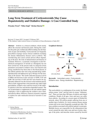
- 1. Vol.:(0123456789) 1 3 Ind J Clin Biochem https://doi.org/10.1007/s12291-023-01127-2 ORIGINAL RESEARCH ARTICLE Long Term Treatment of Corticostreroids May Cause Hepatotoxicity and Oxidative Damage: A Case Controlled Study Priyanka Tiwari1 · Nitika Singh1 · Bechan Sharma1 Received: 23 August 2022 / Accepted: 23 February 2023 © The Author(s), under exclusive licence to Association of Clinical Biochemists of India 2023 Abstract Arthritis is a clinical condition, which mainly affects the structure and function joints. During this condi- tion the joints gets swelled and stiffed resulting into develop- ment of pain and morbidity. Corticosteroids are frequently prescribed to manage various clinical conditions including the chronic inflammatory diseases such as arthritis. The steroidal drug also causes certain adverse effects depend- ing on the dose, the route of administration and duration of treatment. However, a systematic investigation on the bio- chemical consequences of steroids as a therapeutic has not been carried out. In the present study we analyzed certain parameters associated to oxidative stress, liver function and energy metabolism has been done in the blood plasma of the arthritis patients who were using steroidal drugs (meth- ylprednisolone and deflazacort) up to 168 days for the treat- ment of the disease. The results indicated increase in level of MDA and decrease in the activities of SOD, CAT and LDH. The activities of AST and ALT were found to be sig- nificantly enhanced over the increase in the treatment period. These results suggested that corticosteroids may induce lipid peroxidation, oxidative stress and liver toxicity in the arthri- tis patients in the dose and duration dependent manner. The use of antioxidants as supplements to the anti-arthritis agents could play a role in suppressing the oxidative stress medi- ated adverse effects. However, extensive research is required to explore for safer medication devoid of steroids to cure arthritis. Graphical Abstract Keywords Antioxidant system · Corticosteroids · Oxidative stress · Energy metabolism · Hepatotoxicity Introduction The word arthritis is a combination of two words: the Greek arthron means “joint” and the Latin itis means “inflamma- tion”. There are more than 100 types of arthritis and related conditions. People of all ages, sexes and races can suffer from arthritis. According to a recent report of Global RA network, about 350 million people suffering from one or another type of arthritis. It is most common disease among women and occurs more frequently as people get older. The basic problem associated with arthritic patients is joint pain. Multiple factors are responsible for the inflammation that occurs around the joint, daily wear and tear of joints, dam- age of the joints, and muscular strains caused by forceful * Bechan Sharma sharmabi@yahoo.com 1 Department of Biochemistry, Faculty of Science, University of Allahabad, Allahabad 211002, India
- 2. Ind J Clin Biochem 1 3 movements of joints which develops pain and fatigue [1]. There are different types of arthritis [2]. The most common type of joint disorders are osteoarthritis (OA) or degenera- tive joint, rheumatoid arthritis (RA), gouty arthritis (GA), calcium pyrophosphate deposition disease (CPPD), psoriatic arthritis (PA), infectious arthritis (IA) and spondyloarthritis (SA) [2, 3]. Causes of arthritis may vary according to the type of arthritis. In osteoarthritis, obesity, advancing age and dis- comfort in joints show a contributory role. Some genetic modification such as mutations in genes encoding types II, IV, V, and VI collagens have also been found to cause osteoarthritis. On the other hand, RA is an autoimmune disorder. Some studies showed that exposure of air pollu- tion may increase the risk of RA [4]. Some gases such as SO2, NO2, carbon monoxide (CO), and O3 are the main pollutants that induce occurrence of RA. Further, the results from the epidemiological studies have indicated that expo- sure of an individual to many other environmental factors including dioxin, cigarette smoke, noise, and air pollutants [5]. Some other factors such as viral diseases (CoVID-19) and prolonged medication may also cause development of arthritis [6, 7]. In another study carried out by Gong et al. [3], it was found that plant based lectins may be involved in the pathogenesis of some inflammatory disorders in arthritis via activation of NLRP3 inflammasome [8]. Tsuda et al. [9] has reported that cross reaction of serum ACPA of RA patients’ citrullinated proteins derived from viruses (EBNA-1), bacteria (Micrococcus luteus, Mycobacterium gastri, Nocardia brasiliensis), fungi (Aspergillus fumiga- tus, Candida albicans, Cryptococcus neoformans) and plants (Oryza sativa, Solanum lycopersicum, Glycine max, with tissue antigens may induce RA through cross-reactivity [10]. In addition, the excess consumption of high-fructose corn syrup and fruit drinks may cause three-fold higher risk of development arthritis [11]. The corticosteroids, the steroidal hormones, possess anti- inflammatory, vasoconstrictive and immunosuppressive properties [12]. They are prescribed to treat chronic arthritis patients to combat emergence of any serious clinical compli- cations [13–16]. The influence of steroids on inflammatory processes has long been established since the interaction of glucocorticoids and their receptors were found to inhibit several inflammatory pathways. Inflammatory processes are known to be accompanied by an increased oxidative burden followed by a depletion of antioxidants [17]. The oxidative stress is found to be associated with major health complica- tions [18–20]. Some workers have previously shown that RA induces oxidative stress. However, it is not well known that the intake of steroids by arthritis patients may further augment this process [21]. Chronic usage of corticosteroids has been linked to the negative side effects such as weight gain, osteoporosis, hypertension, diabetes mellitus, cataract, dyspepsia, and psychiatric issues [12, 22]. The usage of corticosteroids is considered to be safe, in terms of hepatotoxicity, however, the clinical impact of this not well known. Some workers have shown liver injury in those related to high-dose methylprednisolone [23]. How- ever, the implication of steroidal drugs in generating oxida- tive stress and inducing hepatotoxicity has not been system- atically investigated in human system. In the present study, an attempt has been made to evaluate the effect of steroidal drugs (deflazacort and methylpredniso- lone) on the levels of antioxidant defense system and hepatic markers in the blood plasma of the arthritis patients using these drugs for a long duration. The results from present investigation suggested that the long term treatment with steroidal drugs may induce imbalance in the endogenous antioxidant system and develop hepatotoxicity. Materials and Methods Chemicals Ethylene diaminetetraacetic acid (EDTA), potassium dichro- mate, pyrogallol, sodium bicarbonate (NaHCO3), sodium dihydrogen phosphate (NaH2PO4), disodium hydrogen phosphate (Na2HPO4), sodium acetate, sodium carbonate (Na2CO3), sodium hydroxide, sulfuric acid, Merk Limited (Mumbai, India),1-Chloro 2, 4 dinitrobenzene (CDNB), Fol- lin’s reagent, glutathione (GSH),5, 5′-Dithiobis (2-nitroben- zoic acid) (DTNB), glycerol, NADH, sodium pyruvate, sodium chloride, and Tris-base were from SRL. Pvt. Ltd. (India). Bovine serum albumin (BSA) was from LobaChe- mie (LobaChemie Pvt. Ltd., Mumbai, India). All other chemicals used were analytical grade. Subjects This study included blood samples from 30 patients suf- fering from arthritis, reactive arthritis, and joint pain and receiving steroidal therapies using deflazacort and methyl- prednisolone for different durations such as 7, 14, 28, 32, 84 and 168 days. The blood samples of these subjects were col- lected from different hospitals/ clinics. These patients were prescribed to take short-term and long-term therapies with corticosteroids i.e., deflazacort and methylprednisolone for the improvement of their clinical conditions. The deflazacort is prescribed in 12–18 mg divided dose in a day whereas methylprednisolone is prescribed as 16-24 mg in a day. The blood is collected into the heparinized sterile vials and kept inside the ice box.
- 3. Ind J Clin Biochem 1 3 Inclusion and Exclusion Criteria The inclusion criteria followed by the patients included in this study contained age 30–55 years, suffering from arthritis for at least one month or more, taking steroids (methylpred- nisolone or deflazacort) for therapy, inhabiting in any social structure, and agreeing to extend informed consent for the said purpose. The exclusion criteria for the recruitment of patients in this study were subjects intolerant or allergic to any therapeutic agents, diagnosed positive for viral, bacte- rial, fungal infections or parasite infestation, uncontrolled diabetes mellitus (DM), gastrointestinal problems or gastro- intestinal bleeding (GIB) history, heart failure (HF), active malignancies and received any immune-suppressor agents. The study also excluded pregnant or lactating women. Sample Preparation The blood samples collected in the sterile heparinized tubes, centrifuged at 10,000 rpm for 15 min using refrigerated cen- trifugation machine (Sigma 2-16KL). After centrifugation, the supernatant (plasma) was collected and stored at 4 °C for future uses. Biochemical Assays Determination of Protein Content The total protein content in different samples was deter- mined by the method of Lowry et al. [24] using BSA as a standard. Estimation of Malondialdehyde (MDA) Level Lipid peroxidation (LPO) in terms of level of malondialde- hyde (MDA) was measured in the blood plasma samples fol- lowing the method of Niehaus and Samuelsson (1968) and the results were expressed as n mol MDA/mg protein using 1.56× 10⁵ M−1 cm−1 as an extinction coefficient. Determination of GSH Content The Glutathione (GSH) content in blood plasma was esti- mated according to the method describe Ellman et al. (1959) modified by Sedlak and Lindsay (1968). The optical density of the yellow color complex developed in the reaction mix- ture was measured at 412 nm. The reaction mixture con- taining 50 mM Tris-HCl buffer pH 8.2, 0.06 mM DTNB, absolute methanol, and suitable amount of plasma was incu- bated with intermittent shaking and centrifuged at 800×g for 15 min. The results were expressed as microgram (µg) of GSH/mg protein. The GSH was used as a standard. LDH Activity Assay The activity of LDH in blood plasma was measured by the method of Horecker and Kornberg (1948). Briefly, The total reaction mixture volume (3 ml) contained 50 mM Tris-HCl buffer, pH 7.4, 3 mM KCl (0.15 ml), 1 mM sodium pyruvate, 0.16 mM NADH and suitable amount of enzyme protein. The enzyme activity was monitored at 340 nm for 3 min as a decrease in its absorbance at the regular interval of 30 s. The absorbance of the reaction mixture recorded in absence of enzyme protein was considered as a control. The spe- cific activity of the enzyme was calculated using the extinc- tion coefficient of NADH (6.22× 10³ μmol−1 L−1 min) and denoted as IU/ mg protein. The assays were performed on UV visible double beam spectrophotometer (Spectroscan UV 2700). SOD Activity Assay The activity of superoxide dismutase (SOD: E.C. 1.15.1.1) in plasma was measured by the method of Marklund and Marklund (1974). The method allows measuring the optical density of colored complex involving the autooxidation of pyrogallol at 412 nm for 3 min at the 30 s interval on spec- trophotometer. The unit for enzyme activity was expressed as 50% inhibition of pyrogallol autooxidation per min. CAT Activity Assay The activity of catalase (CAT: E.C.1.11.1.6) in blood plasma was measured according to Beers and Sizer (1952) method. The decrease in absorbance for H2O2 consumption was mon- itored at 240 nm for 3 min with an interval of 30 s. The unit for CAT activity was expressed in terms of μmol of H2O2 hydrolyzed per min. The enzyme activity was calculated by using molar extinction coefficient (43.6 M−1 cm−1 ). Assay for Liver Function Enzymes The activities of transaminases such as aspartate transami- nase (AST: EC 2.6.1.1) and alanine transaminase (ALT: EC 2.6.1.2), also known as SGOT and SGPT, respectively, were monitored in the blood samples using the method of Reitman and Frankel (1957). A suitable amount of blood plasma was incubated in 0.25 ml of substrate (200 mM aspartate with 2 mM α-ketoglutarate) for AST and (200 mM Malanine with 2.0 mM α-ketoglutarate) for ALT in 0.1 M phosphate buffer, pH 7.0, for 30 min. An aliquot of 0.25 ml of DNPH (1 mM) was used to stop the reaction and allowed incubation for 20 min at room temperature (about 26 °C). The reaction
- 4. Ind J Clin Biochem 1 3 mixture was added with 2.5 ml of 0.4 N NaOH followed by gentle vortex. The optical density of the colored complex was monitored at 510 nm. The total reaction mixture volume (TRMV; 3 ml) in the absence of enzyme protein was used as a control in each case. Statistical Analysis The Graph Pad Prism version 5.01 (San Diego, CA) was used for analyzing the data using one way analysis of vari- ance (ANOVA) and calculating the values as mean±standard deviation. Results Level of Blood Plasma Protein in the Arthritis Patients The results obtained after analysis of protein concentration in the blood plasma from the patients suffering from arthritis indicated almost no difference from the normal subjects. Also, the level of blood plasma protein from the arthritis patients taking steroids (methylprednisolone and deflaza- cort) did not reflect any change in the values of protein (data not shown). Effect of Deflazacort and Methylprednisolone on the Level of MDA and GSH in the Plasma of Human Blood Samples The effect of steroidal drugs on the levels of MDA and GSH were determined and the results are shown in Table 1. It was found that there is a significant increase (about two- fold) in the level of MDA in the patients suffering from arthritis in comparison to the normal subjects. The level of MDA was also higher in the steroid treated arthritis patients as compared to controls but these values were lower than those from patients not using steroids. The level of GSH was found to be significantly decreased in the patients tak- ing steroidal drugs. However, there was no change in the level of GSH in the arthritis patients as compared to the normal control. Effect of Deflazacort and Methylprednisolone on the Level of LDH Activity on the plasma of Human Blood Samples The effect of deflazacort and methylprednisolone was evalu- ated on the activity of LDH in the blood plasma of arthritis patients. The results shown in Fig. 1 indicated that there was a slight increase in the LDH activity both in the patients suf- fering from arthritis as well as those using (A) deflazacort or (B) methylprednisolone as therapeutic agents for longer duration. Effect of Deflazacort and Methylprednisolone on the Activity of SOD on the Plasma of Human Blood Samples The effect of deflazacort and methylprednisolone was evaluated on the activity of SOD in the blood plasma of arthritis patients. The results shown in Fig. 2 indicated that there was a decrease in SOD activity by 22% in arthri- tis patients in comparison to the control. After treatment with methylprednisolone and deflazacort, there was no significant alteration was seen with respect to the arthritis patients. However, the slight increment in the activity of enzyme was seen in case of deflazacort treatment. Effect of Deflazacort and Methylprednisolone on the Activity of Catalase on the Plasma of Human Blood Samples The effect of deflazacort and methylprednisolone was evaluated on the activity of catalase in the blood plasma of arthritis patients. The results shown in Fig. 3a indicated that there was only mild decrease (22%) in CAT activity in the arthritis patients. When the arthritis patients received deflazacort for 7 days, the activity of catalase was found to be near normal subjects. There was almost no further change in the activity of catalase when the patients received deflazacort up to 168 days. In contrast, the treatment of Table 1 Effect of steroids on the levels of MDA and GSH in the human blood plasma The data presented in the table reflect the values as mean ±SEM of three independent experiments Treatment Time duration Plasma MDA level Plasma GSH level Control – 3.19±0.16 1.01±0.012 Arthritis patients – 6.14±0.20 1.09±0.13 Arthritis patients using deflaza- cort 7 Days 5.78±0.18 1±0.97 14 Days 5.71±.155 0.80±0.065 28 Days 5.09±0.53 0.78±0.031 84 Days 5.0±0.39 0.77±0.05 168 Days 5.79±0.049 0.72±0.019 Arthritis patients using methyl- prednisolone 7 Days 3.24±0.198 0.77±0.037 14 Days 4.59±0.27 0.76±0.036 28 Days 5.8±0.31 0.72±0.041 84 Days 6.33±0.36 0.70±0.026 168 Days 6.49±0.38 0.70±0.037
- 5. Ind J Clin Biochem 1 3 Fig. 1 Effect of deflazacort (a) and methylprednisolone (b) on the activity of lactate dehydrogenase (LDH) in human blood plasma. The effect was observed for different treatment durations (7–168 days) as mentioned in the Materials and Methods section. The IU represents µmoles of substrate converted into product by the enzyme in one min. The data has been displayed as mean±SD of three independ- ent experiments. C=normal blood plasma, C1=blood plasma from arthritis patients. Treatment durations in days reflect the intake of steroids by arthritis patients (C1) for varying periods Fig. 2 Effect of deflazacort (a) and methylprednisolone (b) was evaluated on the activity of superoxide dismutase (SOD) in human blood plasma. The effect was observed for different treatment dura- tions (7–168 days) as mentioned in the Materials and Methods sec- tion. The IU represents µmoles of substrate converted into product by the enzyme in one min. The data has been displayed as mean±SD of two independent experiments. C=normal blood plasma, C1=blood plasma from arthritis patients. Treatment durations in days reflect the intake of steroids by arthritis patients (C1) Fig. 3 Effect of deflazacort (a) and methylprednisolone (b) on the activity of catalase (CAT) in human blood plasma. The effect was observed for different treatment durations (7–168 days) as mentioned in the Materials and Methods section. The IU represents µmoles of substrate converted into product by the enzyme in one min. The data has been displayed as mean±SD of three independent experiments. C=normal blood plasma, C1=blood plasma from arthritis patients. Treatment durations in days reflect the intake of steroids by arthritis patients (C1)
- 6. Ind J Clin Biochem 1 3 patients with methylprednisolone from 7 to 168 days dem- onstrated a continuous decrease in the catalytic activity of catalase ranging from 30 to 45% in comparison to the control (Fig. 3b) Effect of Deflazacort and Methylprednisolone on the Activity of Alanine Transaminase (ALT) and Aspartate Aminotransferse (AST) on the Plasma of Human Blood Samples The effect of deflazacort and methylprednisolone was eval- uated on the level of ALT and AST in the blood plasma of arthritis patients. The results shown in Table 2 indicated that there was no significant change in the level of ALT in arthritis patients in comparison to the control. The level of ALT recorded on day zero remained unchanged over the period up to 168 days in comparison to the control. However, after treatment with steroids (methylpredniso- lone and deflazacort), the level of ALT was found to be increased by 30–35% up to 168 days of treatment of arthri- tis patients. Similar to ALT, the level of AST was also found to be unchanged in arthritis patients from day 0 to 168 days. After treatment with the steroids (methylpred- nisolone and deflazacort), the level of AST increased by 1.5-fold up to 0–168 days of treatment period in compari- son to the control. There was significant increase in the ratio of AST: ALT as observed from day 28 to 168 after the treatment with deflazacort. This ratio increased from the day 84 to 168 after the treatment with methylpredni- solone (Table 2). Discussion Arthritis, a very common chronic autoimmune disease, needs long term treatment with the therapeutics involving steroids, especially the methylprednisolone and deflazacort. The treatment of arthritis patients with higher doses of drugs combined with steroids for the extended period of time has been shown to develop certain adverse effects [25–27]. In some previous studies, the applications of steroids have been demonstrated to induce oxidative stress and to develop alterations in clinical parameters including hepatotoxicity [22, 28]. In the present investigation, a biochemical analy- sis of different clinical parameters including the markers of oxidative stress and liver function tests has been carried out in the blood plasma of arthritis patients. The results from the present study indicated significant perturbations in the levels of oxidative indices, protein and the activities of antioxidant enzymes, transaminases as well as lactate dehydrogenase, a key enzyme of energy metabolism, in the blood plasma of arthritis patients using steroids (methylprednisolone and deflazacort) with other therapeutics up to 168 days. The significant decrease in the level of GSH and increase in the level of MDA as observed in the arthritis patients indicated the development of oxidative stress due to chronic treatment of patients with steroidal drugs up to 168 days. Similar observation has been recorded by other workers who have studied on the arthritis patients and found perturba- tions in the levels of GSH and MDA [29]. The Hassan et al., (2011) have observed increased level of oxidative species and drastic reduction in the level of anti-oxidative factors in Table 2 Effect of deflazacort (D) and methylprednisolone (M) on the level of Alanine transaminase (ALT) and Aspartate transaminase (AST) [U/mg of protein] in human blood plasma The effect was observed for different treatment durations (0–168 days) as mentioned in the Materials and Methods section. The data has been displayed as mean±SEM of three independent experiments. C=nor- mal blood plasma, C1=blood plasma from arthritis patients. Treatment durations in days reflect the period of intake of steroids by arthritis patients (C1) Groups Treatment duration (days) 0 7 14 28 84 168 AST (C) 32.71±0.10 33.50±0.09 36.09±0.10 34.86±0.15 33.98±0.09 35.70±0.18 ALT (C) 45.08±0.50 47.01±0.01 49.90±0.10 47.48±0.08 45.09±0.13 45.34±0.18 AST/ALT (C) 0.71±0.30 0.712±0.15 0.723±0.09 0.734±0.12 0.753±0.13 0.787±0.09 AST (C1) 34.56±0.22 36.08±0.19 35.78±0.25 34.45±0.21 35.68±0.24 36.90±0.30 ALT (C1) 42.09±0.48 45.60±0.20 43.09±0.30 43.75±0.15 43.98±0.19 46.01±0.20 AST/ALT (C1) 0.81±0.35 0.80±0.16 0.79±0.30 0.78±0.27 0.81±0.31 0.80±0.19 AST (C1+D) 33.61±0.30 34.75±0.25 52.25±0.15 59.95±0.22 61.65±0.17 63.6±0.19 ALT (C1+D) 45.05±0.23 47.31±0.30 53.5±0.05 54.8±0.50 55.7±0.50 61.05±0.51 AST/ALT (C1+D) 0.75±0.15 0.73±0.026 0.976±0.010 1.09±0.36* 1.11±0.03* 1.04±0.35* AST (C1+M) 34.45±0.05 49.95±0.02 51.75±0.04 54.05±0.02 77.1±0.05 80.75±0.20 ALT (C1+M) 42.28±0.07 55.05±0.30 54.0±0.09 59.1±0.03 66.3±0.02 66.95±0.07 AST/ALT (C1+M) 0.81±0.02 0.90±0.16 0.95±0.020 0.91±0.01 1.16±0.04* 1.20±0.01*
- 7. Ind J Clin Biochem 1 3 the patients suffering from rheumatoid arthritis and systemic lupus erythematosus and taking treatment with methoxarate [29]. Some authors have studied the effect of methylpredni- solone in the experimental animal models and reported sig- nificant perturbations in the levels of oxidative indices [7]. The patients under this treatment conditions indicated differential response of deflazacort and methylprednisolone on the activities of antioxidant enzymes i.e. SOD and CAT. These drugs caused significant decrease in the activities of SOD and CAT in blood plasma samples; the effect with the administration of deflazacort being less than that of meth- ylprednisolone. In a study conducted by Sunitiparpluacha (2018) et al., the development of oxidative stress in the oste- oarthritis patients using glucocorticoid has been reported [30]. Some workers have reported the excess production of ROS and reduction in the activities of ROS scavenging enzymes in the patients suffering from eye diseases and tak- ing corticosteroids as a therapeutic agent [31, 32]. Tansaminases (AST and ALT) are key marker enzymes of liver involved in biotransformation of xenobiotics and detox- ification process [3]. The increased level of these enzymes has been shown to be associated with the liver toxicity [3]. The significant increase in the level of transaminases (AST/ ALT) observed in the arthritis patients taking steroids (meth- ylprednisolone and deflazacort) in the present study suggests the adverse impact of these drugs on the liver function of patients. Corticosteroids also have major effects on the liver, particularly when given for long term and in higher than physiologic doses [23, 26]. High doses and long-term use has been associated with the development or exacerbation of nonalcoholic steatohepatitis with elevations in serum ami- notransferase levels and liver histology resembling alcoholic hepatitis with steatosis, chronic inflammation, centrolobular ballooning degeneration [23, 26]. In one of such study con- ducted by Gutkowski et al. (2011) the high dose of meth- ylprednisolone has been reported the cause hepatotoxicity [28]. Conclusion The results from the present study reflected that the long- term treatment of arthritis involving steroids elicit marked perturbations in some key biochemical parameters in the patients. The significant increase in the level of MDA and decrease in the anti-oxidative indices such as GSH and activities of SOD and CAT indicated the development of oxidative stress. The significant alterations in the levels AST and ALT as observed in the blood plasma indicated hepatic damage in the patients taking steroids with the anti-arthritis therapeutics. The mild change in the activity of LDH in the blood plasma of such patients is an indicative of impact of these therapeutic agents on the energy metabolism. These results suggest that the application of steroids either should be avoided or minimized during treatment of arthritis. The use of antioxidants as supplements to the anti-arthritis agents could play a role in suppressing the oxidative stress medi- ated adverse effects. However extensive research is required to explore for safer medication devoid of steroids to cure arthritis. The detail illustration of present work is presented in graphical abstract. Acknowledgements PT and NS are grateful to the University Grant Commission, New Delhi, for providing financial assistance in the form of a Research Fellowship. Authors acknowledge UGC-SAP and DST- FIST for support to Department of Biochemistry, University of Alla- habad, Prayagraj India. Funding No funding was received for conducting this study. Declarations Conflict of interest Corresponding author on behalf of all authors declare no conflict of interest. Ethical Approval Vide office no. IERB/2016/03 by institute of eth- ics committee of population resource and research centre Allahabad. Consent to Participate Consent was taken from the patients. Consent for Publication Consent was obtained from the patients for publication of this case. References 1. Iagnocco A, Meenagh G, Riente L, Filippucci E, et al. Ultrasound imaging for the rheumatologist XXIX. Sonographic assessment of the knee in patients with osteoarthritis. Clin Exp Rheumatol. 2010;28(5):643–6. 2. Chhem RK, Kaplan PA, Dussault RG. Ultrasonography of mus- culoskeletal system. Radiol Clin North Am. 1994;32(2):275–893. 3. Hua C, Buttgereit F, Combe B. Glucocorticoids in rheu- matoid arthritis: current status and future studies. RMD Open. 2020;6(1):e000536. https://doi.org/10.1136/rmdop en-2017-000536. 4. Sun G, Hazlewood G, Bernatsky S, Kaplan GG, Eksteen B, Barnabe C. Association between air pollution and the develop- ment of rheumatic disease: a systematic review. Int J Rheumatol. 2016;10:53–6. https://doi.org/10.1155/2016/5356307. 5. Sadowska AM, Klebe B, Germonpré P, De Backer WA. Glu- cocorticoids as antioxidants in treatment of asthma and COPD. New application for an old medication? Steroids. 2007;72(1):1–6. https://doi.org/10.1016/j.steroids.2006.10.007. 6. Jessar RA, Hollander JL. Types of arthritis and their medical treat- ment. Am J Nurs. 1955;55(4):426–9. 7. Almutairi K, Nossent J, Preen D, Keen H, Inderjeeth C. The global prevalence of rheumatoid arthritis: a meta-analysis based on a systematic review. Rheumatol Int. 2021;41(5):863–77. https://doi. org/10.1007/s00296-020-04731-0. 8. Tsuda R, Ozawa T, Kobayashi E. Monoclonal antibody against citrullinated peptides obtained from rheumatoid arthritis patients
- 8. Ind J Clin Biochem 1 3 reacts with numerous citrullinated microbial and food proteins. Arthritis Rheumatol. 2015;67(8):2020–31. https://doi.org/10. 1002/art.39161. 9. Gong T, Wang X, Yang Y. Plant lectins activate the NLRP3 inflammasome to promote inflammatory disorders. J Immunol. 2017;98(5):2082–92. https://doi.org/10.4049/jimmunol.1600145. 10. De Christopher LR, Uribarri J, Tucker KL. Intake of high-fructose corn syrup sweetened soft drinks, fruit drinks and apple juice is associated with prevalent arthritis in US adults, aged 20–30years. Nutr Diabetes. 2016;6(3):199. https://doi.org/10.1038/nutd.016.7. 11. Solus JF, Chung CP, Oeser A, Li C, Rho YH, Bradley KM, Kawai VK, Smith JR, Stein CM. Genetics of serum concentration of IL-6 and TNFα in systemic lupus erythematosus rheumatoid arthritis: a candidate gene analysis. Clin Rheumatol. 2015;34:1375–82. 12. The nobel prize in physiology and medicine 1950. Nobelprize.org. http://www.nobelprize.org/nobel_prizes/medicine/laureates/1950/ hench 13. Glyn J. The discovery and early use of cortisone. J R Soc Med. 1998;91:513–7. 14. Rhen T, Cidlowski J. Anti-inflammatory action of glucocorticoids new mechanisms for old drugs. N Engl J Med. 2005;353:1711–23. 15. Baschant U, Tuckermann J. The role of the glucocorticoid recep- tor in inflammation and immunity. J Steroid Biochem Mol Biol. 2010;120:69–75. 16. Beers RF, SizerI W. A spectrophotometric method for measur- ing the breakdown of hydrogen peroxide by catalase. J Chem. 1952;195:133–40. 17. Monteserín L, Jiménez M, Linares P, Rodríguez-Martín L, Álva- rez-Cuenllas B, Álvarez-Cañas C, Jorquera CF. Acute hepatitis secondary to high-dose intravenous methylprednisolone. Gas- troenterol Hepatol. 2018;41(8):508–9. https://doi.org/10.1111/j. 1756-185X.2011.01630.x. 18. Gupta VK, Siddiqi NJ, Ojha AK, Sharma B. Hepatoprotective effect of Aloevera against cartap and malathion induced toxicity in Wistar rats. J Cell Physiol. 2019;25:1–15. 19. Agnes EC, Karen EC. The anti-inflammatory and immunosup- pressive effects of Glucocorticoids, recent developments and mechanistic insights. Mol Cell Endocrinol. 2009;335:2–35. 20. Quiñonez-Flores CM, González-Chávez SA, Del RíoNájera D, Pacheco-Tena C. oxidative stress relevance in the pathogenesis of the rheumatoid arthritis: a systematic review. Biomed Res Int. 2016;1–14. 21. Oztürk HS, Cimen MY, Cimen OB, Kaçmaz M, Durak I. Oxidant/ antioxidant status of plasma samples from patients with rheuma- toid arthritis. Rheumatol Int. 1999;19(1–2):35–7. https://doi.org/ 10.1007/s002960050097. 22. Aljebab F, Choonara I, Conroy S. Systematic review of the tox- icity of short-course oral corticosteroids in children. Arch Dis Child. 2016;10(4):365–70. https://doi.org/10.1136/archdischi ld-2015-309522). 23. Reuß R, Retzlaff K, Vogel S, Franke FE, Oschmann P. Autoim- mune hepatitis after high-dose intravenous methylprednisolone pulse in RR-MS. CEJ Med. 2007;2(3):356–9. 24. Marklund S, Marklund G. Involvement of superoxide anion radi- cal in the autoxidation of pyrogallol and a convenient assay for superoxide dismutase. Eur J Biochem. 1974;47:67–474. 25. NICE. Corticosteroids-oral. https://cks.nice.org.uk/corticosteroids oral. Accessed Apr 2017 26. Itoh S, Igarashi M, Tsukada Y, Ichinoe A. Non alcoholic fatty liver with alcoholichyaline after long-term glucocorticoid therapy. Acta Hepato-Gastroenterol. 1977;24(6):415–8. 27. Mateen S, Moin S, Zafar A, Khan AQ. Redox signaling in rheu- matoid arthritis and the preventive role of polyphenols. Clin Chim Acta. 2016;46(3):4–10. 28. Hitchon CA, El-Gabalawy HS. Oxidation in rheumatoid arthritis. Arthritis Res Ther. 2004;6:265–78. 29. Herman S, Zurgil N. Deutsch M Low dose methotrexate induces apoptosis with Reactive oxygen species involve mentin T lym- phocytic cell lines to a greater extent than in monocytic lines. Inflamm Res. 2005;54(7):273–80. https://doi.org/10.1007/ s00011-005-1355-8). 30. Suntiparpluacha M, Tammachote N, Tammachote R. Triamci- nolone acetonide reducesviability, induces oxidative stress, and alters gene expressions of human chondrocytes. Eur Rev Med Pharmacol Sci. 2016;20(23):4985–92. 31. Whitehouse MW, Cleland LG. Reactive oxygen species and drug therapy for inflammatory diseases. Agents Act Suppl. 1985;17:177–218. https://doi.org/10.1007/978-3-0348-7720-6_ 22. 32. Youssef AA, Baron DN. Leucocyte superoxide dismutase in rheu- matoid arthritis. Rheum Dis. 1983;42(5):558–62. 33. Lowry OH, Rosebrough NJ, Farr AL, Reandel RJ. Pro- tein measurement with folin phenol reagent. J Biol Chem. 1951;192:265–75. 34. Mundell L, Lindemann R, Douglas J. Monitoring long-termoral corticosteroids. BMJ Open Quality. 2017;6:1–9. 35. Bethesda (MD): National institute of diabetes and digestive and kidney diseases. 2012. Clinical and Research Information on Drug Induced Liver Injury [Internet]. 36. Ito K, Jazrawi E, Cosio B, Barnes PJ, Adcock IM. p 65-activated histone acetyltransferase activity is repressed by glucocorticoids: mifepristone fails to recruit HDAC2 to the p65-HAT complex. J Biol Chem. 2001;276(32):30208–15. https://doi.org/10.1074/jbc. M103604200. 37. Topal F, Ozaslan E, Akbulut S, Kucukazman M, Yuksel O, Alti- parmak E. Methylprednisolone-induced toxic hepatitis. Ann Phar- macother. 2006;40(10):1868–71. 38. Weissel M, Hauff W. Fatal liver failure after high-dose glucocor- ticoid pulse therapy in a patient with severe thyroid eye disease. Thyroid. 2000;10(6):521. 39. Gutkowski K, Chwist A, Hartleb M. Liver injury induced by high- dose methylprednisolone therapy: a case report and brief review of the literature. Hepat Mon. 2011;11(8):656–61. https://doi.org/ 10.5812/kowsar.1735143X.713. 40. Kiziltunc A, Coğalgil S, Cerrahoğlu L. Carnitine and antioxidants levels in patients with rheumatoid arthritis. Scand J Rheumatol. 1998;27(6):441–5. https://doi.org/10.1080/030097498442271. 41. Cimen MY, Cimen OB, Kaçmaz M, Oztürk HS, Yorgancioğlu R, Durak I. Oxidant/antioxidant status of theery throcytes from patients with rheumatoid arthritis. Clin Rheumatol. 2000;19(4):275–7. https://doi.org/10.1007/pl00011172). 42. Ozgocmen S, Ozyurt H, Sogut S, et al. Antioxidant status, lipid peroxidation and nitric oxide in fibromyalgia: etiologic and thera- peutic concerns. Rheumatol Int. 2006;26:598–603. https://doi.org/ 10.1007/s00296-005-0079-y. 43. Taysi S, Polat F, Gul M, Sari RA, Bakan E. Lipid peroxida- tion, some extracellular antioxidants, and antioxidant enzymes in serum of patients with rheumatoid arthritis. Rheumatol Int. 2002;21(5):200–4. https://doi.org/10.1007/s00296-001-0163-x. 44. Karatas F, Ozates I, Canatan H, Halifeoglu I. Antioxidant status & lipid peroxidation in patients with rheumatoid arthritis. Indian J Med Res. 2003;118:178–81. 45. Kamanli A, Naziroğlu M, Aydilek N, Hacievliyagil C. Plasma lipid peroxidation and Antioxidant levels in patients with rheu- matoid arthritis. Cell Biochem Funct. 2004;22(1):53–7. https:// doi.org/10.1002/cbf.1055. 46. Sarban S, Kocyigit A, Yazar M, Isikan UE. Plasma total anti- oxidant capacity, lipid peroxidation, anderythrocyte antioxidant enzyme activities in patients with rheumatoid arthritis and osteo- arthritis. Clin Biochem. 2005;38(11):981–6. 47. Altindag O, Karakoc M, Kocyigit A, Celik H, Soran N. Increased DNA damageand Oxidative stress in patients with rheumatoid
- 9. Ind J Clin Biochem 1 3 arthritis. Clin Biochem. 2007;40(3–4):167–71. https://doi.org/10. 1016/j.clinbiochem.2006.10.006. 48. Ozkan Y, Yardým-Akaydýn S, Sepici A, Keskin E, Sepici V, Sim- sek B. Oxidative status in rheumatoid arthritis. Clin Rheumatol. 2007;26(1):64–8. https://doi.org/10.1007/s10067-006-0244-z. 49. Vasanthi P, Nalini G, Rajasekhar G. Status of oxidative stress in rheumatoid arthritis. Int J Rheum Dis. 2009;12(1):29–33. https:// doi.org/10.1111/j.1756-185X.2009.01375.x. 50. Tetik S, Ahmad S, Alturfan AA, Fresko I, Disbudak M, Sahin Y, et al. Determination of oxidant stress in plasma of rheumatoid arthritis and primary osteoarthritis patients. Indian J Biochem Biophys. 2010;47(6):353–8. 51. Kapoor M, Martel-Pelletier J, Lajeunesse D, Pelletier JP, Fahmi H. Role of proinflammatory cytokines in the pathophysiology of osteoarthritis. Nat Rev Rheumatol. 2011;7(1):33–42. https://doi. org/10.1038/nrrheum.2010.96. 52. Bijlsma JW, Berenbaum F, Lafeber FP. Osteoarthritis: an update with relevance for clinical practice. Lancet. 2011;377(9783):2115– 26. https://doi.org/10.1016/S0140-6736(11)60243-2. 53. Granner DK, Wang JC, Yamamoto KR. Regulatory actions of glucocorticoid hormones: from organisms to mechanisms. Adv Exp Med Biol. 2015;872:3–31. https://doi.org/10.1007/ 978-1-4939-2895-8_1. 54. Laffleur F, Keckeis V. Advances in drug delivery systems: work in progresss till needed? Int J Pharm. 2020;59:199–221. https:// doi.org/10.1016/j.ijpharm.2020.119912. 55. Piancino MG, Tortarolo A, Polimeni A, Cannavale R, Tonni I, Deregibus A. Adverse effects of the bite-raised condition in animal studies: a systematic review. Arch Oral Biol. 2019;107:104516. https://doi.org/10.1016/j.archoralbio.2019.104516. 56. Saag K, Koehnke R, Caldwell J, Brasington R, Burmeister L, Zimmerman B, Kohler J, Furst D. Lowdoselong-term corticoster- oid therapy in rheumatoid arthritis: an analysis of serious advers events. Am J Med. 1994;96(2):115–23. 57. Topal F, Özaslan E, Akbulut S, Küçükazman M, Yüksel O, Altıparmak E. Methylprednisolone-induced toxic hepatitis. Ann Pharmacother. 2006;40(10):1868–71. 58. Lepetsos P, Papavassiliou AG. ROS/oxidative stress signaling in osteoarthritis. Biochim Biophys Acta. 2016;1862(4):576–91. https://doi.org/10.1016/j.bbadis.2016.01.003). 59. Hassan SZ, Gheita TA, Kenawy SA, Fahim AT, El-Sorougy IM, Manal S. Oxidative Stress in systemic lupus erythematosus and rheumatoid arthritis patients: relationship to disease manifesta- tions andactivity. Int J Rheum Dis. 2011;14(4):325–31. https:// doi.org/10.1111/j.1756-185X.2011.01630.x. 60. Shen L, Zhang H, Zhou, Liu R. Association between polymor- phisms of interleukin 12 61. Ghanei M, Solaymani-Dodaran M, Qazvini A, et al. The effi- cacy of corticosteroids therapy in patients with moderate to severe SARS-CoV-2 infection: a multicenter, randomized, open- label trial. Respir Res. 2021;22(1):245. https://doi.org/10.1186/ s12931-021-01833-6. Publisher’s Note Springer Nature remains neutral with regard to jurisdictional claims in published maps and institutional affiliations. Springer Nature or its licensor (e.g. a society or other partner) holds exclusive rights to this article under a publishing agreement with the author(s) or other rightsholder(s); author self-archiving of the accepted manuscript version of this article is solely governed by the terms of such publishing agreement and applicable law.