
Recommended
More Related Content
What's hot
What's hot (20)
Similar to Scoliosis
Similar to Scoliosis (20)
Recently uploaded
Recently uploaded (20)
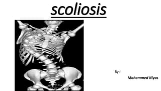
Scoliosis
- 2. Scoliosis • Scoliosis is a lateral (toward the side) curvature in the normally straight vertical line of the spine. • When viewed from the side, the spine should show a mild roundness in the upper back and shows a degree of swayback (inward curvature) in the lower back.
- 3. • Dextroscoliosis describes a spinal curve to the right ("dextro" = right). Usually occurring in the thoracic spine, this is the most common type of curve. It can occur on its own (forming a "C" shape) or with another curve bending the opposite way in the lower spine (forming an "S"). • Levoscoliosis describes a spinal curve to the left ("levo" = left). While common in the lumbar spine, the rare occurrence of levoscoliosis in the thoracic spine indicates a higher probability that the scoliosis may be secondary to a spinal cord tumor
- 4. Types of scoliosis: • Idiopathic • Most common type (65%) • Idiopathic scoliosis which has no definite cause. Idiopathic scoliosis is broken down by age group: • infant (0 to 3 years) • juvenile (4 to 10 years) • adolescent (11 to 18 years) • adult (18+ years)
- 5. Congenital: • Failure of formation - This may be either partial (wedge vertebra) or complete (hemivertebra) • Top left: anterior central defect. • Top right: incarcerated hemivertebra. • Bottom left to right: free hemivertebra, wedge vertebra, and multiple hemivertebrae.
- 6. • Failure of segmentation - This may be either unilateral (unilateral unsegmented bar) or bilateral (block vertebra) • Left: block vertebra. • Right: unilateral unsegmented bar.
- 7. • Mixed (see the third image below) - This type includes elements of both failure of formation and failure of segmentation • Mixed vertebral deformity involving the thoracolumbar spine.
- 8. Neuromuscular • The neuropathic conditions subdivided into those with upper and lower motor neuron lesions. • The upper motor neuron lesions includes diseases such as cerebral palsy, syringomyelia, and spinal cord trauma; • The lower motor neuron lesions includes poliomyelitis and spinal muscular atrophy. • The myopathic conditions • Arthrogryposis, muscular dystrophy, and other forms of myopathy.
- 9. Degenerative scoliosis. • This may result from traumatic (from an injury or illness) bone collapse, previous major back surgery, osteoporosis (thinning of the bones).
- 10. Signs and symptoms • Diminishing lung capacity, • Pressure exerted on the heart, • Restricted physical activities, • Uneven musculature on one side of the spine • A rib prominence or a prominent shoulder blade, caused by rotation of the ribcage in thoracic scoliosis • Uneven hips, arms or leg lengths • Slow nerve action
- 11. Physical examination • For the examination, thepatient should be undressed to the waist or wear abathing suit and a routine should be followed. • Theshoulders and iliac crest are inspected to determinewhether they are at the same level. • The scapulae,ribcage and flanks are then observed for symmetry. • The spinous processes are palpated to determine their alignment. • Rib hump or abnormal paraspinal muscular prominence indicates spinal rotation. • Rib hump leads to asymmetry of the trunk and is called angle trunk rotation (ATR). It is measured by using a scoliometer. • The patient is then made to bend forward to see for the disappearance of the curve (Adam’s test).
- 12. • Imaging • X-ray: uses radiation to create a picture of the spine • MRI: uses radio and magnetic waves to get a detailed picture of bones and tissue surrounding them • CT scan: X-rays taken at a variety of angles to get a 3D picture of the skeleton • Bone scan: a solution that is radioactive is injected into your blood. It will be concentrated in area of increased circulation, making spinal abnormalities easier.
- 13. Treatment • Scoliosis treatment decisions are primarily based on two factors: • The skeletal maturity of the patient (or rather, how much more growth can be expected) • The degree of spinal curvature. • There are three main scoliosis treatment options for adolescents: • Observation • Back braces • Scoliosis surgery
- 14. Cobb method • The upper and lower vertebrae are identified. The upper end vertebra is the highest one whose superior border converges towards the concavity of the curve and the lower end vertebra is the one whose inferior border converges towards the concavity. • Intersecting perpendicular line from the superior surface of the superior end vertebrae and from the inferior surface of the inferior end vertebrae is drawn. • The angle of deviation of these perpendiculars from a straight line is the ‘angle of the curve’.
- 15. • Reisser’s sign: This is a classification of the ossification of the iliac epiphysis, which usually starts from the anterior superior iliac spine and progresses posteriorly towards the posterior iliac spine. • Reisser’s stage 4 corresponds with cessation of spine growth and stage 5 correlates with cessation of height increase. Reisser’s classification It uses ossification of iliac apophysis to grade the remaining skeletal growth. The ossification progresses from lateral to medial: Type I — Ossification of lateral 25 percent Type II — Ossification of lateral 50 percent Type III — Ossification of lateral 75 percent Type IV— Ossification of lateral 100 percent Type V — Fusion of ilium
- 16. Braces • Made of polypropylene. • Contoured to size and shape of body. • Curved to oppose specific points of scoliosis curvature. • Flexible and comfortable. • Worn under clothing. • Nighttime/daytime use.
- 17. Bracing • Duration and time in brace • 23 hours per day • Wear until skeletally mature • Types • Milwaukee • Underarm orthosis • Electrical stimulation
- 18. Surgery • Failed bracing • Curves >45 degrees • Unbalanced curves >40 degrees • Surgery is fusion with instrumentation
- 19. Surgical Options: • Infantile and juvenile scoliosis: <8 yrs- instrumentation without fusion. After 8 years- anterior and posterior spinal fusion. After 11 years- posterior spinal fusion. • Adolescent scoliosis: Posterior spinal fusion with instrumentation. Anterior spinal fusion if younger than 11 years and with open triradiate cartilage.
- 20. References: • http://www.webmd.com/osteoarthritis/guide/arthritis- scoliosis?page=2 • http://www.spine-health.com/conditions/scoliosis/scoliosis- treatment • http://www.healthline.com/symptom/scoliosis • https://www.uichildrens.org/treating-scoliosis