6. Content of CS
Artery inside CS
The internal carotid artery enters the
sinus from its base, runs forward and
superiorly and then exits at the superior
wall of the sinus.
Nerve related to CS
CNs: III, IV, V1,V2, VI, Sympathetic
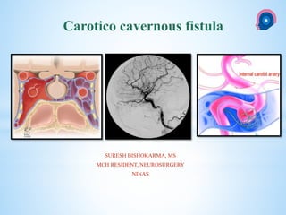
7. * The caroticocavernous fistula is a specific type of dural
arteriovenousfistula characterized by abnormal arteriovenous shunting
within the cavernous sinus.
* A caroticocavernous fistula results in high-pressure arterial blood entering
the low-pressure venous cavernous sinus.
* This interferes with normal venous drainage patterns and compromises
blood flow within the cavernous sinus and the orbit.
INTRODUCTION
8. • Caroticocavernous fistulas represent approximately 12% of all dural
arteriovenous fistulas.
• Direct CCFs are often secondary to trauma: head trauma: Youngs:
• Presentation: acute/rapid.
• indirect CCFs : Post menopause: insidious.
Epidemiology
9. • Two main types:
1. Direct
2. Indirect
CLASSIFICATION
10. CLASSIFICATION
• Another method is to classify according to four main types:
• Type A
• Type B
• Type C
• Type D
Barrow's Classification of CCF s.
11. 1. Type A: direct connection between the intracavernous ICA and CS
2. Type B: dural shunt between intracavernous branches of the ICA
and CS
3. Type C: dural shunts between meningeal branches of the ECA and
CS
4. Type D: B + C
CLASSIFICATION
12. • Traumatic or spontaneous fistulas.
• Flow:
• Direct high flow
• Indirect low flow fistula
OTHER CLASSIFICATION
14. Direct: type A: ICACS
Indirect: Br of ICA/ECS CS; types B, C, D
The most frequent among indirect is type C, with meningeal branches of
the ECA forming the fistula.
Pathophysiology
Direct CCF:
• Trauma
• Ruptured intracavernous
carotid aneurysms
• Collagen deficiency
syndromes arterial
dissection
• Fibromuscular dysplasia
• Direct surgical trauma
Indirect CCF:
Cause often unknown:
• Pregnancy
• Sinusitis
• Trauma
• Surgical procedures
• Cavernous sinus thrombosis
They are postulated to occur
secondary to cavernous sinus
thrombosis with revascularisation
15. • Their symptoms range from benign to extremely severe ophthalmologic
or neurologic complications.
• Clinical presentation is consequence of the elevated intracavernous
pressure.
• In direct, high-flow CCF ́s, symptoms appear suddenly.
• Symptoms caused by CCFs are related to their size, duration, location,
adequacy and route of venous drainage, and presence of arterial and
venous collaterals
Clinical presentation
17. • Moreover, other factors like dominant pattern of venous drainage the size
and location of CCF and the presence of collateral vessels (arterial or
venous) are important in this setting.
• Diplopia, pain, cephalic bruit, ophtalmoplegia, visual loss (Ophth. vein)
• Intracranial haemorrhage : sphenoparietal sinus and deep middle cerebral
vein)
• External haemorrhage: Otorrhagia, epistaxis (Pterygoid plexus)
18. CT
• Proptosis Enlarged superior ophthalmic veins
• Extraocular muscles may be enlarged
• Orbital oedema
• May show SAH/ICH from a ruptured cortical vein
Angiography (DSA)
• Rapid shunting from ICA to CS
• Enlarged draining veins
• Retrograde flow from CS, most commonly into the ophthalmic veins
Ultrasound
• Arterialised ophthalmic veins may be seen on Doppler study
Radiographic features
21. DSA
a. Digital angiogram of carotid circulation confirming carotid-cavernous fistula
b. Digital angiogram of vertebral circulation showing right ophthalmic vein
ingurgitated.
c. Digital angiogram with final image after treatment of the traumatic CCF
22. • Treatment and prognosis
• The natural history of CCF is highly varied, ranging from spontaneous
closure to rapidly progressive symptoms.
• Poor treatment outcome indicators include feeding vessel aneurysms
(indirect CCF) and retrograde filling of cortical veins (increased risk of
haemorrhage).
• Direct fistulas have a relatively high spontaneous rate of haemorrhage
(8.4%).
• subarachnoid, intracerebral or external haemorrhage (epistaxis, or
otorrhagia).
• Subconjunctival haemorrhage is also common but does not carry the
same poor prognosis
23. • Direct CCF: Occlude the tear between ICAand CS , preserving the
patency of ICA
• Indirect CCF : Interrupt fistulous communications/reduce CS pressure
GOAL OF TREATMENT
25. • Contralateral hand: 10sec: 4-6/hr: Reduces AV shunting + Increase outlet
venous pressures Thrombosis.
• Most useful in the treatment of indirect fistulas resulting in spontaneous
closure in up to 30% of cases.
Carotid compression therapy
26. Options:
• Ligation of the CC
• Surgical trapping of the fistula, and
• Surgical transvenous packing.
Both direct and indirect CCFs:
Disadv: Cranial nerve deficits and residual fistulous communications.
Indications for surgical repair include
1. Compromised proximal arterial access that prevents endovascular
repair or causes it to fail.
2. Salvage:failed endovascular treatments.
Surgery
27. • Arterial sacrifice may be required as a life-saving emergency treatment
• Indication: Difficult case:
• Extensive traumatic vessel wall damage
• Active hemorrhage or
• A rapidly expanding hematoma of the soft tissues
PARENT ARTERY OCCLUSION
28. • TOC: Symptomatic direct CCF.
• If not possible, detachable coils may be use
• Both arterial and venous access (including superior ophthalmic vein)
• Indirect fistulas typically require a combined transarterial (closure of
feeders) and transvenous (closure of cavernous sinus) approach.
• Indirect types are more difficult to treat and have a higher rate of
spontaneous closure
Transarterial balloon embolisation
29. • This procedure requires that the CS must be large enough to put the
balloon for embolization and the size of fistula must be smaller than the
inflated balloon, but large enough to allow a deflated balloon.
• The balloon has the advantage of being able to be flow-directed through
the fistula and CS, and must be inflated to a volume larger than the fistula
orifice to prevent its retrograde migration into ICA.
• Angiography is repeated to ensure closure of the fistula and patency of
the ICA.
Balloon Occlusion
30. • Mainstay of treatment in high-flow direct CCF ́s.
• It's an alternative when residual AV shunt remains in dural CCF.
• Embolization can be made with detachable platinum coils and liquid
embolic agents (n-butyl cyanoacrylate, ethylene-vinyl alcohol
copolymer);
• Coils are preferred because of their reliable and controlled deployment
into CS.
• Complications of this procedure includes thromboembolus and ICA
dissection
Transarterial embolization
31. • Recent Advance: poly flurotetraethylene-covered stents
• Traumatic arterial damage
• immediate obliteration of a direct CCF, while preserving ICA patency
• Disadv:
• Longitudinal flexibility: difficult navigation: tortuosity of the
intracranial vasculature.
• Vasospasms: Intra-arterial nimodipine and papaverine infusion
• Endoleak, coverage of vital perforators, dissection and rupture
Covered stent graft placement
32. • Is the current method of choice in treatment of indirect CCF’s.
• The goal of this technique is to catheterize the abnormal CS
superselectively and occlude the fistula without re-routing venous
drainage to cortical structures..
• Several routes: Most: inferior petrosal sinus (IPS
Transvenous embolization
33. • Indirect CCFs.
• Gamma knife radiosurgery can be used either alone or as an adjunct
therapy before/after endovascular intervention.
• Preliminary data : safe and effective alternative treatment
• Drawback: 22-mo average lag
RADIOSURGERY
34. Fistulous point located at left CS, with ICA supply by meningo-hipofisary trunks (red arrow) and ECA supply
by middle meningeal artery (blue arrow)and clivus branches from ascendent pharyngeal artery. Venous drainage
to superior ophtalmic vein (yellow arrow) and to inferior petrous sinus.
36. Thank you
NATIONAL INSTITUTE OF NEUROLOGICAL AND ALLIED SCIENCES, BANSBARI, KATHMANDU
NATIONAL INSTITUTE OF NEUROLOGICAL AND ALLIED SCIENCES, BANSBARI, KATHMANDU
CAROTICO CAVERNOUS FISTULA