SlideShare a Scribd company logo
1 of 19
Download to read offline
An Overview on Mechanical, Medicinal, Physical,
and Physiological Spinal Applications
BME 167
Introduction to Engineering Biomechanics
Spring 2016
Mrudula Vemuri
Thuy O
Miles Quaife
1
Table of Contents
Abstract . . . . . . . . . . . . . . . . . . . . . . . . . . . . . . . . . . . . . . . . . . . . . . . . . . . . . . . . . 2
1.0 Introduction . . . . . . . . . . . . . . . . . . . . . . . . . . . . . . . . . . . . . . . . . . . . . . . . . . . 3
2.0 Composition and Structure . . . . . . . . . . . . . . . . . . . . . . . . . . . . . . . . . . . . . . . 3
3.0 Constitutive Behavior. . . . . . . . . . . . . . . . . . . . . . . . . . . . . . . . . . . . . . . . . . . . 9
4.0 Mechanical Characteristics . . . . . . . . . . . . . . . . . . . . . . . . . . . . . . . . . . . . . . . 10
5.0 Physiological Function and Loading Conditions . . . . . . . . . . . . . . . . . . . . . . 13
6.0 Disease and Medical Condition. . . . . . . . . . . . . . . . . . . . . . . . . . . . . . . . . . . . 14
7.0 Treatment Options . . . . . . . . . . . . . . . . . . . . . . . . . . . . . . . . . . . . . . . . . . . . 15
8.0 Conclusion . . . . . . . . . . . . . . . . . . . . . . . . . . . . . . . . . . . . . . . . . . . . . . . . . . . 16
9.0 References . . . . . . . . . . . . . . . . . . . . . . . . . . . . . . . . . . . . . . . . . . . . . . . . . . . 16
10.0 Images . . . . . . . . . . . . . . . . . . . . . . . . . . . . . . . . . . . . . . . . . . . . . . . . . . . . . 18
11.0 Tables . . . . . . . . . . . . . . . . . . . . . . . . . . . . . . . . . . . . . . . . . . . . . . . . . . . . . . 19
12.0 Equations . . . . . . . . . . . . . . . . . . . . . . . . . . . . . . . . . . . . . . . . . . . . . . . . . . 19
13.0 Acknowledgements . . . . . . . . . . . . . . . . . . . . . . . . . . . . . . . . . . . . . . . . . . . 19
2
Abstract
The spinal column is of great interest under many areas of study, specifically vertical
compressional load capacity, physiological structure, decrease in vertebral disc height due to
time subjected fatigue, as well as many other interesting physical strains and stress adaptations.
The spine has the ability to resist work done on it though many different angles, and is composed
of materials such that allow it to compress many times cyclically without failing, as long as the
load is such that plastic deformation is not observed inside the spinal column and surrounding
tissues. The adult spinal column is made of approximately 24 vertebral bones that have the
ability to act together as a single ball and joint socket, yet still contain the main connecting
pathways between the brain with surrounding tissues, and allow simple flexion and extension
without impinging on these signal pathways. The spine is important in the functioning capacity
of the human body and how any form of injury can be largely incapacitating even with a small
amount of experienced trauma. The spine acts a protective housing for the nerves traversed
through it, it also is thoroughly intertwined with nerve endings, various tendons and muscles
adding support, which further increase the complexity of the body’s dependence on this
structure. In a body’s free standing nature, the spine acts as a column that supports the weight of
the upper torso, as well as stabilizes any planar movement generated by the body while serving
as an anchoring point for a majority of muscles that create great work movements. The
summation of these interactions and our ability as engineers to model, predict, and prevent
debilitating injuries. An immediate application is the medical condition involving the spine is
scoliosis, which is a musculoskeletal disorder that involves a sideways curvature of the spine.
The majority of scoliosis cases has unknown causes. Patients with scoliosis experience the same
axial compression on their curved spines without balanced support from the musculoskeletal
system. The lack of balanced support causes an asymmetric loading of the vertebral axis, which
results in the development and progression of spinal deformity. Many treatments are available
for patients with scoliosis, including exercised-based physical therapy, brace treatment, surgery,
and drug therapy.
3
1.0 Introduction
The spine plays a central role in the human body. Holding up the upper body and
allowing for a wide range of motion. The spine has several important functions in the body. It
provides structural integrity to the body and houses the spinal cord. The spinal cord relays
information between the brain and the rest of the body. Injury to this particular structure can be
devastating and result in a total lack of movement. Understanding how each component of the
spine works will create a better understanding of the spine and therefore allow for faster and
more complete recoveries from spinal injuries.
2.0 Composition and Structure
The spinal column is the main load bearing assembly in living vertebrate organism that
physically disperses superior loads to the inferior physiological assemblies. It also acts as a
protecting column to the central nervous system axon passageways interconnecting the brain
with the peripheral nervous system and the rest of the body. These tissues consist of composite
blends of various cell types, constituting the summation of functions producing organ systems
such as vertebral bone, intervertebral discs and connecting ligaments, and other connective
tissues such as cartilaginous structures and substances. The spinal column bones are individually
termed as “vertebrae,” which consist of the general molecular makeup as that of most load
bearing bones and are numbered both by cranial to sacral direction and region of spine.
Bones are formed from mesenchymal stem cells that are clustered together and begin
formation in either one of two ways. Of interest in spinal formation, is the “Endochondral
Ossification” method, in which these mesenchymal stem cells are crowded together in the
general pattern of the bone in which they are going to form, and then differentiate into
chondroblasts, which secrete cartilage matrix around the clump of now differentiated
chondrocytes, creating a cartilage model of what will become bones covered in hyaline cartilage,
which is totally surrounded by a membrane called the perichondrium. As this model grows, the
chondroblasts are further buried interiorly to the perichondrium, being called chondrocytes. This
model continues to grow until chondrocytes in the middle of the bone burst, causing
calcifications by an increase in pH. This calcification starts on the surface and proceeds inward.
At this time, a nutrient artery pierces the superficial perichondrium protective surface and the
calcifying cartilage model which then feeds and stimulates osteogenic cells in the model to
differentiate into osteoblasts. Various differences occur depending on specific bone function, but
vertebrae follow this model to varying degrees with time in each stage regulated by vertebra size
(Tortora, 2005). Figure 1 below shows the variations in vertebrae regions. Listed are: the atlas
and axis which give direct mobility to the head as special cervical vertebra, which give mobility
to the neck; the thoracic vertebrae which give limited mobility and maximum stability to the
upper torso, and the lumbar vertebrae (lower torso region), which have the largest area per
vertebra, which contributes to supporting the greatest pressures applied to the spine.
4
Figure 1. Vertebral Regional Representation [1]
The vertebral composition, in its final state, is a composite porous and spiny structure that
is both rigid in nature as well as saturated with a gel like substance with an entire superficial
hyaline cartilage covering. The gel like substance contributes to the structural ability to
elastically deform to a certain extent without permanent plastic deformation. The porous
structure and the gel like substances are classified as either organic matrix or inorganic matrix.
Bone cells are part of the organic matrix and once the method of ossification is complete, are
osteoblasts, osteocytes, and osteoclasts, Osteoblasts build up bone tissue by secreting the matrix
tissue of bone, being calcium phosphate salts). Osteocytes are osteoblasts fully surrounded in
bone matrix and lie within the chambers (lacunae) of formed matrix. Osteoclasts break down
bone tissue by breaking down the matrix tissue of bone which is necessary for maintaining
proper blood levels of calcium. The rest of the organic matrix is a mixture of collagen fibers and
a gel-like osteoid tissue. This osteoid tissue primarily contains proteoglycans, which are proteins
covalently attached to glycosaminoglycans, which are tissue organizing molecules that deal with
cellular regulations and activities, and are composed primarily of chondroitin sulfate molecules
and glucosamine. The high saturation ability of proteoglycans contributes to a high water
saturation level of 25% (Muscilino, 2005). The inorganic bone matrix is composed of
hydroxyapatite crystals (calcium phosphate crystalline salts) and is the structurally rigid aspect of
the spine. Figure 2 shows below standard bone scaffolding structure (inorganic tissue only) from
a low power scanning electron microscope, where the porous voids are the locations of the gel
like organic material. This image is taken from the third lumbar vertebra of a deceased 30 year
old woman. It is easy to see the interconnectedness of both the organic and inorganic tissue
matrices as the organic secretes the inorganic (Muscilino, 2005).
5
Figure 2. Electron Scanning Microscope 3rd Lumbar (x20) [2]
The vertebra structures are grouped by region and similar structure, which differ as load
and function change in the spine itself. Directly beneath the skull is the cervical spine, whose
general features are shown below in Figure 3. It has 7 vertebrae and are smaller in size than
either the thoracic or lumbar, as it supports the least amount of pressure. Special attention is
focused to the spinous process on the posterior side, as well as the transverse processes on either
side, where later discussed ligaments attach. These processes are the consistent attaching
locations for the spinal connective tissues. The vertebral foramen creates nerve bundle
passageway from the brain to the peripheral nervous system. The articular facet joints are the
locations where the inferior vertebral spiny process rest, on top of fibrocartilage and held in place
by more later discussed ligaments. Differing in the first 2 cervical spine vertebra, are both
structure and functions. Atlas, is the title for C1 and is named after the mythological god who
supported the world on his shoulders. It is a ring of bone with anterior/posterior arches, and does
not have a body with spinous processes. Directly beneath is C2, also known as the axis, which
does have a body. The noticeable differences are the dens, which is a spike-like projection out of
the superior face of the vertebrae. The dens is a pivoting point for which the head rests and
pivots on. The axis and atlas are both shown in illustrative form below in Figure 4.
Figure 3. Typical Cervical Spine Vertebra [3]
6
Figure 4. Illustrative Figure of Atlas and Axis Cervical Vertebra [4]
The thoracic vertebrae are directly inferior to C7, and consist of 12 vertebrae bones that
are larger than the superior cervical vertebra. As the distance increases vertically from the head,
pressure increases, relating to an obvious increase in pressure on the spine and associated joints,
requiring an increase in structural performance. The thoracic vertebrae also have notches in the
posterior side where the ribs connect via the vertebrocostal joints, as seen below in Figure 5.
These vertebrocostal joints limit the mobility of the thoracic region (Tortora, 2005).
Figure 5. Thoracic Vertebra [5]
Inferior to the thoracic region are the lumbar vertebrae, which are the largest and
strongest vertebrae. The projections of these vertebrae are short and thick, with associated
articular processes directed medially. The Spinous processes are thick and broad, and project
almost perfectly posteriorly. Because of their size, they are quite suited for the attachment of the
large back muscles (Tortora, 2005), as shown below in Figure 6.
7
Figure 6. Common Lumbar Vertebra [5]
The most inferior portions of the spine are the Sacrum and the Coccyx. In children, these
vertebrae are not fused together, yet begin to fuse by the age 16, and are generally completely
fused by the age of 30. The sacrum is triangular in shape located medial to either hip bone, and
serves as a strong anchoring place for the pelvic girdle. Females have a shorter sacrum and is
more curved than their male counterpart. The sacral canal in the sacrum is the continuance of the
vertebral canal, and is shown below along with the coccyx in Figure 7 (Tortora, 2005).
Figure 7. Sacrum and Coccyx Spinal Vertebrae [5]
The coccyx is the most inferior portion of the spine and is formed like the sacrum, by the
fusion of vertebrae between the ages 20 and 30 years old. The superior face of the coccyx 1 is
connected to the sacrum by 2 ligaments, and articulates superiorly with the sacrum.
Generally, the structure and function of each vertebra is similar to that of the one above
and below it. Slight differences obviously occur as the pressure load increases, or the places for
the ribs to attach. However, the structure of the spine dictates its major functions, being to resist
vertical deforming forces, and protect the central nervous systems axon pathways.
In addition to the vertebral aspect of the spine, are associated connective and stabilizing
tissues, as shown below in the animated Figure 8. A total of 9 ligament types connect the stacked
8
vertebrae in order to limit excessive flexion or extension. Ligaments are classified as short bands
of tough fibrous connective tissues that connect bone to bone, cartilage to cartilage, or hold
together a joint structure. On the anterior side is the “Anterior longitudinal ligament (1),” which
is a three layer ligament which covers both vertebral bone bodies and intervertebral discs. The
superficial layer covers 3-4 vertebrae, the intermediate secondary layer covers 2-3 vertebrae, and
the deepest layer only the anterior face of one vertebra. The posterior side has a similar ligament,
known as the “Posterior longitudinal ligament (2)” and is also shown below on the posterior side
of the vertebral canal. The Posterior longitudinal ligament is thicker in the thoracic region, yet
broader in the cervical and lumbar regions. Other ligaments, as shown below, are the facet
capsulary ligaments (3), which connect the spiny process on the posterior side of the vertebrae
and function so stabilize the facet joints and limit the extreme motions of the spine except
extension and inferior translation/compression; the supraspinous ligament (4), which connects
the posterior sides of the spiny processes of the vertebrae and limits flexion on the spinal joints;
the annulus fibrosus (laminated layers of fibrocartilage) of the disc joint (5), which stabilizes
each disc joint and limits the motion extremities of all spinal motions; 2 ligamentum flava (6) are
located on the either sagittal side of the spinal column, which also contribute to the limiting
flexion of the spine, the interspinous ligaments (7) which are short separate ligaments that are
interconnected between adjacent spiny processes, which also limit the flexion of spinal joints; the
intertransverse ligaments which are located between adjacent processes of the vertebrae whose
function is to limit contralateral-lateral (opposite sided) flexion of the spinal joints, and finally
the nuchal ligament found from the spinous process on cervical vertebra 7 (C7) to the external
occipital protuberance of the skull (Summarized from Muscilino, 2005).
Generally, the structure and function of each vertebra is similar to that of the one above
and below it. Slight differences obviously occur as the pressure load increases, or the differences
pertaining to places for the ribs or connective tissues to attach. However, the structure of the
spine dictates its major functions, being to resist vertical deforming forces, and protect the
central nervous systems axon pathways. With that as focus, it is easy to see why there are so
many similarities across these 26 vertebral bones and associated connective tissues.
Figure 8. Animated Image of Spinal Connective Tissue [5]
3.0 Constitutive Behavior
Constitutive Laws rate the stress and strain fields. These laws consist of a set of
mathematical idealizations based on observed behavior and they can be used to characterize
9
mechanical behavior. Constitutive equations or relations can be established specifically for a
material to describe its response to external stimuli such as applied forces or fields. These
equations can be used to calculate the material’s behavior in fluid mechanics, solid state physics,
structural analysis, or deformation problems. Some general equations used to determine
constitutive behavior include general stress or pressure, strain, elastic modulus, Young’s
modulus, shear modulus, and compressibility. The table below summarizes these equations.
Table #1: Equations used to Characterize the Constitutive Behavior of a Material. [1]
Name Symbol Defining Equation
Stress
Strain
Elastic Modulus
Young’s Modulus Y
Shear Modulus G
Bulk Modulus B
Compressibility C
The deformation of solids is defined by the constitutive behavior of the material. Outside
stimuli such as collisions, solid-state deformations, stress strain, and friction all contribute to
deformation.
The constitutive behavior of the spine can be broken up into the constitutive behavior of
the vertebrae and the soft tissue surrounding the vertebrae. The bone elements in the spine are
assumed to have a constitutive behavior of an elastic and perfectly plastic material. The elastic
constant is transversely isotropic while the yield strength is isotropic (Silvia, 1998). To model the
soft tissue of the spine, it needs to be broken up into two parts. The first part is the energy
contributions from the matrix and the second part will be the energy contributions from the fiber.
This break-up is important because of the fiber-matrix shear interaction determines the response
of the annulus fibrosis or soft tissue around the intervertebral disk (Guo, 1962).
According to Guo, the matrix is modeled as an incompressible neo-Hookean material
while the fiber is modeled as the same with the stiffness dependent on the stretch of the fiber. A
neo-Hookean model is a model for a hyperelastic material and is similar to Hooke’s law. This
model can predict the nonlinear stress-strain behavior of materials that are subject to large
deformations. The stress-strain curve of this model is not completely linear, but is instead linear
at the beginning with a plateau after a certain point.
The energy contributions of the fiber and the matrix can be quantified by mathematical
equations. Equation 1 below quantifies the energy contributions from the fibers.
Equation 1 [1]
10
The equation for the energy contribution of the shear interaction of the fiber-matrix is
defined by Equation 2 below.
Equation 2 [1]
These equations are derived from the neohookean model. This model uses a
multiplicative decomposition to split the deformation gradient into two parts. These two parts are
the uniaxial deformation in the direction of the fiber and the shear deformations. Extending
effective stiffness formula for linear elastic composites into a large deformation range will
provide an expression for strain energy function for the model (Guo, 1962). This model has
limitations and areas for further study to make a more accurate model. It assumes that the soft
tissue can be modeled similarly to a composite material. The mechanical properties however are
different and to appropriately model the tissue in this way, a reference for the mechanical
properties of the tissues must be investigated. This will allow a more accurate strain energy
function to be developed.
4.0 Mechanical Characteristics
4.1 The Spine as a Single Unit
Due to the total summation of all tissues utilized in the contribution of movement and
load stabilization, the spinal column has been studied in light of several functional units. First
considered, is the spinal column as a whole functional unit. Secondly is the functional spinal
unit, which is an analysis of two vertebrae succinctly joined together by two joints and 9
connecting ligaments. Lastly is the analysis on the vertebra itself, as a single functional unit
which is a hard, porous, and gel filled composite material.
In its totality, the spine has the ability to act as a mixture between a spring or shock
absorber, and a static truss through load dispersal. When analyzing this model on an adult
species from the lateral point of view, it is easy to see curves in the thoracic, cervical, lumbar,
and sacrococcygeal regions. When viewed from the posterior side, the spine should be as straight
as possible. As shown below in Figure 9 below, the primary curves of the spine are the thoracic
and sacrococcygeal curves which are described as kyphotic. This means they are curved concave
anteriorly, and curved convex posteriorly. Considered as secondary curves, the spine also curves
in the cervical and lumbar regions, described as lordotic (curved concavely posteriorly and
convex anteriorly) (Muscolino, 2005).
11
Figure 9. Lateral View Spinal Curvature [5]
When analyzing the spine as a single unit, it is easy to imagine the types of forces that
are endured during various aspects of loading. It is also easy to the ability of the spine to use its
ability to compress without deforming to a certain maximum, as well as withstand tangential
forces that may cause shearing resistance. Mechanically, it has also been observed that spinal
stability, although greatly assisted by connective ligament tissues, an even greater amount of
stability is used from musculature connecting to the spine, and inserting at other areas (Oxland,
2015). Future developments are seeking to ascertain the true meaning of a newly developed term
being “sagittal balance,” as well as further understand predetermined ideas and observations
through mathematical model developments. This term newly designates an idea of the ability of
the spine to be sagittally imbalanced, which is giving rise to various angles of shift for the pelvis
relative the spinal column in what could be considered a physical equilibrium. Increases in
angular deviations from observed equilibrium values cause increased intervertebral pressures, as
well as degradation and material fatigue (Melnyk, 2016).
The spine in its totality functions as a single ball and joint socket (Ozkaya, 2012). With
this mechanical analogy, the spine has 3 degrees of freedom, limited only in range onset
shortening by extended time spans of decreased motion (Jaumard, N. V., 2011). Because of this
structure, the spine has an associated degrees of motion. These are: 135 Degrees flexion, 120
degrees extension, 90 degrees right lateral flexion, 90 degrees left lateral flexion, 120 degrees
right rotation, and 120 degrees left rotation. Obviously an impingement or injury to the spine
decreases these values. Also, when analyzing the different segments of the spine, the associated
range of motion, greatly diminishes. It is only in the spines entirety that it has such free range of
motion.
4.2 Vertebrae
12
The spiny nature of the vertebra contribute to movement and stability of the vertebral
column, and are analyzed in what is considered the functional spinal unit (FSU) (Oxland, 2015).
Each vertebra has 3 vertically connecting joints, with two being facet joints between the spiny
processes on the posterior side connected between the spiny processes in two vertically adjacent
vertebra. The major joint is the inferior body face connected to superior body face of the vertebra
in column through the intervertebral disc, separated by the intervertebral discs. However, its
molecular structures also contribute an anisotropic characteristic by type of osteoblast secretion.
Internally, softer sponge bone is surrounded by compact bone which is harder. Based on the
molecular order of these cells, applied directional loads may be withstood in greater numbers in
some directions relative to others (Bankoff, 2012).
Loading the FSU in various directions will generate reaction forces that can either resist
the load without creating injury to the spine, or create a deforming load in which some aspect of
shearing/straining is observed intervertebrally in either the vertebral unit, or some connective
ligament. The FSU couples all three joints into its possible achieved motions for flexion and
extension (summarized and images taken from Muscilino, 2005).
The FSU also allows flexion and extension within the sagittal plane around the
mediolateral axis, which is a midline axis of separation between vertebras, as shown below. Left
and right rotation are also allowed within the transverse plane of vertebra and the one directly
inferior to it. Finally, translational motions are also allowed, as long as shearing or strain is not
observed.
The limiting factor in these motions are the elastic ability of the intervertebral disc and
supporting ligaments to accept the applied forces causing the movement without straining
(Schmidt, 2016). Under laboratory experiment, it was observed that the median critical shearing
force in the posterior-anterior direction was 2877 Newtons (an equivalent mass of 293.27 kg
under the influence of gravitational acceleration), and lateral side to lateral side of 3199 Newtons
(an equivalent mass of 326.1 kg under the influence of gravitational acceleration) (Schmidt,
2016). Compressive failures occur typically at the vertebral endplate by the high pressure of the
intervertebral disc nucleus (nucleus pulpous) (Bankoff, 2012).
4.3 Individual Vertebral Bone
For final considerations, is the individual vertebral bone, or simply, the vertebra. Typical
anatomical features are given in the above text, with composition being primarily cortical (rigid)
bone, and the inner matrix a region of cancellous (spongy) bone. The superficial characteristics,
such as the spiny processes, are anchoring points for the attachment of tendons of movement
producing musculature, and stabilizing ligaments to create the FSU. Mechanical loading
capabilities of the vertebra increase as bone size increase from superior to inferior placement in
the spine correlating with the increase in cross-sectional area (as pressure dependence dictates),
with the load applied to both the rigid outer shell and inner spongy tissue (Bankoff, 2012).
Mean failure loads (N) were measured and tabulated in analyzing axial compression, with
finding the posterior side of the vertebra were roughly 2.5 times stronger than most central
regions, with an . In analyzing postural loads, it is was found that flexed, unextended postures
result in an increased loading on the vertebral body, while cervically extended postures attribute
more loading to the process attachment points, called the neural arch. With advancing age, the
load passing through this arch increases, specifically after the age of 60. This is due largely to
decreasing the height of the intervertebral disk by a lifetime of repetitive cyclic loading
generating intervertebral disk fatigue (Adams, et al 2006). It was also shown that the increased
13
neural arch loading as age also increases, by the decrease in anterior loading, leads to a decrease
bone in vertebral density. This was thought to be a contributing factor to elderly intervertebral
fractures (Adams, et al 2006).
4.4 Supplementary Materials
Biological materials are very complex, not only their ability to self-replicate and self-
heal, but also their near perfect order with which they perform their function. Many tissue
materials type fatigue, are rejected by the body, or cause some type of reaction through their
lifetime. Bone represents a composite that is perfectly balanced between osteocytes and bone
matrix, and changing the composition of either can have drastic side effects on the functional
support of the body. If too spongy, rigid support for the body is not physically possible, and if
too brittle, bones do not have any elastic behavior and will deform at much lower pressures than
currently observed. Limiting factors in designing artificial bone, are finding materials that body
will not reject, as well as being able to do many work loading cycles without fatigue. Bone is
very porous which allows it to be a solid structure and handle compressible forces, yet still allow
blood vessels to penetrate to keep it regenerating itself and health. In the spine, because of
synovial fluid, and pressure, materials can both fatigue physically and chemically. For vertebral
replacement currently, truss/column like scaffolds are bridged into the spine as well as titanium
foam bars (Kubosch, 2016). These have variable success rates, and most experienced bony
integration to the implant (Lange, 2006).
5.0 Physiological Function and Loading Behavior
It is easy to imagine the importance of the spine. It is central to the body and all of its
movements. The spine provides structure and support to hold the body up while still being
flexible enough to allow the body to move. The spine does all of this while also performing the
invaluable task of housing and protecting a column of nerves that feed from the brain to the rest
of the body. This amazing structure is made up of smaller subsets called vertebrae. These
vertebrae are individual bones that make up the building blocks of the spinal column. The
combined vertebrae create a hollow tube that allows the spinal cord to pass through. Each
vertebrae has a strong and hard outer bone called the cortical bone and a spongy inner bone
called the cancellous bone. Intervertebral disks serve as shock absorbers between each vertebra
of the spine. There is a disk between each vertebrae and each disk has an outer ring of fibers
called the annulus fibers. Annulus fibers are the strongest area of the disk and helps keep the
center of the disk intact and connects the vertebrae.
Different activities result in different loading conditions load the spine differently.
Examples of activities that change the loading of the spine include running and weightlifting.
The spine is subject to many different kinds of loading in any activity. These types of loading
include loading by gravity, by change in motion, muscle activity, and external forces or work.
Exercising will usually involve all of the above types of loading. The four elemental forces of the
spine are compression, tension, torque, and shear forces. These forces are defined as being any
external stimuli that causes a change in a free body or creates internal stress through attempted
deformation. The most perpetual, constant, and understandable example of such a force is
gravity. Compression and tension forces act along the axis of the spine. Compressive forces
flatten the disks and vertebrae of the spine. Tension forces act to elongate those spinal
components. Large loads may result in spinal shrinkage. Spinal shrinkage occurs when the
compressive load is larger than the interstitial osmotic pressure of the tissues. When the
14
interstitial osmotic pressure is exceeded, water is extruded from the disk wall and the disk height
reduces resulting in spinal shrinkage and loss of stature (Leatt, 1986). Compression and Tension
forces can both be found in simple movements such as a jump. When an individual begins a
jump, the spine experiences a compressive force. At the height of the jump when all body leaves
the ground, the compressive force becomes a tension force. The momentum of the jump pulls the
spine up while the gravitational forces pull it down.
Shear forces are forces applied perpendicular to the spinal axis. Shear forces in the spine
are associated with spinal injury. They are not desirable because they work to slide the spinal
components away from their natural axis. Shear forces contribute to the increase of internal
stress in the body. Large shear forces lead to ligament and disk tears as well as vertebral
fractures.
Torsional forces are also applied perpendicular to the spinal axis. Unlike shear forces,
however, they are not in the plane of the spine. They exist, instead as a rotational force.
Torsional forces rotate the components of the spine along its natural axis. Another name for this
force is torque. A wide range of torsional forces fall within the normal use of the spine, however,
excess torsional force may cause disk and ligament tears or torsional vertebrae fractures.
6.0 Disease and Medical Condition
A medical condition that involves the spine is scoliosis, which is a musculoskeletal
disorder that involves a sideways curvature of the spine. In the majority of cases, scoliosis is
under mild condition, and treatment is not required. The natural curve of the spine allows
individuals without scoliosis to balance, move, and walk normally. Individuals with scoliosis
have vertebrae that rotate, resulting in the sideway curvature of the spine. The medical condition
usually develops rapidly over time, commonly affecting the thoracic spine or lumbar spine. The
cervical spine is rarely affected. In several cases, there is a second curvable of the spine in the
opposite direction, resulting in a S-shaped spine.
Kalichman et al. (2016) stated that several different types of scoliosis exist with
adolescent idiopathic scoliosis (AIS) being the most common. Anand et al. (2011) mentioned
that AIS is diagnosed when the exact cause for the spine curvature is unknown, and this
condition appears in more than 80 percent of diagnosed scoliosis cases. Congenital and
neuromuscular are two other types of scoliosis, and these types comprises from 5% to 7% of
scoliosis cases. Hawes et al. stated that about 20 percent of AIS patients has one or more family
members with scoliosis, indicating that an inherited factor may be one of the causes. Recent
investigations of the causes of scoliosis have been focused on structural elements of the spine,
collagenous structures, spinal musculature, central nervous system, endocrine system, and
genetics. However, currently, no mechanism is established as the causes of AIS.
Zavatsky et al. (2015) studied the influence of race and economic status on disease
severity and treatment in adolescent idiopathic scoliosis. Zavatsky et al. found that 1% to 2% of
the general population has AIS. All children, regardless of race and socioeconomic levels, have a
possibility of developing scoliosis. Scoliosis affects about 3 percent of children and adolescents
over 10 years old. Older children between the ages of 10 and 12 and adolescents are more prone
to develop AIS since the medical condition usually develops during a growth spurt. In the
majority of cases, the curvature of the spine is not severe, and treatment is not required. Both
boys and girls are as likely to develop scoliosis, but a higher number of treatments is needed for
girls. Children with a family history of scoliosis have a higher chance of developing the
condition. In addition, the condition occurs more frequently in children with other medical
15
problems, such as connective tissue disorders, neuromuscular diseases, and rheumatic diseases.
Black patients have a higher curve magnitude and a greater percentage of patients with curves in
the surgical range than white patients. Across all racial groups, no difference was observed in
age on presentation or treatment offered. White patients are less likely to choose surgery as their
initial treatment than black patients.
Kalichman et al. (2016) mentioned that many patients with AIS have a misalignment of
the body, a loss of balance and shrinking height, a leg and buttocks pain, and many hip, bladder
and bowel problems. When the spine is deformed, the shoulders, waist, and hips are also not
aligning properly since the bones in the spine had rotated. Patients with scoliosis have
diminished overall height because of the misalignment of the body. Leg and buttocks pain occurs
due to arthritic pain or nerve compression originated from the spine. Hip problems occur because
the ribs are more apparent on one side of the spine. Scoliosis can also causes a loss of control of
bladder and bowel function.
The pathological loading conditions are different from normal physiological loading
conditions. For instance, how the body response to physiologically relevant axial compression
differently among individuals with and without scoliosis. The spine experiences axial
compression due to the body weight in the upright position. Hawes et al. stated that the bony axis
of a normal spine can tolerate a weight of approximately more than 10 kilograms without
buckling. This bony axis depends on the balanced support from the musculoskeletal system,
which is controlled by the central nervous system. Patients with scoliosis experience the same
axial compression on their curved spines without balanced support from the musculoskeletal
system. The lack of balanced support causes an asymmetric loading of the vertebral axis, which
results in the development and progression of spinal deformity.
7.0 Treatment Options
Many treatments are available for patients with scoliosis, including exercised-based
physical therapy, brace treatment, surgery, and drug therapy. Treatment of scoliosis is depended
on the level of severity of the curvature of the spine and the possibility of the curve from
becoming worse. Kalichman et al. also mentioned that two conservative treatments of AIS are
exercised-based physical therapy and brace treatment. Current physical therapy for patients with
scoliosis is specific exercises that are unique for each individual patient. Specific exercises are
designed to reduce the effects of spinal deformity, including scoliosis specific exercises, the
Dobosiewicz technique, the schroth method, the side-shift program, and SEAS. Bracing
treatment, on the other hand, may reduce the need for surgery, restore the sagittal profile and
affect vertebral rotation. Braces are usually worn full-time for about two to four years until the
termination of bone growth. Cunin (2015) stated that another major treatment option is surgery,
and these surgical methods include growing rods, guided growth systems, and compression-
based systems. Growing rods place a distraction force to the spine, specifically the concavity of
the curve, to stabilize the curvature of the spine and the growth of the T1-S1 segment. Several
surgical procedures are required for growing rods treatment to lengthen the rod, to maintain the
result during the index surgery, and to follow spine growth. The rods can also be lengthened
noninvasively via a magnetic mechanism. Guided growth systems are surgical devices that are
placed along the vertebral column to maintain the spine in its reduced position while allowing
the spine to grow. Convexity compression devices place a compressive force on the convexity of
the spine curvature to restrict its growth. Hui et al. investigated the efficacy of a combined
Traditional Chinese Medicine (TCM) therapy, including acupotomology, Daoyin, and Tuina Hui
16
et al. (2015) found that TCM combined therapy has the ability to prevent the progression of
spinal deformity by targeting the muscular imbalance between spinal disorder and scoliosis
sides.
Kalichman et al. (2016) stated that the most effective conservative treatment for AIS is
bracing. Exercise-based physical therapy can prevent a worsening of the curvature of the spine,
which may reduce the need for bracing. Exercise-based physical therapy is also the only
treatment that improves respiratory function. However, limited evidence is available to specify
physical exercises that are effective treatment methods for AIS. The combination of bracing and
exercising increases treatment efficacy in comparison to treatment efficacy of a single treatment.
Cunin mentioned that out of all three surgical treatments, growing rods is the most common
since it provides better control over the Cobb angle during maturation. Current available
instrumentation for growth guidance systems is highly invasive and less common than growing
rods. Compared with other methods of treatment, TCM combined therapy accounts for patient
activeness, which does not affect daily life nor causes any adverse effects on physical and
psychological development.
8.0 Conclusion
The spinal column is the main load bearing assembly in living vertebrate organism. The
spine interconnects the brain with the peripheral nervous system and the rest of the body. The
spinal column bones are formed from mesenchymal stem cells that are clustered together and
begin formation. Constitutive laws rate the stress and strain fields and can be used to calculate
the material’s behavior in fluid mechanics, solid state physics, structural analysis, or deformation
problems. These general equations are used to determine constitutive behavior include general
stress or pressure, strain, elastic modulus, Young’s modulus, shear modulus, and compressibility.
The spine has the ability to act as a mixture between a spring or shock absorber, and a static truss
through load dispersal. The spine in its totality functions as a single ball and joint socket, which
has 3 degrees of freedom, limited only in range onset shortening by extended time spans of
decreased motion. Compressive forces flatten the disks and vertebrae of the spine whereas
tension forces act to elongate those spinal components. Shear forces are forces applied
perpendicular to the spinal axis whereas torsional forces are also applied perpendicular to the
spinal axis. A medical condition that involves the spine is scoliosis, with AIS being the most
common type. AIS is diagnosed when the exact cause for the spine curvature is unknown.
Patients with scoliosis experience the same axial compression on their curved spines without
balanced support from the musculoskeletal system. The lack of balanced support causes an
asymmetric loading of the vertebral axis, which results in the development and progression of
spinal deformity. Many treatments are available for patients with scoliosis, including exercised-
based physical therapy, brace treatment, surgery, and drug therapy.
17
9.0 References
1. Adams, M.A.,Pollintine,P.,Tobias,J.H.,Wakley,G.K.,Dolan,P.,(2006).Intervertebral disc
degeneration can predispose to anterior vertebral fractures in the thoracolumbar spine
.J.BoneMin.Res.21(9),1409–1416.
2. Anand, N., & Baron, E. M. (2011). Minimally invasive treatment of adult scoliosis.
Seminars in Spine Surgery,23(1), 66-71.
doi:http://dx.doi.org/10.1053/j.semss.2010.12.007
3. Bankoff, A. D. P. (2012). Biomechanical characteristics of the bone. INTECH Open
Access Publisher.
4. Cunin, V. (2015). Early-onset scoliosis – Current treatment.Orthopaedics &
Traumatology: Surgery & Research,101(1, Supplement), S109-S118.
doi:http://dx.doi.org/10.1016/j.otsr.2014.06.032
5. Guo, Z. Y., Peng, X. Q., & Moran, B. (2006). A composites-based hyperelastic
constitutive model for soft tissue with application to the human annulus fibrosus. Journal
of the Mechanics and Physics of Solids, 54(9), 1952-1971.
6. Hawes, M. C., & O’Brien, J. P. (2006). The transformation of spinal curvature into spinal
deformity: pathological processes and implications for treatment.Scoliosis, 1, 3.
http://doi.org/10.1186/1748-7161-1-3
7. Hui, W., Jinyuan, X., Zhong, J., Shuliang, Y., Hongquan, S., Xitao, N., . . . Honggen, D.
(2015). Effect of a traditional chinese medicine combined therapy on adolescent
idiopathic scoliosis: A randomized controlled trial. Journal of Traditional Chinese
Medicine, 35(5), 514-519. doi:http://dx.doi.org/10.1016/S0254-6272(15)30133-3
8. Jaumard, N. V., Welch, W. C., & Winkelstein, B. A. (2011). Spinal Facet Joint
Biomechanics and Mechanotransduction in Normal, Injury and Degenerative Conditions.
Journal of Biomechanical Engineering, 133(7), 71010–NaN.
http://doi.org/10.1115/1.4004493
9. Kalichman, L., Kendelker, L., & Bezalel, T. (2016). Bracing and exercise-based
treatment for idiopathic scoliosis. Journal of Bodywork and Movement Therapies, 20(1),
56-64. doi:http://dx.doi.org/10.1016/j.jbmt.2015.04.007
10. Kubosch, D., Kubosch, E. J., Gueorguiev, B., Zderic, I., Windolf, M., Izadpanah, K., …
Strohm, P. C. (2016). Biomechanical investigation of a minimally invasive posterior
spine stabilization system in comparison to the Universal Spinal System (USS). BMC
Musculoskeletal Disorders, 17, 134. http://doi.org/10.1186/s12891-016-0983-1
11. Leatt, P., Reilly, T., & Troup, J. G. (1986). Spinal loading during circuit weight-training
and running. British Journal of Sports Medicine, 20(3), 119-124.
12. Lange, U., Edeling, S., Knop, C., Bastian, L., Oeser, M., Krettek, C., & Blauth, M.
(2007). Anterior vertebral body replacement with a titanium implant of adjustable height:
a prospective clinical study. European Spine Journal, 16(2), 161–172.
http://doi.org/10.1007/s00586-005-0015-6
13. Melnyk, Angela, et al. "Characterization Of The Behavior Of A Novel Low-Stiffness
Posterior Spinal Implant Under Anterior Shear Loading On A Degenerative Spinal
Model." European Spine Journal 24.4 (2015): 775-782. Academic Search Complete.
Web. 10 Mar. 2016.
14. Muscolino, J. E. (2005). Kinesiology: The Skeletal System and Muscle Function. St.
Louis, Mo.: Mosby/Elsevier.
18
15. Oxland, Thomas R..(2015). Fundamental biomechanics of the spine—What we have
learned in the past 25 years and future directions Journal of Biomechanics , Volume 49 ,
Issue 6 , 817 - 832
16. Ö zkaya, N., & Ö zkaya, N. (2012). Fundamentals of biomechanics: Equilibrium, motion,
and deformation. New York, NY: Springer.
17. Schmidt, H., Häußler, K., Wilke, H. J., & Wolfram, U. (2015). Structural behavior of
human lumbar intervertebral disc under direct shear. Journal of applied biomaterials &
functional materials, 13(1).
18. Silva, M. J., Brodt, M. D., & Hucker, W. J. (2005). Finite element analysis of the mouse
tibia: Estimating endocortical strain during three‐point bending in SAMP6 osteoporotic
mice. The Anatomical Record Part A: Discoveries in Molecular, Cellular, and
Evolutionary Biology, 283(2), 380-390.
19. Tortora, G. J. (2005). Principles of Human Anatomy. New York: John Wiley & Sons.
20. Zavatsky, J. M., Peters, A. J., Nahvi, F. A., Bharucha, N. J., Trobisch, P. D., Kean, K. E.,
Lonner, B. S. (2015). Disease severity and treatment in adolescent idiopathic scoliosis:
The impact of race and economic status. The Spine Journal, 15(5), 939-943.
doi:http://dx.doi.org/10.1016/j.spinee.2013.06.043
10.0 Images
1. http://www.ectsoc.org/gallery/#myGallery-picture(2)
2. http://www.slideshare.net/hermizan84/35-cervical-vertebra
3. http://www.spineuniverse.com/conditions/back-pain/upper-back-pain/anatomy-upper-
back
4. http://emedicine.medscape.com/article/1899031-overview#a2
5. Tortora, G. J. (2005). Principles of Human Anatomy. New York: John Wiley & Sons
11.0 Tables
1. Ö zkaya, N., & Ö zkaya, N. (2012). Fundamentals of biomechanics: Equilibrium, motion,
and deformation. New York, NY: Springer.
12.0 Equations
1. Ö zkaya, N., & Ö zkaya, N. (2012). Fundamentals of biomechanics: Equilibrium, motion,
and deformation. New York, NY: Springer.
13.0 Acknowledgments
Thuy O: Disease and Medical Condition, Treatment Options, Conclusion
Mrudula V.: Introduction, Constitutive Behavior, Physiological Function and Loading
Conditions
Miles Q.: Abstract, Composition and Structure, Mechanical Characteristics, Personal References

More Related Content

What's hot

Dr. B Ch 05_lecture_presentation
Dr. B Ch 05_lecture_presentationDr. B Ch 05_lecture_presentation
Dr. B Ch 05_lecture_presentationTheSlaps
 
The musculoskeletal system SSA+S
The musculoskeletal system SSA+SThe musculoskeletal system SSA+S
The musculoskeletal system SSA+Scalderonscience
 
163 ch 06_lecture_presentation
163 ch 06_lecture_presentation163 ch 06_lecture_presentation
163 ch 06_lecture_presentationgwrandall
 
Im chapter20
Im chapter20Im chapter20
Im chapter20Franz Uy
 
Dr. Ali Selk Ghafari: Biomechanics of Musculoskeletal System: Components of t...
Dr. Ali Selk Ghafari: Biomechanics of Musculoskeletal System: Components of t...Dr. Ali Selk Ghafari: Biomechanics of Musculoskeletal System: Components of t...
Dr. Ali Selk Ghafari: Biomechanics of Musculoskeletal System: Components of t...a_selkghafari
 
Lecture 4 (bone growth and remodeling)
Lecture 4 (bone growth and remodeling)Lecture 4 (bone growth and remodeling)
Lecture 4 (bone growth and remodeling)Ayub Abdi
 
Musculoskeletal system
Musculoskeletal systemMusculoskeletal system
Musculoskeletal systemKerrie O'Bryan
 
Musculoskeletal System Lecture
Musculoskeletal System LectureMusculoskeletal System Lecture
Musculoskeletal System LectureJofred Martinez
 
Anatomy unit 5 skeletal and muscular systems bone types and composition
Anatomy unit 5 skeletal and muscular systems bone types and compositionAnatomy unit 5 skeletal and muscular systems bone types and composition
Anatomy unit 5 skeletal and muscular systems bone types and compositionrozeka01
 
Comparison Of Joints And Cartilage
Comparison Of  Joints And  CartilageComparison Of  Joints And  Cartilage
Comparison Of Joints And Cartilagejustinbieberlover
 
Skeletal System Notes
Skeletal System NotesSkeletal System Notes
Skeletal System Notesrachelkucera
 
Medical Surgical Nursing - Musculoskeletal Disorders
Medical Surgical Nursing - Musculoskeletal DisordersMedical Surgical Nursing - Musculoskeletal Disorders
Medical Surgical Nursing - Musculoskeletal DisordersMa Inna Paulina Palaña
 

What's hot (17)

Easy skeletalsystem
Easy skeletalsystemEasy skeletalsystem
Easy skeletalsystem
 
Dr. B Ch 05_lecture_presentation
Dr. B Ch 05_lecture_presentationDr. B Ch 05_lecture_presentation
Dr. B Ch 05_lecture_presentation
 
PMS 1 intro
PMS 1 introPMS 1 intro
PMS 1 intro
 
The musculoskeletal system SSA+S
The musculoskeletal system SSA+SThe musculoskeletal system SSA+S
The musculoskeletal system SSA+S
 
163 ch 06_lecture_presentation
163 ch 06_lecture_presentation163 ch 06_lecture_presentation
163 ch 06_lecture_presentation
 
Skeletal System
Skeletal SystemSkeletal System
Skeletal System
 
Im chapter20
Im chapter20Im chapter20
Im chapter20
 
Dr. Ali Selk Ghafari: Biomechanics of Musculoskeletal System: Components of t...
Dr. Ali Selk Ghafari: Biomechanics of Musculoskeletal System: Components of t...Dr. Ali Selk Ghafari: Biomechanics of Musculoskeletal System: Components of t...
Dr. Ali Selk Ghafari: Biomechanics of Musculoskeletal System: Components of t...
 
Anatomy skeletal muscular
Anatomy skeletal muscularAnatomy skeletal muscular
Anatomy skeletal muscular
 
Lecture 4 (bone growth and remodeling)
Lecture 4 (bone growth and remodeling)Lecture 4 (bone growth and remodeling)
Lecture 4 (bone growth and remodeling)
 
Musculoskeletal system
Musculoskeletal systemMusculoskeletal system
Musculoskeletal system
 
Musculoskeletal System Lecture
Musculoskeletal System LectureMusculoskeletal System Lecture
Musculoskeletal System Lecture
 
Anatomy unit 5 skeletal and muscular systems bone types and composition
Anatomy unit 5 skeletal and muscular systems bone types and compositionAnatomy unit 5 skeletal and muscular systems bone types and composition
Anatomy unit 5 skeletal and muscular systems bone types and composition
 
Comparison Of Joints And Cartilage
Comparison Of  Joints And  CartilageComparison Of  Joints And  Cartilage
Comparison Of Joints And Cartilage
 
Ch 5 skeletal system
Ch 5   skeletal systemCh 5   skeletal system
Ch 5 skeletal system
 
Skeletal System Notes
Skeletal System NotesSkeletal System Notes
Skeletal System Notes
 
Medical Surgical Nursing - Musculoskeletal Disorders
Medical Surgical Nursing - Musculoskeletal DisordersMedical Surgical Nursing - Musculoskeletal Disorders
Medical Surgical Nursing - Musculoskeletal Disorders
 

Viewers also liked

Vishal_SQL_DBA_2exp
Vishal_SQL_DBA_2expVishal_SQL_DBA_2exp
Vishal_SQL_DBA_2expvishal thapa
 
Presentation bearddrop eng 2
Presentation bearddrop eng 2Presentation bearddrop eng 2
Presentation bearddrop eng 2Bearddrop_nation
 
Sistema Nacional de Bachillerato
Sistema Nacional de BachilleratoSistema Nacional de Bachillerato
Sistema Nacional de Bachilleratojorge armando
 
Robust region of interest determination based on user attention model through...
Robust region of interest determination based on user attention model through...Robust region of interest determination based on user attention model through...
Robust region of interest determination based on user attention model through...Hongwei Huang
 
Evaluating the Selectivity of Growth Media as a Function of pH – A Comparison...
Evaluating the Selectivity of Growth Media as a Function of pH – A Comparison...Evaluating the Selectivity of Growth Media as a Function of pH – A Comparison...
Evaluating the Selectivity of Growth Media as a Function of pH – A Comparison...Paul Pasco
 
Bu Si - (WSW) 13 - RZ
Bu Si - (WSW) 13 - RZBu Si - (WSW) 13 - RZ
Bu Si - (WSW) 13 - RZStripovi Klub
 
論文輪読(Effective LSTMs for Target-Dependent Sentiment Classification, Duyu Tang...
論文輪読(Effective LSTMs for Target-Dependent Sentiment Classification, Duyu Tang...論文輪読(Effective LSTMs for Target-Dependent Sentiment Classification, Duyu Tang...
論文輪読(Effective LSTMs for Target-Dependent Sentiment Classification, Duyu Tang...Masatoshi Kurihara
 

Viewers also liked (11)

Vishal_SQL_DBA_2exp
Vishal_SQL_DBA_2expVishal_SQL_DBA_2exp
Vishal_SQL_DBA_2exp
 
Presentation bearddrop eng 2
Presentation bearddrop eng 2Presentation bearddrop eng 2
Presentation bearddrop eng 2
 
Sistema Nacional de Bachillerato
Sistema Nacional de BachilleratoSistema Nacional de Bachillerato
Sistema Nacional de Bachillerato
 
Robust region of interest determination based on user attention model through...
Robust region of interest determination based on user attention model through...Robust region of interest determination based on user attention model through...
Robust region of interest determination based on user attention model through...
 
Evaluating the Selectivity of Growth Media as a Function of pH – A Comparison...
Evaluating the Selectivity of Growth Media as a Function of pH – A Comparison...Evaluating the Selectivity of Growth Media as a Function of pH – A Comparison...
Evaluating the Selectivity of Growth Media as a Function of pH – A Comparison...
 
Bu Si - (WSW) 13 - RZ
Bu Si - (WSW) 13 - RZBu Si - (WSW) 13 - RZ
Bu Si - (WSW) 13 - RZ
 
Vascular access devices_setting%0D%0A your organization up for success
Vascular access devices_setting%0D%0A your organization up for successVascular access devices_setting%0D%0A your organization up for success
Vascular access devices_setting%0D%0A your organization up for success
 
Elisa de captura
Elisa de capturaElisa de captura
Elisa de captura
 
論文輪読(Effective LSTMs for Target-Dependent Sentiment Classification, Duyu Tang...
論文輪読(Effective LSTMs for Target-Dependent Sentiment Classification, Duyu Tang...論文輪読(Effective LSTMs for Target-Dependent Sentiment Classification, Duyu Tang...
論文輪読(Effective LSTMs for Target-Dependent Sentiment Classification, Duyu Tang...
 
The failure list
The failure listThe failure list
The failure list
 
Metodos elaborar curvas
Metodos elaborar curvasMetodos elaborar curvas
Metodos elaborar curvas
 

Similar to An In-Depth Look at Spinal Composition, Structure, Function and Treatment

Literature Review of State of the Art Numerical and Analytical Approaches to ...
Literature Review of State of the Art Numerical and Analytical Approaches to ...Literature Review of State of the Art Numerical and Analytical Approaches to ...
Literature Review of State of the Art Numerical and Analytical Approaches to ...Christine Reubi, CAPM
 
Osteopathic Manual Therapy and Pilates in the Rehabilitation of Disc Herniati...
Osteopathic Manual Therapy and Pilates in the Rehabilitation of Disc Herniati...Osteopathic Manual Therapy and Pilates in the Rehabilitation of Disc Herniati...
Osteopathic Manual Therapy and Pilates in the Rehabilitation of Disc Herniati...London College of Osteopathy
 
Restoration of Bone's Elastic Response Using Osteopathic Techniques
Restoration of Bone's Elastic Response Using Osteopathic TechniquesRestoration of Bone's Elastic Response Using Osteopathic Techniques
Restoration of Bone's Elastic Response Using Osteopathic TechniquesLondon College of Osteopathy
 
Biomechanics Assignment
Biomechanics AssignmentBiomechanics Assignment
Biomechanics AssignmentLiz Sims
 
Desgin and Analysis of Human Knee Cartilage
Desgin and Analysis of Human Knee CartilageDesgin and Analysis of Human Knee Cartilage
Desgin and Analysis of Human Knee CartilageShaunak Chandwadkar
 
Locomotor system
Locomotor systemLocomotor system
Locomotor systemKamal Deen
 
Histological changes in dentofacial orthopaedics1 /certified fixed orthodont...
Histological changes in dentofacial orthopaedics1  /certified fixed orthodont...Histological changes in dentofacial orthopaedics1  /certified fixed orthodont...
Histological changes in dentofacial orthopaedics1 /certified fixed orthodont...Indian dental academy
 
The Importance Of Stem Cell Research
The Importance Of Stem Cell ResearchThe Importance Of Stem Cell Research
The Importance Of Stem Cell ResearchCrystal Alvarez
 
Fundamentals of biomechanics
Fundamentals of biomechanicsFundamentals of biomechanics
Fundamentals of biomechanicsSpringer
 
Behaviour of tendons and ligaments towards Load
Behaviour of tendons and ligaments towards LoadBehaviour of tendons and ligaments towards Load
Behaviour of tendons and ligaments towards LoadDr. Raj Maheshwari
 
Antomy and physiology (1355p)
Antomy and physiology (1355p)Antomy and physiology (1355p)
Antomy and physiology (1355p)Kiệm Phan
 
Anatomy and Physiology - Openstax College.pdf
Anatomy and Physiology - Openstax College.pdfAnatomy and Physiology - Openstax College.pdf
Anatomy and Physiology - Openstax College.pdfPaolo Naguit
 
MORPHOLOGICAL STUDY OF THE MUSCLE- BONE INTERFACE IN MAN Master thesis 29.9.2004
MORPHOLOGICAL STUDY OF THE MUSCLE- BONE INTERFACE IN MAN Master thesis 29.9.2004MORPHOLOGICAL STUDY OF THE MUSCLE- BONE INTERFACE IN MAN Master thesis 29.9.2004
MORPHOLOGICAL STUDY OF THE MUSCLE- BONE INTERFACE IN MAN Master thesis 29.9.2004Prof. Hesham N. Mustafa
 
Morphological study of the musclebone interface in man 29.9.2004
Morphological study of the musclebone interface in man 29.9.2004Morphological study of the musclebone interface in man 29.9.2004
Morphological study of the musclebone interface in man 29.9.2004Prof. Hesham N. Mustafa
 
Surgical Review Lumbar Spinal Fusion
Surgical Review Lumbar Spinal FusionSurgical Review Lumbar Spinal Fusion
Surgical Review Lumbar Spinal FusionElijah Walker
 

Similar to An In-Depth Look at Spinal Composition, Structure, Function and Treatment (20)

Literature Review of State of the Art Numerical and Analytical Approaches to ...
Literature Review of State of the Art Numerical and Analytical Approaches to ...Literature Review of State of the Art Numerical and Analytical Approaches to ...
Literature Review of State of the Art Numerical and Analytical Approaches to ...
 
Osteopathic Manual Therapy and Pilates in the Rehabilitation of Disc Herniati...
Osteopathic Manual Therapy and Pilates in the Rehabilitation of Disc Herniati...Osteopathic Manual Therapy and Pilates in the Rehabilitation of Disc Herniati...
Osteopathic Manual Therapy and Pilates in the Rehabilitation of Disc Herniati...
 
Restoration of Bone's Elastic Response Using Osteopathic Techniques
Restoration of Bone's Elastic Response Using Osteopathic TechniquesRestoration of Bone's Elastic Response Using Osteopathic Techniques
Restoration of Bone's Elastic Response Using Osteopathic Techniques
 
Biomechanics Assignment
Biomechanics AssignmentBiomechanics Assignment
Biomechanics Assignment
 
Skeletal System Essay
Skeletal System EssaySkeletal System Essay
Skeletal System Essay
 
Desgin and Analysis of Human Knee Cartilage
Desgin and Analysis of Human Knee CartilageDesgin and Analysis of Human Knee Cartilage
Desgin and Analysis of Human Knee Cartilage
 
Locomotor system
Locomotor systemLocomotor system
Locomotor system
 
Histological changes in dentofacial orthopaedics1 /certified fixed orthodont...
Histological changes in dentofacial orthopaedics1  /certified fixed orthodont...Histological changes in dentofacial orthopaedics1  /certified fixed orthodont...
Histological changes in dentofacial orthopaedics1 /certified fixed orthodont...
 
The Importance Of Stem Cell Research
The Importance Of Stem Cell ResearchThe Importance Of Stem Cell Research
The Importance Of Stem Cell Research
 
Skeletal System
Skeletal SystemSkeletal System
Skeletal System
 
Fundamentals of biomechanics
Fundamentals of biomechanicsFundamentals of biomechanics
Fundamentals of biomechanics
 
Behaviour of tendons and ligaments towards Load
Behaviour of tendons and ligaments towards LoadBehaviour of tendons and ligaments towards Load
Behaviour of tendons and ligaments towards Load
 
Antomy and physiology (1355p)
Antomy and physiology (1355p)Antomy and physiology (1355p)
Antomy and physiology (1355p)
 
Skeletal system
Skeletal systemSkeletal system
Skeletal system
 
Anatomy and Physiology - Openstax College.pdf
Anatomy and Physiology - Openstax College.pdfAnatomy and Physiology - Openstax College.pdf
Anatomy and Physiology - Openstax College.pdf
 
Anatomy of athlete
Anatomy of athleteAnatomy of athlete
Anatomy of athlete
 
MORPHOLOGICAL STUDY OF THE MUSCLE- BONE INTERFACE IN MAN Master thesis 29.9.2004
MORPHOLOGICAL STUDY OF THE MUSCLE- BONE INTERFACE IN MAN Master thesis 29.9.2004MORPHOLOGICAL STUDY OF THE MUSCLE- BONE INTERFACE IN MAN Master thesis 29.9.2004
MORPHOLOGICAL STUDY OF THE MUSCLE- BONE INTERFACE IN MAN Master thesis 29.9.2004
 
Morphological study of the musclebone interface in man 29.9.2004
Morphological study of the musclebone interface in man 29.9.2004Morphological study of the musclebone interface in man 29.9.2004
Morphological study of the musclebone interface in man 29.9.2004
 
Surgical Review Lumbar Spinal Fusion
Surgical Review Lumbar Spinal FusionSurgical Review Lumbar Spinal Fusion
Surgical Review Lumbar Spinal Fusion
 
Hierarchical Bone
Hierarchical BoneHierarchical Bone
Hierarchical Bone
 

An In-Depth Look at Spinal Composition, Structure, Function and Treatment

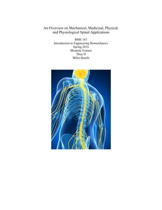
  • 1. An Overview on Mechanical, Medicinal, Physical, and Physiological Spinal Applications BME 167 Introduction to Engineering Biomechanics Spring 2016 Mrudula Vemuri Thuy O Miles Quaife
  • 2. 1 Table of Contents Abstract . . . . . . . . . . . . . . . . . . . . . . . . . . . . . . . . . . . . . . . . . . . . . . . . . . . . . . . . . 2 1.0 Introduction . . . . . . . . . . . . . . . . . . . . . . . . . . . . . . . . . . . . . . . . . . . . . . . . . . . 3 2.0 Composition and Structure . . . . . . . . . . . . . . . . . . . . . . . . . . . . . . . . . . . . . . . 3 3.0 Constitutive Behavior. . . . . . . . . . . . . . . . . . . . . . . . . . . . . . . . . . . . . . . . . . . . 9 4.0 Mechanical Characteristics . . . . . . . . . . . . . . . . . . . . . . . . . . . . . . . . . . . . . . . 10 5.0 Physiological Function and Loading Conditions . . . . . . . . . . . . . . . . . . . . . . 13 6.0 Disease and Medical Condition. . . . . . . . . . . . . . . . . . . . . . . . . . . . . . . . . . . . 14 7.0 Treatment Options . . . . . . . . . . . . . . . . . . . . . . . . . . . . . . . . . . . . . . . . . . . . 15 8.0 Conclusion . . . . . . . . . . . . . . . . . . . . . . . . . . . . . . . . . . . . . . . . . . . . . . . . . . . 16 9.0 References . . . . . . . . . . . . . . . . . . . . . . . . . . . . . . . . . . . . . . . . . . . . . . . . . . . 16 10.0 Images . . . . . . . . . . . . . . . . . . . . . . . . . . . . . . . . . . . . . . . . . . . . . . . . . . . . . 18 11.0 Tables . . . . . . . . . . . . . . . . . . . . . . . . . . . . . . . . . . . . . . . . . . . . . . . . . . . . . . 19 12.0 Equations . . . . . . . . . . . . . . . . . . . . . . . . . . . . . . . . . . . . . . . . . . . . . . . . . . 19 13.0 Acknowledgements . . . . . . . . . . . . . . . . . . . . . . . . . . . . . . . . . . . . . . . . . . . 19
  • 3. 2 Abstract The spinal column is of great interest under many areas of study, specifically vertical compressional load capacity, physiological structure, decrease in vertebral disc height due to time subjected fatigue, as well as many other interesting physical strains and stress adaptations. The spine has the ability to resist work done on it though many different angles, and is composed of materials such that allow it to compress many times cyclically without failing, as long as the load is such that plastic deformation is not observed inside the spinal column and surrounding tissues. The adult spinal column is made of approximately 24 vertebral bones that have the ability to act together as a single ball and joint socket, yet still contain the main connecting pathways between the brain with surrounding tissues, and allow simple flexion and extension without impinging on these signal pathways. The spine is important in the functioning capacity of the human body and how any form of injury can be largely incapacitating even with a small amount of experienced trauma. The spine acts a protective housing for the nerves traversed through it, it also is thoroughly intertwined with nerve endings, various tendons and muscles adding support, which further increase the complexity of the body’s dependence on this structure. In a body’s free standing nature, the spine acts as a column that supports the weight of the upper torso, as well as stabilizes any planar movement generated by the body while serving as an anchoring point for a majority of muscles that create great work movements. The summation of these interactions and our ability as engineers to model, predict, and prevent debilitating injuries. An immediate application is the medical condition involving the spine is scoliosis, which is a musculoskeletal disorder that involves a sideways curvature of the spine. The majority of scoliosis cases has unknown causes. Patients with scoliosis experience the same axial compression on their curved spines without balanced support from the musculoskeletal system. The lack of balanced support causes an asymmetric loading of the vertebral axis, which results in the development and progression of spinal deformity. Many treatments are available for patients with scoliosis, including exercised-based physical therapy, brace treatment, surgery, and drug therapy.
  • 4. 3 1.0 Introduction The spine plays a central role in the human body. Holding up the upper body and allowing for a wide range of motion. The spine has several important functions in the body. It provides structural integrity to the body and houses the spinal cord. The spinal cord relays information between the brain and the rest of the body. Injury to this particular structure can be devastating and result in a total lack of movement. Understanding how each component of the spine works will create a better understanding of the spine and therefore allow for faster and more complete recoveries from spinal injuries. 2.0 Composition and Structure The spinal column is the main load bearing assembly in living vertebrate organism that physically disperses superior loads to the inferior physiological assemblies. It also acts as a protecting column to the central nervous system axon passageways interconnecting the brain with the peripheral nervous system and the rest of the body. These tissues consist of composite blends of various cell types, constituting the summation of functions producing organ systems such as vertebral bone, intervertebral discs and connecting ligaments, and other connective tissues such as cartilaginous structures and substances. The spinal column bones are individually termed as “vertebrae,” which consist of the general molecular makeup as that of most load bearing bones and are numbered both by cranial to sacral direction and region of spine. Bones are formed from mesenchymal stem cells that are clustered together and begin formation in either one of two ways. Of interest in spinal formation, is the “Endochondral Ossification” method, in which these mesenchymal stem cells are crowded together in the general pattern of the bone in which they are going to form, and then differentiate into chondroblasts, which secrete cartilage matrix around the clump of now differentiated chondrocytes, creating a cartilage model of what will become bones covered in hyaline cartilage, which is totally surrounded by a membrane called the perichondrium. As this model grows, the chondroblasts are further buried interiorly to the perichondrium, being called chondrocytes. This model continues to grow until chondrocytes in the middle of the bone burst, causing calcifications by an increase in pH. This calcification starts on the surface and proceeds inward. At this time, a nutrient artery pierces the superficial perichondrium protective surface and the calcifying cartilage model which then feeds and stimulates osteogenic cells in the model to differentiate into osteoblasts. Various differences occur depending on specific bone function, but vertebrae follow this model to varying degrees with time in each stage regulated by vertebra size (Tortora, 2005). Figure 1 below shows the variations in vertebrae regions. Listed are: the atlas and axis which give direct mobility to the head as special cervical vertebra, which give mobility to the neck; the thoracic vertebrae which give limited mobility and maximum stability to the upper torso, and the lumbar vertebrae (lower torso region), which have the largest area per vertebra, which contributes to supporting the greatest pressures applied to the spine.
  • 5. 4 Figure 1. Vertebral Regional Representation [1] The vertebral composition, in its final state, is a composite porous and spiny structure that is both rigid in nature as well as saturated with a gel like substance with an entire superficial hyaline cartilage covering. The gel like substance contributes to the structural ability to elastically deform to a certain extent without permanent plastic deformation. The porous structure and the gel like substances are classified as either organic matrix or inorganic matrix. Bone cells are part of the organic matrix and once the method of ossification is complete, are osteoblasts, osteocytes, and osteoclasts, Osteoblasts build up bone tissue by secreting the matrix tissue of bone, being calcium phosphate salts). Osteocytes are osteoblasts fully surrounded in bone matrix and lie within the chambers (lacunae) of formed matrix. Osteoclasts break down bone tissue by breaking down the matrix tissue of bone which is necessary for maintaining proper blood levels of calcium. The rest of the organic matrix is a mixture of collagen fibers and a gel-like osteoid tissue. This osteoid tissue primarily contains proteoglycans, which are proteins covalently attached to glycosaminoglycans, which are tissue organizing molecules that deal with cellular regulations and activities, and are composed primarily of chondroitin sulfate molecules and glucosamine. The high saturation ability of proteoglycans contributes to a high water saturation level of 25% (Muscilino, 2005). The inorganic bone matrix is composed of hydroxyapatite crystals (calcium phosphate crystalline salts) and is the structurally rigid aspect of the spine. Figure 2 shows below standard bone scaffolding structure (inorganic tissue only) from a low power scanning electron microscope, where the porous voids are the locations of the gel like organic material. This image is taken from the third lumbar vertebra of a deceased 30 year old woman. It is easy to see the interconnectedness of both the organic and inorganic tissue matrices as the organic secretes the inorganic (Muscilino, 2005).
  • 6. 5 Figure 2. Electron Scanning Microscope 3rd Lumbar (x20) [2] The vertebra structures are grouped by region and similar structure, which differ as load and function change in the spine itself. Directly beneath the skull is the cervical spine, whose general features are shown below in Figure 3. It has 7 vertebrae and are smaller in size than either the thoracic or lumbar, as it supports the least amount of pressure. Special attention is focused to the spinous process on the posterior side, as well as the transverse processes on either side, where later discussed ligaments attach. These processes are the consistent attaching locations for the spinal connective tissues. The vertebral foramen creates nerve bundle passageway from the brain to the peripheral nervous system. The articular facet joints are the locations where the inferior vertebral spiny process rest, on top of fibrocartilage and held in place by more later discussed ligaments. Differing in the first 2 cervical spine vertebra, are both structure and functions. Atlas, is the title for C1 and is named after the mythological god who supported the world on his shoulders. It is a ring of bone with anterior/posterior arches, and does not have a body with spinous processes. Directly beneath is C2, also known as the axis, which does have a body. The noticeable differences are the dens, which is a spike-like projection out of the superior face of the vertebrae. The dens is a pivoting point for which the head rests and pivots on. The axis and atlas are both shown in illustrative form below in Figure 4. Figure 3. Typical Cervical Spine Vertebra [3]
  • 7. 6 Figure 4. Illustrative Figure of Atlas and Axis Cervical Vertebra [4] The thoracic vertebrae are directly inferior to C7, and consist of 12 vertebrae bones that are larger than the superior cervical vertebra. As the distance increases vertically from the head, pressure increases, relating to an obvious increase in pressure on the spine and associated joints, requiring an increase in structural performance. The thoracic vertebrae also have notches in the posterior side where the ribs connect via the vertebrocostal joints, as seen below in Figure 5. These vertebrocostal joints limit the mobility of the thoracic region (Tortora, 2005). Figure 5. Thoracic Vertebra [5] Inferior to the thoracic region are the lumbar vertebrae, which are the largest and strongest vertebrae. The projections of these vertebrae are short and thick, with associated articular processes directed medially. The Spinous processes are thick and broad, and project almost perfectly posteriorly. Because of their size, they are quite suited for the attachment of the large back muscles (Tortora, 2005), as shown below in Figure 6.
  • 8. 7 Figure 6. Common Lumbar Vertebra [5] The most inferior portions of the spine are the Sacrum and the Coccyx. In children, these vertebrae are not fused together, yet begin to fuse by the age 16, and are generally completely fused by the age of 30. The sacrum is triangular in shape located medial to either hip bone, and serves as a strong anchoring place for the pelvic girdle. Females have a shorter sacrum and is more curved than their male counterpart. The sacral canal in the sacrum is the continuance of the vertebral canal, and is shown below along with the coccyx in Figure 7 (Tortora, 2005). Figure 7. Sacrum and Coccyx Spinal Vertebrae [5] The coccyx is the most inferior portion of the spine and is formed like the sacrum, by the fusion of vertebrae between the ages 20 and 30 years old. The superior face of the coccyx 1 is connected to the sacrum by 2 ligaments, and articulates superiorly with the sacrum. Generally, the structure and function of each vertebra is similar to that of the one above and below it. Slight differences obviously occur as the pressure load increases, or the places for the ribs to attach. However, the structure of the spine dictates its major functions, being to resist vertical deforming forces, and protect the central nervous systems axon pathways. In addition to the vertebral aspect of the spine, are associated connective and stabilizing tissues, as shown below in the animated Figure 8. A total of 9 ligament types connect the stacked
  • 9. 8 vertebrae in order to limit excessive flexion or extension. Ligaments are classified as short bands of tough fibrous connective tissues that connect bone to bone, cartilage to cartilage, or hold together a joint structure. On the anterior side is the “Anterior longitudinal ligament (1),” which is a three layer ligament which covers both vertebral bone bodies and intervertebral discs. The superficial layer covers 3-4 vertebrae, the intermediate secondary layer covers 2-3 vertebrae, and the deepest layer only the anterior face of one vertebra. The posterior side has a similar ligament, known as the “Posterior longitudinal ligament (2)” and is also shown below on the posterior side of the vertebral canal. The Posterior longitudinal ligament is thicker in the thoracic region, yet broader in the cervical and lumbar regions. Other ligaments, as shown below, are the facet capsulary ligaments (3), which connect the spiny process on the posterior side of the vertebrae and function so stabilize the facet joints and limit the extreme motions of the spine except extension and inferior translation/compression; the supraspinous ligament (4), which connects the posterior sides of the spiny processes of the vertebrae and limits flexion on the spinal joints; the annulus fibrosus (laminated layers of fibrocartilage) of the disc joint (5), which stabilizes each disc joint and limits the motion extremities of all spinal motions; 2 ligamentum flava (6) are located on the either sagittal side of the spinal column, which also contribute to the limiting flexion of the spine, the interspinous ligaments (7) which are short separate ligaments that are interconnected between adjacent spiny processes, which also limit the flexion of spinal joints; the intertransverse ligaments which are located between adjacent processes of the vertebrae whose function is to limit contralateral-lateral (opposite sided) flexion of the spinal joints, and finally the nuchal ligament found from the spinous process on cervical vertebra 7 (C7) to the external occipital protuberance of the skull (Summarized from Muscilino, 2005). Generally, the structure and function of each vertebra is similar to that of the one above and below it. Slight differences obviously occur as the pressure load increases, or the differences pertaining to places for the ribs or connective tissues to attach. However, the structure of the spine dictates its major functions, being to resist vertical deforming forces, and protect the central nervous systems axon pathways. With that as focus, it is easy to see why there are so many similarities across these 26 vertebral bones and associated connective tissues. Figure 8. Animated Image of Spinal Connective Tissue [5] 3.0 Constitutive Behavior Constitutive Laws rate the stress and strain fields. These laws consist of a set of mathematical idealizations based on observed behavior and they can be used to characterize
  • 10. 9 mechanical behavior. Constitutive equations or relations can be established specifically for a material to describe its response to external stimuli such as applied forces or fields. These equations can be used to calculate the material’s behavior in fluid mechanics, solid state physics, structural analysis, or deformation problems. Some general equations used to determine constitutive behavior include general stress or pressure, strain, elastic modulus, Young’s modulus, shear modulus, and compressibility. The table below summarizes these equations. Table #1: Equations used to Characterize the Constitutive Behavior of a Material. [1] Name Symbol Defining Equation Stress Strain Elastic Modulus Young’s Modulus Y Shear Modulus G Bulk Modulus B Compressibility C The deformation of solids is defined by the constitutive behavior of the material. Outside stimuli such as collisions, solid-state deformations, stress strain, and friction all contribute to deformation. The constitutive behavior of the spine can be broken up into the constitutive behavior of the vertebrae and the soft tissue surrounding the vertebrae. The bone elements in the spine are assumed to have a constitutive behavior of an elastic and perfectly plastic material. The elastic constant is transversely isotropic while the yield strength is isotropic (Silvia, 1998). To model the soft tissue of the spine, it needs to be broken up into two parts. The first part is the energy contributions from the matrix and the second part will be the energy contributions from the fiber. This break-up is important because of the fiber-matrix shear interaction determines the response of the annulus fibrosis or soft tissue around the intervertebral disk (Guo, 1962). According to Guo, the matrix is modeled as an incompressible neo-Hookean material while the fiber is modeled as the same with the stiffness dependent on the stretch of the fiber. A neo-Hookean model is a model for a hyperelastic material and is similar to Hooke’s law. This model can predict the nonlinear stress-strain behavior of materials that are subject to large deformations. The stress-strain curve of this model is not completely linear, but is instead linear at the beginning with a plateau after a certain point. The energy contributions of the fiber and the matrix can be quantified by mathematical equations. Equation 1 below quantifies the energy contributions from the fibers. Equation 1 [1]
  • 11. 10 The equation for the energy contribution of the shear interaction of the fiber-matrix is defined by Equation 2 below. Equation 2 [1] These equations are derived from the neohookean model. This model uses a multiplicative decomposition to split the deformation gradient into two parts. These two parts are the uniaxial deformation in the direction of the fiber and the shear deformations. Extending effective stiffness formula for linear elastic composites into a large deformation range will provide an expression for strain energy function for the model (Guo, 1962). This model has limitations and areas for further study to make a more accurate model. It assumes that the soft tissue can be modeled similarly to a composite material. The mechanical properties however are different and to appropriately model the tissue in this way, a reference for the mechanical properties of the tissues must be investigated. This will allow a more accurate strain energy function to be developed. 4.0 Mechanical Characteristics 4.1 The Spine as a Single Unit Due to the total summation of all tissues utilized in the contribution of movement and load stabilization, the spinal column has been studied in light of several functional units. First considered, is the spinal column as a whole functional unit. Secondly is the functional spinal unit, which is an analysis of two vertebrae succinctly joined together by two joints and 9 connecting ligaments. Lastly is the analysis on the vertebra itself, as a single functional unit which is a hard, porous, and gel filled composite material. In its totality, the spine has the ability to act as a mixture between a spring or shock absorber, and a static truss through load dispersal. When analyzing this model on an adult species from the lateral point of view, it is easy to see curves in the thoracic, cervical, lumbar, and sacrococcygeal regions. When viewed from the posterior side, the spine should be as straight as possible. As shown below in Figure 9 below, the primary curves of the spine are the thoracic and sacrococcygeal curves which are described as kyphotic. This means they are curved concave anteriorly, and curved convex posteriorly. Considered as secondary curves, the spine also curves in the cervical and lumbar regions, described as lordotic (curved concavely posteriorly and convex anteriorly) (Muscolino, 2005).
  • 12. 11 Figure 9. Lateral View Spinal Curvature [5] When analyzing the spine as a single unit, it is easy to imagine the types of forces that are endured during various aspects of loading. It is also easy to the ability of the spine to use its ability to compress without deforming to a certain maximum, as well as withstand tangential forces that may cause shearing resistance. Mechanically, it has also been observed that spinal stability, although greatly assisted by connective ligament tissues, an even greater amount of stability is used from musculature connecting to the spine, and inserting at other areas (Oxland, 2015). Future developments are seeking to ascertain the true meaning of a newly developed term being “sagittal balance,” as well as further understand predetermined ideas and observations through mathematical model developments. This term newly designates an idea of the ability of the spine to be sagittally imbalanced, which is giving rise to various angles of shift for the pelvis relative the spinal column in what could be considered a physical equilibrium. Increases in angular deviations from observed equilibrium values cause increased intervertebral pressures, as well as degradation and material fatigue (Melnyk, 2016). The spine in its totality functions as a single ball and joint socket (Ozkaya, 2012). With this mechanical analogy, the spine has 3 degrees of freedom, limited only in range onset shortening by extended time spans of decreased motion (Jaumard, N. V., 2011). Because of this structure, the spine has an associated degrees of motion. These are: 135 Degrees flexion, 120 degrees extension, 90 degrees right lateral flexion, 90 degrees left lateral flexion, 120 degrees right rotation, and 120 degrees left rotation. Obviously an impingement or injury to the spine decreases these values. Also, when analyzing the different segments of the spine, the associated range of motion, greatly diminishes. It is only in the spines entirety that it has such free range of motion. 4.2 Vertebrae
  • 13. 12 The spiny nature of the vertebra contribute to movement and stability of the vertebral column, and are analyzed in what is considered the functional spinal unit (FSU) (Oxland, 2015). Each vertebra has 3 vertically connecting joints, with two being facet joints between the spiny processes on the posterior side connected between the spiny processes in two vertically adjacent vertebra. The major joint is the inferior body face connected to superior body face of the vertebra in column through the intervertebral disc, separated by the intervertebral discs. However, its molecular structures also contribute an anisotropic characteristic by type of osteoblast secretion. Internally, softer sponge bone is surrounded by compact bone which is harder. Based on the molecular order of these cells, applied directional loads may be withstood in greater numbers in some directions relative to others (Bankoff, 2012). Loading the FSU in various directions will generate reaction forces that can either resist the load without creating injury to the spine, or create a deforming load in which some aspect of shearing/straining is observed intervertebrally in either the vertebral unit, or some connective ligament. The FSU couples all three joints into its possible achieved motions for flexion and extension (summarized and images taken from Muscilino, 2005). The FSU also allows flexion and extension within the sagittal plane around the mediolateral axis, which is a midline axis of separation between vertebras, as shown below. Left and right rotation are also allowed within the transverse plane of vertebra and the one directly inferior to it. Finally, translational motions are also allowed, as long as shearing or strain is not observed. The limiting factor in these motions are the elastic ability of the intervertebral disc and supporting ligaments to accept the applied forces causing the movement without straining (Schmidt, 2016). Under laboratory experiment, it was observed that the median critical shearing force in the posterior-anterior direction was 2877 Newtons (an equivalent mass of 293.27 kg under the influence of gravitational acceleration), and lateral side to lateral side of 3199 Newtons (an equivalent mass of 326.1 kg under the influence of gravitational acceleration) (Schmidt, 2016). Compressive failures occur typically at the vertebral endplate by the high pressure of the intervertebral disc nucleus (nucleus pulpous) (Bankoff, 2012). 4.3 Individual Vertebral Bone For final considerations, is the individual vertebral bone, or simply, the vertebra. Typical anatomical features are given in the above text, with composition being primarily cortical (rigid) bone, and the inner matrix a region of cancellous (spongy) bone. The superficial characteristics, such as the spiny processes, are anchoring points for the attachment of tendons of movement producing musculature, and stabilizing ligaments to create the FSU. Mechanical loading capabilities of the vertebra increase as bone size increase from superior to inferior placement in the spine correlating with the increase in cross-sectional area (as pressure dependence dictates), with the load applied to both the rigid outer shell and inner spongy tissue (Bankoff, 2012). Mean failure loads (N) were measured and tabulated in analyzing axial compression, with finding the posterior side of the vertebra were roughly 2.5 times stronger than most central regions, with an . In analyzing postural loads, it is was found that flexed, unextended postures result in an increased loading on the vertebral body, while cervically extended postures attribute more loading to the process attachment points, called the neural arch. With advancing age, the load passing through this arch increases, specifically after the age of 60. This is due largely to decreasing the height of the intervertebral disk by a lifetime of repetitive cyclic loading generating intervertebral disk fatigue (Adams, et al 2006). It was also shown that the increased
  • 14. 13 neural arch loading as age also increases, by the decrease in anterior loading, leads to a decrease bone in vertebral density. This was thought to be a contributing factor to elderly intervertebral fractures (Adams, et al 2006). 4.4 Supplementary Materials Biological materials are very complex, not only their ability to self-replicate and self- heal, but also their near perfect order with which they perform their function. Many tissue materials type fatigue, are rejected by the body, or cause some type of reaction through their lifetime. Bone represents a composite that is perfectly balanced between osteocytes and bone matrix, and changing the composition of either can have drastic side effects on the functional support of the body. If too spongy, rigid support for the body is not physically possible, and if too brittle, bones do not have any elastic behavior and will deform at much lower pressures than currently observed. Limiting factors in designing artificial bone, are finding materials that body will not reject, as well as being able to do many work loading cycles without fatigue. Bone is very porous which allows it to be a solid structure and handle compressible forces, yet still allow blood vessels to penetrate to keep it regenerating itself and health. In the spine, because of synovial fluid, and pressure, materials can both fatigue physically and chemically. For vertebral replacement currently, truss/column like scaffolds are bridged into the spine as well as titanium foam bars (Kubosch, 2016). These have variable success rates, and most experienced bony integration to the implant (Lange, 2006). 5.0 Physiological Function and Loading Behavior It is easy to imagine the importance of the spine. It is central to the body and all of its movements. The spine provides structure and support to hold the body up while still being flexible enough to allow the body to move. The spine does all of this while also performing the invaluable task of housing and protecting a column of nerves that feed from the brain to the rest of the body. This amazing structure is made up of smaller subsets called vertebrae. These vertebrae are individual bones that make up the building blocks of the spinal column. The combined vertebrae create a hollow tube that allows the spinal cord to pass through. Each vertebrae has a strong and hard outer bone called the cortical bone and a spongy inner bone called the cancellous bone. Intervertebral disks serve as shock absorbers between each vertebra of the spine. There is a disk between each vertebrae and each disk has an outer ring of fibers called the annulus fibers. Annulus fibers are the strongest area of the disk and helps keep the center of the disk intact and connects the vertebrae. Different activities result in different loading conditions load the spine differently. Examples of activities that change the loading of the spine include running and weightlifting. The spine is subject to many different kinds of loading in any activity. These types of loading include loading by gravity, by change in motion, muscle activity, and external forces or work. Exercising will usually involve all of the above types of loading. The four elemental forces of the spine are compression, tension, torque, and shear forces. These forces are defined as being any external stimuli that causes a change in a free body or creates internal stress through attempted deformation. The most perpetual, constant, and understandable example of such a force is gravity. Compression and tension forces act along the axis of the spine. Compressive forces flatten the disks and vertebrae of the spine. Tension forces act to elongate those spinal components. Large loads may result in spinal shrinkage. Spinal shrinkage occurs when the compressive load is larger than the interstitial osmotic pressure of the tissues. When the
  • 15. 14 interstitial osmotic pressure is exceeded, water is extruded from the disk wall and the disk height reduces resulting in spinal shrinkage and loss of stature (Leatt, 1986). Compression and Tension forces can both be found in simple movements such as a jump. When an individual begins a jump, the spine experiences a compressive force. At the height of the jump when all body leaves the ground, the compressive force becomes a tension force. The momentum of the jump pulls the spine up while the gravitational forces pull it down. Shear forces are forces applied perpendicular to the spinal axis. Shear forces in the spine are associated with spinal injury. They are not desirable because they work to slide the spinal components away from their natural axis. Shear forces contribute to the increase of internal stress in the body. Large shear forces lead to ligament and disk tears as well as vertebral fractures. Torsional forces are also applied perpendicular to the spinal axis. Unlike shear forces, however, they are not in the plane of the spine. They exist, instead as a rotational force. Torsional forces rotate the components of the spine along its natural axis. Another name for this force is torque. A wide range of torsional forces fall within the normal use of the spine, however, excess torsional force may cause disk and ligament tears or torsional vertebrae fractures. 6.0 Disease and Medical Condition A medical condition that involves the spine is scoliosis, which is a musculoskeletal disorder that involves a sideways curvature of the spine. In the majority of cases, scoliosis is under mild condition, and treatment is not required. The natural curve of the spine allows individuals without scoliosis to balance, move, and walk normally. Individuals with scoliosis have vertebrae that rotate, resulting in the sideway curvature of the spine. The medical condition usually develops rapidly over time, commonly affecting the thoracic spine or lumbar spine. The cervical spine is rarely affected. In several cases, there is a second curvable of the spine in the opposite direction, resulting in a S-shaped spine. Kalichman et al. (2016) stated that several different types of scoliosis exist with adolescent idiopathic scoliosis (AIS) being the most common. Anand et al. (2011) mentioned that AIS is diagnosed when the exact cause for the spine curvature is unknown, and this condition appears in more than 80 percent of diagnosed scoliosis cases. Congenital and neuromuscular are two other types of scoliosis, and these types comprises from 5% to 7% of scoliosis cases. Hawes et al. stated that about 20 percent of AIS patients has one or more family members with scoliosis, indicating that an inherited factor may be one of the causes. Recent investigations of the causes of scoliosis have been focused on structural elements of the spine, collagenous structures, spinal musculature, central nervous system, endocrine system, and genetics. However, currently, no mechanism is established as the causes of AIS. Zavatsky et al. (2015) studied the influence of race and economic status on disease severity and treatment in adolescent idiopathic scoliosis. Zavatsky et al. found that 1% to 2% of the general population has AIS. All children, regardless of race and socioeconomic levels, have a possibility of developing scoliosis. Scoliosis affects about 3 percent of children and adolescents over 10 years old. Older children between the ages of 10 and 12 and adolescents are more prone to develop AIS since the medical condition usually develops during a growth spurt. In the majority of cases, the curvature of the spine is not severe, and treatment is not required. Both boys and girls are as likely to develop scoliosis, but a higher number of treatments is needed for girls. Children with a family history of scoliosis have a higher chance of developing the condition. In addition, the condition occurs more frequently in children with other medical
  • 16. 15 problems, such as connective tissue disorders, neuromuscular diseases, and rheumatic diseases. Black patients have a higher curve magnitude and a greater percentage of patients with curves in the surgical range than white patients. Across all racial groups, no difference was observed in age on presentation or treatment offered. White patients are less likely to choose surgery as their initial treatment than black patients. Kalichman et al. (2016) mentioned that many patients with AIS have a misalignment of the body, a loss of balance and shrinking height, a leg and buttocks pain, and many hip, bladder and bowel problems. When the spine is deformed, the shoulders, waist, and hips are also not aligning properly since the bones in the spine had rotated. Patients with scoliosis have diminished overall height because of the misalignment of the body. Leg and buttocks pain occurs due to arthritic pain or nerve compression originated from the spine. Hip problems occur because the ribs are more apparent on one side of the spine. Scoliosis can also causes a loss of control of bladder and bowel function. The pathological loading conditions are different from normal physiological loading conditions. For instance, how the body response to physiologically relevant axial compression differently among individuals with and without scoliosis. The spine experiences axial compression due to the body weight in the upright position. Hawes et al. stated that the bony axis of a normal spine can tolerate a weight of approximately more than 10 kilograms without buckling. This bony axis depends on the balanced support from the musculoskeletal system, which is controlled by the central nervous system. Patients with scoliosis experience the same axial compression on their curved spines without balanced support from the musculoskeletal system. The lack of balanced support causes an asymmetric loading of the vertebral axis, which results in the development and progression of spinal deformity. 7.0 Treatment Options Many treatments are available for patients with scoliosis, including exercised-based physical therapy, brace treatment, surgery, and drug therapy. Treatment of scoliosis is depended on the level of severity of the curvature of the spine and the possibility of the curve from becoming worse. Kalichman et al. also mentioned that two conservative treatments of AIS are exercised-based physical therapy and brace treatment. Current physical therapy for patients with scoliosis is specific exercises that are unique for each individual patient. Specific exercises are designed to reduce the effects of spinal deformity, including scoliosis specific exercises, the Dobosiewicz technique, the schroth method, the side-shift program, and SEAS. Bracing treatment, on the other hand, may reduce the need for surgery, restore the sagittal profile and affect vertebral rotation. Braces are usually worn full-time for about two to four years until the termination of bone growth. Cunin (2015) stated that another major treatment option is surgery, and these surgical methods include growing rods, guided growth systems, and compression- based systems. Growing rods place a distraction force to the spine, specifically the concavity of the curve, to stabilize the curvature of the spine and the growth of the T1-S1 segment. Several surgical procedures are required for growing rods treatment to lengthen the rod, to maintain the result during the index surgery, and to follow spine growth. The rods can also be lengthened noninvasively via a magnetic mechanism. Guided growth systems are surgical devices that are placed along the vertebral column to maintain the spine in its reduced position while allowing the spine to grow. Convexity compression devices place a compressive force on the convexity of the spine curvature to restrict its growth. Hui et al. investigated the efficacy of a combined Traditional Chinese Medicine (TCM) therapy, including acupotomology, Daoyin, and Tuina Hui
  • 17. 16 et al. (2015) found that TCM combined therapy has the ability to prevent the progression of spinal deformity by targeting the muscular imbalance between spinal disorder and scoliosis sides. Kalichman et al. (2016) stated that the most effective conservative treatment for AIS is bracing. Exercise-based physical therapy can prevent a worsening of the curvature of the spine, which may reduce the need for bracing. Exercise-based physical therapy is also the only treatment that improves respiratory function. However, limited evidence is available to specify physical exercises that are effective treatment methods for AIS. The combination of bracing and exercising increases treatment efficacy in comparison to treatment efficacy of a single treatment. Cunin mentioned that out of all three surgical treatments, growing rods is the most common since it provides better control over the Cobb angle during maturation. Current available instrumentation for growth guidance systems is highly invasive and less common than growing rods. Compared with other methods of treatment, TCM combined therapy accounts for patient activeness, which does not affect daily life nor causes any adverse effects on physical and psychological development. 8.0 Conclusion The spinal column is the main load bearing assembly in living vertebrate organism. The spine interconnects the brain with the peripheral nervous system and the rest of the body. The spinal column bones are formed from mesenchymal stem cells that are clustered together and begin formation. Constitutive laws rate the stress and strain fields and can be used to calculate the material’s behavior in fluid mechanics, solid state physics, structural analysis, or deformation problems. These general equations are used to determine constitutive behavior include general stress or pressure, strain, elastic modulus, Young’s modulus, shear modulus, and compressibility. The spine has the ability to act as a mixture between a spring or shock absorber, and a static truss through load dispersal. The spine in its totality functions as a single ball and joint socket, which has 3 degrees of freedom, limited only in range onset shortening by extended time spans of decreased motion. Compressive forces flatten the disks and vertebrae of the spine whereas tension forces act to elongate those spinal components. Shear forces are forces applied perpendicular to the spinal axis whereas torsional forces are also applied perpendicular to the spinal axis. A medical condition that involves the spine is scoliosis, with AIS being the most common type. AIS is diagnosed when the exact cause for the spine curvature is unknown. Patients with scoliosis experience the same axial compression on their curved spines without balanced support from the musculoskeletal system. The lack of balanced support causes an asymmetric loading of the vertebral axis, which results in the development and progression of spinal deformity. Many treatments are available for patients with scoliosis, including exercised- based physical therapy, brace treatment, surgery, and drug therapy.
  • 18. 17 9.0 References 1. Adams, M.A.,Pollintine,P.,Tobias,J.H.,Wakley,G.K.,Dolan,P.,(2006).Intervertebral disc degeneration can predispose to anterior vertebral fractures in the thoracolumbar spine .J.BoneMin.Res.21(9),1409–1416. 2. Anand, N., & Baron, E. M. (2011). Minimally invasive treatment of adult scoliosis. Seminars in Spine Surgery,23(1), 66-71. doi:http://dx.doi.org/10.1053/j.semss.2010.12.007 3. Bankoff, A. D. P. (2012). Biomechanical characteristics of the bone. INTECH Open Access Publisher. 4. Cunin, V. (2015). Early-onset scoliosis – Current treatment.Orthopaedics & Traumatology: Surgery & Research,101(1, Supplement), S109-S118. doi:http://dx.doi.org/10.1016/j.otsr.2014.06.032 5. Guo, Z. Y., Peng, X. Q., & Moran, B. (2006). A composites-based hyperelastic constitutive model for soft tissue with application to the human annulus fibrosus. Journal of the Mechanics and Physics of Solids, 54(9), 1952-1971. 6. Hawes, M. C., & O’Brien, J. P. (2006). The transformation of spinal curvature into spinal deformity: pathological processes and implications for treatment.Scoliosis, 1, 3. http://doi.org/10.1186/1748-7161-1-3 7. Hui, W., Jinyuan, X., Zhong, J., Shuliang, Y., Hongquan, S., Xitao, N., . . . Honggen, D. (2015). Effect of a traditional chinese medicine combined therapy on adolescent idiopathic scoliosis: A randomized controlled trial. Journal of Traditional Chinese Medicine, 35(5), 514-519. doi:http://dx.doi.org/10.1016/S0254-6272(15)30133-3 8. Jaumard, N. V., Welch, W. C., & Winkelstein, B. A. (2011). Spinal Facet Joint Biomechanics and Mechanotransduction in Normal, Injury and Degenerative Conditions. Journal of Biomechanical Engineering, 133(7), 71010–NaN. http://doi.org/10.1115/1.4004493 9. Kalichman, L., Kendelker, L., & Bezalel, T. (2016). Bracing and exercise-based treatment for idiopathic scoliosis. Journal of Bodywork and Movement Therapies, 20(1), 56-64. doi:http://dx.doi.org/10.1016/j.jbmt.2015.04.007 10. Kubosch, D., Kubosch, E. J., Gueorguiev, B., Zderic, I., Windolf, M., Izadpanah, K., … Strohm, P. C. (2016). Biomechanical investigation of a minimally invasive posterior spine stabilization system in comparison to the Universal Spinal System (USS). BMC Musculoskeletal Disorders, 17, 134. http://doi.org/10.1186/s12891-016-0983-1 11. Leatt, P., Reilly, T., & Troup, J. G. (1986). Spinal loading during circuit weight-training and running. British Journal of Sports Medicine, 20(3), 119-124. 12. Lange, U., Edeling, S., Knop, C., Bastian, L., Oeser, M., Krettek, C., & Blauth, M. (2007). Anterior vertebral body replacement with a titanium implant of adjustable height: a prospective clinical study. European Spine Journal, 16(2), 161–172. http://doi.org/10.1007/s00586-005-0015-6 13. Melnyk, Angela, et al. "Characterization Of The Behavior Of A Novel Low-Stiffness Posterior Spinal Implant Under Anterior Shear Loading On A Degenerative Spinal Model." European Spine Journal 24.4 (2015): 775-782. Academic Search Complete. Web. 10 Mar. 2016. 14. Muscolino, J. E. (2005). Kinesiology: The Skeletal System and Muscle Function. St. Louis, Mo.: Mosby/Elsevier.
  • 19. 18 15. Oxland, Thomas R..(2015). Fundamental biomechanics of the spine—What we have learned in the past 25 years and future directions Journal of Biomechanics , Volume 49 , Issue 6 , 817 - 832 16. Ö zkaya, N., & Ö zkaya, N. (2012). Fundamentals of biomechanics: Equilibrium, motion, and deformation. New York, NY: Springer. 17. Schmidt, H., Häußler, K., Wilke, H. J., & Wolfram, U. (2015). Structural behavior of human lumbar intervertebral disc under direct shear. Journal of applied biomaterials & functional materials, 13(1). 18. Silva, M. J., Brodt, M. D., & Hucker, W. J. (2005). Finite element analysis of the mouse tibia: Estimating endocortical strain during three‐point bending in SAMP6 osteoporotic mice. The Anatomical Record Part A: Discoveries in Molecular, Cellular, and Evolutionary Biology, 283(2), 380-390. 19. Tortora, G. J. (2005). Principles of Human Anatomy. New York: John Wiley & Sons. 20. Zavatsky, J. M., Peters, A. J., Nahvi, F. A., Bharucha, N. J., Trobisch, P. D., Kean, K. E., Lonner, B. S. (2015). Disease severity and treatment in adolescent idiopathic scoliosis: The impact of race and economic status. The Spine Journal, 15(5), 939-943. doi:http://dx.doi.org/10.1016/j.spinee.2013.06.043 10.0 Images 1. http://www.ectsoc.org/gallery/#myGallery-picture(2) 2. http://www.slideshare.net/hermizan84/35-cervical-vertebra 3. http://www.spineuniverse.com/conditions/back-pain/upper-back-pain/anatomy-upper- back 4. http://emedicine.medscape.com/article/1899031-overview#a2 5. Tortora, G. J. (2005). Principles of Human Anatomy. New York: John Wiley & Sons 11.0 Tables 1. Ö zkaya, N., & Ö zkaya, N. (2012). Fundamentals of biomechanics: Equilibrium, motion, and deformation. New York, NY: Springer. 12.0 Equations 1. Ö zkaya, N., & Ö zkaya, N. (2012). Fundamentals of biomechanics: Equilibrium, motion, and deformation. New York, NY: Springer. 13.0 Acknowledgments Thuy O: Disease and Medical Condition, Treatment Options, Conclusion Mrudula V.: Introduction, Constitutive Behavior, Physiological Function and Loading Conditions Miles Q.: Abstract, Composition and Structure, Mechanical Characteristics, Personal References