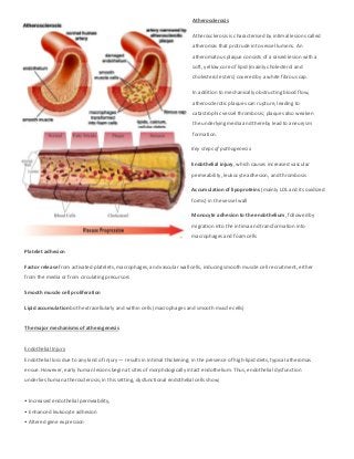
1. Atherosclerosis
Atherosclerosis is characterised by intimal lesions called
atheromas that protrude into vessel lumens. An
atheromatous plaque consists of a raised lesion with a
soft, yellow core of lipid (mainly cholesterol and
cholesterol esters) covered by a white fibrous cap.
In addition to mechanically obstructing blood flow,
atherosclerotic plaques can rupture, leading to
catastrophic vessel thrombosis; plaques also weaken
the underlying media and thereby lead to aneurysm
formation.
Key steps of pathogenesis:
Endothelial injury, which causes increased vascular
permeability, leukocyte adhesion, and thrombosis
Accumulation of lipoproteins (mainly LDL and its oxidized
forms) in the vessel wall
Monocyte adhesion to the endothelium, followed by
migration into the intima and transformation into
macrophages and foam cells
Platelet adhesion
Factor release from activated platelets, macrophages, and vascular wall cells, inducing smooth muscle cell recruitment, either
from the media or from circulating precursors
Smooth muscle cell proliferation
Lipid accumulation both extracellularly and within cells (macrophages and smooth muscle cells)
The major mechanisms of atherogenesis
Endothelial Injury
Endothelial loss due to any kind of injury — results in intimal thickening; in the presence of high-lipid diets, typical atheromas
ensue. However, early human lesions begin at sites of morphologically intact endothelium. Thus, endothelial dysfunction
underlies human atherosclerosis; in this setting, dysfunctional endothelial cells show;
• Increased endothelial permeability,
• Enhanced leukocyte adhesion
• Altered gene expression
2. The specific pathways and factors contributing to endothelial cell dysfunction in early atherosclerosis are not completely
understood, many etiologic culprits are suspected and these can stimulate pro-atherogenic patterns of endothelial cell gene
expression. However, the two most important causes of endothelial dysfunction are hemodynamic disturbances and
hypercholesterolemia.
1. Hemodynamic Disturbances
Plaques tend to occur at Ostia of exiting vessels, branch points, and along the posterior wall of the abdominal aorta, where
there are disturbed flow patterns.
• Non-turbulent flow in the normal vasculature leads to the induction of endothelial genes whose products protect against
atherosclerosis.
2. Lipids
Lipids are typically transported in the bloodstream bound to specific apoproteins (forming lipoproteins). Dyslipoproteinemias
can result from mutations that alter the apoproteins or the lipoprotein receptors on cells, or from other disorders that affect
the circulating levels of lipids.
The mechanisms by which hyperlipidemia contributes to atherogenesis include:
• Hyperlipidemia can directly impair endothelial cell function by increasing local oxygen free radical production; oxygen free
radicals can injure tissues and accelerate nitric oxide decay, reducing its vasodilator activity.
• With chronic hyperlipidemia, lipoproteins accumulate within the intima. These lipids are oxidized through the action of
oxygen free radicals locally generated by macrophages or endothelial cells. Oxidized LDL is ingested by macrophages through a
scavenger receptor, and accumulates in phagocytes, which are then called foam cells. In addition, oxidized LDL stimulates the
release of growth factors, cytokines, and chemokines by endothelial cells and macrophages that increase monocyte
recruitment into lesions. Finally, oxidized LDL is cytotoxic to endothelial cells and smooth muscle cells and can induce
endothelial cell dysfunction.