Journal clubmer09 20-10
•Download as PPS, PDF•
0 likes•297 views

Journal Club Dr. Jim Mark´s Lab. UCSF MER 9-20-10

Recommended
More Related Content
Similar to Journal clubmer09 20-10
Similar to Journal clubmer09 20-10 (20)
Recently uploaded
Recently uploaded (20)
Journal clubmer09 20-10
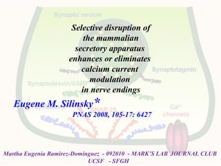
- 1. Selective disruption of the mammalian secretory apparatus enhances or eliminates calcium current modulation in nerve endings Eugene M. Silinsky* PNAS 2008, 105-17: 6427 Martha Eugenia Ramirez-Dominguez - 092010 - MARK’S LAB JOURNAL CLUB UCSF - SFGH
- 2. Docking: specifically attaches vesicles to the active zone; Priming: makes the vesicles competent for Ca2+ -triggered release and may involve a partial fusion reaction; Fusion: final Ca2+ -regulated step that completes fusion. Synaptic vesicle exocytosis occurs in consecutive steps: MarthaEugeniaRamirez-Dominguez-092010-MARK’SLAB
- 3. Microdomains of high [Ca]I occur at the pore exit, because Ca2+ enter the channel faster than they can diffuse into the surrounding cytoplasm. Why do calcium channels (Cavs) interact with proteins thought to be located at the interface between a docked synaptic vesicle and the plasma membrane? Primed fusion-ready vesicles that are not linked to CaVs are much less likely to release their contents when an action potential invades the nerve terminal. It follows on from this hypothesis that regulatory processes which modulate interactions between channels and synaptic core complexes could play an important role in the plasticity of presynaptic events. Molecular links between CaVs and the exocytotic machinery would guarantee that the calcium trigger initiating vesicle fusion is located within a restricted zone where the appropriate [Ca]i is attained upon channel activation. Synaptic vesicle fusion requires relatively high 50-100 uM [Ca]i only attained close to the inner mouth of the activated CaVs. Synprint Site MarthaEugeniaRamirez-Dominguez-092010-MARK’SLAB
- 4. SynaptotagminSynaptotagmin :: Ca2+ -binding protein, essential for Ca2+-triggered release and probably serves as the Ca2+-sensor in fusion. Rab3: limits the number of vesicles that can be fused as a function of Ca2+ in order to allow a temporally limited, repeatable signal. Once membrane depolarization and Ca influx occur, conformational changes in the SNARE complex, along with other less well defined processes, facilitate the fusion of the two membranes and release neurotransmitter into the synapse. MarthaEugeniaRamirez-Dominguez-092010-MARK’SLAB
- 5. Skeletal Neuromuscular Junction G protein coupled Receptors (GPCR) : modulated by Ca2= entrance through Cav- Channels & Interaction with Secretoty Apparatus Adenosine GPCR Inhibit Neurosecretion Neuromuscular depression Agonist 1) A1 – Adenosine Receptor Activation A1R(+) P/Q Cav Neurotransmission Neuromuscular Depression Adenosine effect on Secretory Complex by Cavs ? Anfibia NO Mouse YES 2) By BoNTx assays SNARES important role in mediating effects of Adenosine at mouse motor nerve So, there is an interconection between A1R – P/Q Ca channels mediated by SNAREs ? Ach release
- 6. The secretory apparatus and the cleavage sites for BoNtxs. Cav channels N-type and P/Q-type contain a Synaptic Protein Interaction (synprint) site within the II–III loop that assembles with synaptic proteins such as Syntaxin (Sx), SNAP-25 (SN) and Synaptotagmin (Sg) This association couples synaptic vesicles and the source of extracellular calcium and serves as a regulatory element by which synaptic proteins may control calcium channel function . Synprint MarthaEugeniaRamirez-Dominguez-092010-MARK’SLAB
- 7. Isolated phrenic nerve hemidiaphragm from Rab3A−/− mutant mice Calcium currents were quantified by measuring voltage changes in the perineural space The perineural recording electrodes were filled with normal physiological salt solution (3–10 MΩ) and positioned near small axon bundles at the heminode. The electrophysiological correlates of Ach secretion (EPPs) were also recorded to monitor the elimination of Ach release. MarthaEugeniaRamirez-Dominguez-092010-MARK’SLAB
- 8. The effects of Adenosine in the Rab3A-/- mutant with and w.o. BoNTx treatment * 9 aa Nor required Presynaptic ionic currents End-plate potentials Ach release Sodium current P/Q calcium current Cd++ & ω-agatoxin block Sx Important target x Adenosin inhib SN-25 Coupling A1R to Cav inhib *** 25 aa 17 aa required MarthaEugeniaRamirez-Dominguez-092010-MARK’SLAB
- 9. Increased efficacy of adenosine in the Rab3A-/- mutant after cleavage of the vesicle protein synaptobrevin with Botx/D. Complete inhib of P/Q Cav Reversible Prevent vesicle participate in priming with t-SNARES MarthaEugeniaRamirez-Dominguez-092010-MARK’SLAB
- 10. A model for the enhanced effects of adenosine before and after disruption of vesicle proteins. MarthaEugeniaRamirez-Dominguez-092010-MARK’SLAB
- 11. SV fusion is likely to be initiated by the change in conformation and/or electrostatic potential of a Ca2+ -sensing protein. Similarly, limiting neurotransmitter release to the particular SV that has initiated the fusion process—out of a pool of fusion-competent vesicles— requires a mechanism that excludes a complicated time-consuming cascade of protein- protein-interactions. MarthaEugeniaRamirez-Dominguez-092010-MARK’SLAB
- 13. • what are the clostridial neurotoxins doing that is so deadly? ‘Blocking neurotransmission’ is the answer which prompted many physicians ‘SNARE cleavage’ is the answer that stimulated many cell biologists to use clostridial neurotoxins as research tools for solving fundamental questions on secretion. What will the next answer and application be? How botulinum and tetanus neurotoxins block neurotransmitter releaseHow botulinum and tetanus neurotoxins block neurotransmitter release MarthaEugeniaRamirez-Dominguez-092010-MARK’SLAB
- 14. Jarvis & Zamponi (2005) Cell Calcium37, 5: 483-488 Calcium in the function of the nervous system: New implications MarthaEugeniaRamirez-Dominguez-092010-MARK’SLAB
Editor's Notes
- The secretory apparatus and the cleavage sites for Botxs. The SNAREs syntaxin (red), SNAP-25 (green), and synaptobrevin (darker blue) are shown in their primed state based on the crystal structure of the complex, as are the cleavage sites for Botx/A, C, D, and E (38). Some liberty has been taken with respect to the distance between vesicle and nerve terminal membrane and with the relative sizes of the HABC domains of syntaxin for illustrative pur- poses in this figure and in Fig. 5. For syntaxin, the helical core domain (core, also termed the H3 domain) contributes one helical domain to the parallel four-helix bundle of the primed SNAREs, with SNAP-25 (two helices) and synaptobrevin (one helix) constituting the rest of the bundle. Recent publi- cations provide strong evidence that the Hcore domain is the likely target for GPCR activation (10, 32). The other three N-terminal helices of syntaxin (HA, B, and C) do not contribute to the SNARE complex. The reported interactions of the HA domain (32) and the Hcore (H3) domains (35) with the synprint region (pink) of P/Q- and N-type Ca2 channels are both depicted. The synprint region and the P/Q Ca2 channel structure were drawn in accordance with refs. 28–32. Also shown is GTP-bound Rab3A (lighter blue) interacting with RIM (yellow). Synaptotagmins (20), which are the Ca2 sensors that mediate the action of Ca2 once this divalent cation has entered the nerve ending via voltage gated Ca2 channels, are not shown.
- The effects of adenosine in the Rab3A/ mutant in the absence of botulinum toxin treatment (A) and after cleavage of syntaxin with Botx/C (B) and SNAP-25 with Botx/E (C). (A) Typical effect of supramaximal concentra- tions of adenosine (10 mM) in the absence of botulinum toxins on perineural calcium currents (Ca2, upper traces) and Ach release (EPPs, lower traces) recorded under the same conditions. See Materials and Methods for details of the polarities of the waveforms and description of the perineural currents, which are reflected as voltage changes in the extracellular compartment and are thus calibrated in millivolts. Dose–response relationships for adenosine as an inhibitor of Ach release and calcium currents in both normal and Rab3A/ deletion mutants have been previously published by this laboratory (5, 17). Because the efficacy, not the potency, of adenosine was to be examined, and for comparison with previously published results (see figure in ref. 5), supra- maximal concentrations of adenosine (10 mM) were used in all experiments in this study. (B) Cleavage of syntaxin with Botx/C eliminates the inhibitory effect of adenosine (the averaged peak perineural waveform was 1.1 mV in both control and adenosine traces in A, n 5 responses averaged) but not that of Cd2 on Ca2 currents. (C) Cleavage of SNAP-25 with Botx/E eliminates the inhibitory effect of adenosine (the averaged peak perineural waveform was 1.2 mV in both control and adenosine traces in B, n 9–12 responses averaged), but not that of Cd2, on Ca2 currents.
- Increased efficacy of adenosine in the Rab3A/ mutant after cleavage of the vesicle protein synaptobrevin with Botx/D. (A) Raw traces (each trace is the average of three to six stimuli, 0.017 Hz). Note the complete and reversible inhibition of Ca2 currents by adenosine and its similarity to the effects of Cd2. (B) Inhibition by adenosine of the normalized peak Ca2 current. Each point is the response to a single stimulus. To normalize the peak Ca2 current, the residual outward Cd2-insensitive component (a component that is independent of Ca2 entry via presynaptic Ca2 channels; see refs. 5 and 29) was subtracted from peak currents. This residual outward current after Cd2 is likely to be due to the Na current associated with the action potential, a current that passively repolarizes the part of the nerve ending under the recording electrode or an as-yet-uncharacterized ionic current (4, 5, 29). (C) Effects of adenosine after cleavage of synaptobrevin with Botx/D in Rab3A/ mutant (filled bar) are indistinguishable from those of maximal P/Q-type calcium channel block with Cd2 (shaded bar). In contrast, the effects of Botx/D in the wild-type mouse (wt, open bar) are indistinguishable from the effects of adenosine in the absence of toxin treatment (see Fig. 3 in ref. 19). For further details, see the text.
- A model for the enhanced effects of adenosine before and after disruption of vesicle proteins. ( A ) Same conditions as Fig. 1 , but with the adenosine receptor and G protein included. The depiction of the G protein includes the β propeller region of Gβ that is believed to interact with effectors and the Gγ subunit with its prenylated attachment to the membrane (39). The Gα subunit was drawn in accordance with the crystal structure, with the receptor interaction domain and the GTP-binding region shown as a black circle and a black curving line, respectively. The helical domains are shown as ear-like appendages on the canine-like subunit. This figure also shows the N terminus of RIM interacting with Rab3A and two additional domains (C2 domains) that have been shown to interact with t-SNAREs and Ca 2+ channels (40). For further details of the GPCR activation scheme for adenosine, see the text. ( B ) The situation in the Rab3A −/− mutant. Note the change in conformation in the SNARE complex in this mutant ( Left ) such that the complex has a higher affinity for Gβ. The SNARE- Ca 2+ channel complex ( Right ) is not capable of being modulated by adenosine (no modulation). ( C ) The Rab3A −/− mutant after cleavage of synaptobrevin with Botx/D ( Left ). Note the recruitment of the additional site for modulation by adenosine by changes in conformation of the t-SNARE–Ca 2+ channel complex when synaptobrevin is cleaved in the mutant mouse ( Right , modulation). Recruitment of this site thus increases the level of inhibition of the macroscopic calcium currents. For further details of the synaptic proteins, see Fig. 1 .
- Fig. 1. A model for the regulation of calcium influx by vesicle-associated proteins during the vesicle release cycle. Legend : The presynaptic Ca 2+ channel is shown as only the main pore-forming α 1 subunit with the II–III linker (light brown); syntaxin 1A (blue); SNAP-25 (golden); VAMP2 (pink); CSP (green); nSec-1 (purple). Top panel: syntaxin 1 is open and bound in the SNARE core complex with SNAP-25 and VAMP2. At this point, synaptotagmin (not shown) will also be associated, in addition to CSP, which is associated with the docked vesicle (not shown). At this stage, all potentials channel-inhibitory mechanisms are nullified by virtue of the various proteins being complexed, so potential Ca 2+ is maximal. Also note that potential G-protein interaction with the channel/core complex is maximal at this stage. Second panel: following membrane depolarization, the channel opens, allowing the rapid Ca 2+ influx which triggers vesicle release within 200 μs. Third panel: immediately after fusion, NSF activity (not shown) leads to SNARE core complex disassembly, which then allows syntaxin to adopt its most stable closed conformation, which stabilizes the inactivated state of the channel. Bottom panel: the solitary syntaxin 1 is a prime substrate for nSec-1 which binds following SNARE core complex disassembly. Experiments have shown that a channel expressed with syntaxin 1 and nSec-1 is not inactivated, suggesting that channels in this stage may allow Ca 2+ influx although a vesicle is not docked [27]. nSec-1 binding to syntaxin 1 is followed by munc-13 binding (not shown), which initiates syntaxin 1 priming, prior to SNARE core complex assembly and vesicle docking. Fig. 2. Schematic representation of the interactions between the vesicle release proteins and the N-type calcium channel. Legend : Proteins of the vesicle release machinery mediate Ca 2+ influx through the N-type channel via two distinct processes: (1) directly, by binding to the channel and stabilizing it's inactivated state, and/or (2) indirectly, but promoting the association of an inhibitory G-protein βγ subunit and the channel. Purple arrows indicate direct inactivation-promoting interactions with the channel. Peach arrows indicate indirect inhibition via Gβγ. Black arrows indicate interactions between presynaptic proteins. The green arrow indicates CSPs ability to promote increased channel activity. It should be noted that G-protein inhibition is not necessarily constant, but enhances channel inhibition by activated G-proteins.