
Recommended
More Related Content
What's hot
What's hot (20)
Similar to Enteral Tube Feeding
Similar to Enteral Tube Feeding (20)
Recently uploaded
Recently uploaded (19)
Enteral Tube Feeding
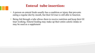
- 1. Enteral tube insertion: • A person on enteral feeds usually has a condition or injury that prevents eating a regular diet by mouth, but their GI tract is still able to function. • Being fed through a tube allows them to receive nutrition and keep their GI tract working. Enteral feeding may make up their entire caloric intake or may be used as a supplement
- 2. Types of enteral tube insertion: • Nasogastric tube feeding • Percutaneous endoscopic gastrostomy • Percutaneous endoscopic jejunostomy
- 3. Nasogastric tube feeding: • By implanting a nasogastric tube, you are gaining access to the stomach through the nose . This enables you to passage gastric contents, decompress the stomach, obtain a sample of the gastric contents, or introduce a passage into the GI tract. This tube will allow you to gastric immobility and bowel obstruction. In trauma settings, NG tubes can be used in the stoppage of vomiting and aspiration, as well as for assessment of GI bleeding. NG tubes can also be used for enteral feeding usually
- 4. Equipment's used during NGT: • All necessary equipment should be prepared, assembled and available at the bedside prior to starting the NG tube. Basic equipment includes: • Personal protective equipment • NG/OG tube • Catheter tip syringe • Water-soluble lubricant, preferably 2% Xylocaine jelly • Adhesive tape • Low powered suction device • Stethoscope • Cup of water (if necessary)/ ice chips • pH indicator strips
- 5. Method • Gather equipment • take non-sterile gloves • Explain the procedure to the patient and show equipment • If possible, sit patient upright for in optimal position • Examine nostrils for deformity/obstructions to determine best side for insertion • Measure tubing from tip of nose to earlobe, then to the point halfway between the end of the sternum and the nave • Mark measured length with a marker or note the distance
- 6. • Lubricate 2-4 inches of tube with lubricant. This procedure is quite uncomfortable for many patients, so a squirt of Xylocaine jelly in the nostril, and a spray of Xylocaine to the back of the throat will help to alleviate the discomfort. • Pass tube via either nose posteriorly, past the pharynx into the esophagus and then into stomach. • rotate tube slowly with downward progression toward closes ear. Do not force. • Withdraw tube instantly if changes occur in patient's respiratory status if tube coils in mouth, if the patient begins to cough or turns pretty colors
- 7. • Check the placement by attaching syringe to free end of the tube, aspirate sample of gastric contents. Dont inject a air bolus, as the best way is to test the pH of the aspirated contents to ensure that contents are acidic. The pH should be below 6. take an x-ray to verify placement before instilling any feedings/medications. • Secure tube with tape or commercially prepare the tube holder
- 8. • for suction remove syringe from free end of tube ;connect to suction; set machine on type of suction and apply pressure as prescribed. • Document the reason for the tube insertion such as type & size of tube the nature and amount of aspirate, the type of suction and pressure setting for suction and the effectiveness of the intervention
- 9. Percutaneous endoscopy gastrostomy: • A percutaneous endoscopic gastrostomy (PEG) is a safe and effective way to provide food, liquids and medications (when appropriate) directly into the stomach. The feeding is done for patients who are feel trouble in swallowing • PEG tube feeding done during; • Esophageal cancer oral surgery Inflammation of the pancreas • Radiation therapy or if inflammatory bowel disease affecting small intestine
- 10. Equipments used during PEG: Equipment's used during PEG are: • PEG tube • Guide wire • Syringe, 5 mL • Snare. • Needle, 22 gauge. • Sterile fenestrated drape. • Lidocaine. • Needle/catheter assembly
- 11. Method: Endoscope ,wire and needle is required for this procedure Before performing this sedative or anesthesia is applied to abdomen Endoscope is passed through your mouth into esophagus stomach and to intestine finally Various pictures are taken through the cameras on the end of the endoscope which helps to find the proper location for insertion of needle
- 12. Needle is inserted through the skin of the abdomen Wire is passed through the needle into stomach Then pull out the endoscope along with wire from the mouth Patient will receive fluids through it for 1 to 2 days after this Patient can receive tube feeding formula if clear liquids are tolerated through PEG tube
- 13. Percutaneous endoscopic jejunostomy: Jejunal feeding is the method of feeding directly into the small bowel. The feeding tube is passed into the stomach, through the pylorus and then into jejunum. Such type of feeding is also called as post-pyloric or trans-pyloric feeding. Method Nasojejunal tubes may be placed with the assistance of endoscopy. Confirm the correct position of a newly inserted tube is mandatory before medication The pH level of the NJT should not be tested The tip of the jejunal tube has potential to migrate back into the stomach. The tube marking at the nostril should be recorded after insertion
- 14. If the patient is experiencing clinical symptoms such as, vomiting, retching, excessive coughing- this may prove that the tube may have migrated to the stomach Do not aspirate the NJT as this can cause breakdown and recoil of the tube The PEJ tube must not be rotated as there is a risk of displacing the jejunal tube by coiling it up in the stomach. As an alternative, the tube should be moved very slightly in and out of the tract approximately one cm
- 15. Parenteral tube insertion: Parenteral tube insertion is the intravenous provision of nutrients without using the gastrointestinal system it should be considered for patients with: Malnutrition Prolonged GI system failure Dysmotility ,fistulae ,surgical resection etc
- 16. Types of parenteral nutrition Central parenteral nutrition(TPN) Peripheral parenteral nutrition (PPN)
- 17. Peripheral parenteral nutrition: your body needs nutrients that offer you with the energy required to go about your daily activities. If you cannot ingest these nutrients or your intestines are not working properly, you must fulfill your nutritional requirements through other means Peripheral parental nutrition (PPN) is administered through the veins outside the superior vena cava.
- 18. Equipment's used during peripheral parenteral nutrition: 10 ml syringe prefilled with normal slime 10-18 gauge venous cannula Sterile gloves Alcohol swabs and 10 percent iodine solution Adhesive tape and self adhesive dressing designed for peripheral cannula
- 19. Method: In this method to decrease the damage during infusing parenteral nutrition we approach peripheral veins with the largest diameter so that the blood flow allow solution to be diluted maximum Veins which are used during peripheral parenteral nutrition are basilic ,cephalic, and median arm veins The solution flows out of the intravenous catheter trip which is placed in the vein with greatest blood flow An iv catheter pump allows the solution to flow at a safe and consistent rate The peripheral solution given in superior vana cava is less cocentrated which fairly increases the pump rate
- 20. this procedure allow more fluid to be infused peripherally to provide more nutrients A filter in the venous catheter tubing prevent the large particles from infusing
- 21. Central parenteral nutrition: Often called as total parenteral nutrition ;delivered into a smaller or peripheral vein Purpose • It is used to provide body fluids ,blood to the patient • To take the blood or fluid of the patient for medical tests
- 22. Method: The skin is applied with the antiseptic properly with the help of cotton the patient should be in tredlenburg position so that the area is flat Apply the stripes around area land marks are made for proper positioning of line clavicle and medial and lateral head of sternocleidomastoid forms the triangle Entry point of the vein is at the apex of triangle, triangle includes external juggler vein and catted artery
- 23. triangle includes external juglar vein and catid artery Palpitate the carotid and insert the needle to the point of triangle and vending slowly and later alto carotid Trickle of blood shows that the needle is sucking blood from jugular
- 24. Withdraw the needle and re enter the cell dinger needle by noting the location Hold the needle ,remove the syringe and start feeding wire upto 20cm marks .remove the needle carefully take a blade with sharp edge and make an incision which facilitates the pathway for catheter Before putting the catheter inside measure that the catheter one end should be at the sternal angle and the other at the point of incision so that catheter is rightly fitted in the patient
- 25. Advance the catheter over the wire and started withdrawing the wire until the wire is seen at the catheter hub uncapping the hub totally removes the wire Put the hub back and now an important check put a syringe containing saline in the hub and make sure that you can both withdraw and infuse fluid