College Call Girls Pune Mira 9907093804 Short 1500 Night 6000 Best call girls...
Oski 3.pdf
1. Original Study
Noninvasive ventilation in cats
Judy E. Brown, DVM, MSc; Alexa M.E. Bersenas, DVM, MSc, DACVECC; Karol A. Mathews, DVM,
DVSc, DACVECC and Carolyn L. Kerr, DVM, PhD, DVSc, DACVA
Abstract
Objective – The primary objective of this study was to assess the feasibility of noninvasive mechanical
ventilation (NIV) in cats. The secondary objective was to determine whether cardiovascular parameters and
anesthetic drug requirements associated with noninvasive ventilation differ from those associated with
invasive ventilation.
Design – Randomized, cross-over design.
Setting – A research laboratory in a veterinary teaching hospital.
Animals – Eight healthy adult cats, 3 intact females and 5 intact males, weighing between 3 and 6kg, were used.
Interventions – Each cat was randomly assigned to NIV via nasal mask, or invasive ventilation using an
endotracheal tube. Mechanical ventilation was performed for 6 hours. Anesthesia was provided using
continuous infusions of propofol and butorphanol. After a minimum 9-day washout period, the procedure
was repeated using the alternate ventilation interface.
Measurements and Main Results – Cardiovascular parameters (heart rate, rectal temperature, direct arterial
blood pressure), arterial blood gases, drug requirements, sedation score, and ventilation parameters, were
monitored throughout the procedures. These values were evaluated using ANCOVA for repeated measures.
All cats were effectively ventilated using NIV. There were no significant differences in cardiovascular
parameters, drug requirements, or sedation scores between groups. Although PaCO2 values did not differ,
PaO2 values were significantly higher in the invasively ventilated group. Inspiratory tidal volumes were
similar between groups, whereas expiratory tidal volumes were significantly lower in the NIV group.
Inspiratory pressures were significantly higher in the NIV group. Respiratory frequency was significantly
higher in the invasively ventilated group.
Conclusions – NIV of cats is possible. However, currently it does not confer any cardiovascular benefit over
invasive ventilation and drug requirements are similar. Use of a correctly fitted mask is essential for successful
NIV as air leaks account for the observed discrepancy between inspiratory and expiratory volumes. Further
investigation into this modality is warranted.
(J Vet Emerg Crit Care 2009; 19(5): 416–425) doi: 10.1111/j.1476-4431.2009.00458.x
Keywords: endotracheal intubation, feline, nasal mask, NIV, NPPV
Introduction
Respiratory distress is a common presenting sign of
cats in the emergency setting. Recognizable signs
of respiratory distress do not usually manifest until
the underlying problem has reached a critical stage be-
cause cats conceal disease well and typically dictate
their own level of physical exertion, masking exercise
intolerance. Some of these patients require general
anesthesia, endotracheal intubation (ETI), and mechan-
ical ventilation for stabilization. However, veterinary
literature provides little information regarding thera-
peutic mechanical ventilation in cats. The few studies
that report outcomes show that, compared with dogs,
cats are less likely to survive mechanical ventilation
and often succumb to complications associated with
ventilation or an underlying disease.1,2
These find-
ings raise the question of whether a less invasive
ventilation modality, requiring minimal pharmacologic
intervention, might improve overall outcome in feline
patients.
This work was presented in abstract form at IVECCS in Pheonix AZ, Sep-
tember 2008.
Funding provided by: Ontario Veterinary College Pet Trust.
None of the authors have conflicts of interest to declare.
Address correspondence and reprint requests to
Dr. Judy Brown, Department of Clinical Studies, Ontario Veterinary College,
University of Guelph, 63-78 College Avenue West, Guelph, ON N1G 4S7,
Canada.
Email: jebrown@uoguelph.ca
From the Department of Clinical Studies, Ontario Veterinary College,
University of Guelph, Guelph, ON, Canada.
Journal of Veterinary Emergency and Critical Care 19(5) 2009, pp 416–425
doi:10.1111/j.1476-4431.2009.00458.x
& Veterinary Emergency and Critical Care Society 2009
416
2. In human medicine, efforts to avoid complications
associated with ETI and general anesthesia have led to
the development of noninvasive ventilation (NIV).3,4
NIV involves the delivery of positive-pressure ventila-
tion using face masks, nasal masks, or nasal prongs,
obviating the need for ETI and general anesthesia.5
Since the late 1980s, the use of NIV in human medicine
has increased dramatically, and applications now span
a broad range of clinical settings. NIV is currently used
in the management of cardiogenic pulmonary edema,
acute exacerbations of chronic obstructive pulmonary
disease and asthma, and in the treatment of chronic
respiratory failure due to neuromuscular disease.6–9
NIV in humans has been shown to decrease rates of
ETI, length of ICU stay, length of hospital stay, mor-
bidity, and mortality.2–5,10
The similarity in disease processes that afflict both
humans and cats suggests that NIV may be applicable
to feline patients in respiratory distress. Cardiogenic
pulmonary edema and inflammatory bronchial disease
(feline asthma) are both common causes of dyspnea in
cats11,12
that are treated successfully with NIV in the
human population. Further, NIV is commonly applied
to neonatal infants6,13,14
whose size is comparable
to, and often smaller, than that of adult cats. Given
the success of NIV in humans, exploration of this
modality in companion animals is warranted. This
study describes the application of NIV to cats. The
primary objective of the investigation was to assess
the feasibility of NIV in healthy cats. Secondary
objectives were to characterize the effects of NIV on
anesthetic drug requirements and cardiovascular pa-
rameters as compared with traditional invasive venti-
lation. It was hypothesized that NIV would be
successful in cats and would require less anesthetic
drugs, thereby improving cardiovascular parameters.
Anticipated complications included limited patient
compliance and air leaks associated with an imperfect
mask-patient interface.
Materials and Methods
Animals
Eight intact, adult cats (6 domestic shorthair cats and 2
domestic longhair cats, 3 females and 5 males) were
used in this study. The age of the cats was unknown but
all cats appeared to be between 6 and 24 months of age,
based on physical examination. All cats weighed be-
tween 3 and 6 kg. Each cat was determined to be
healthy based on physical examination, CBC, serum
biochemistry profile, seronegative FeLV and FIV status,
and thoracic radiographs. Following arrival at the
facility, each cat had at least a 2-week acclimatization
period. The study was approved by the University of
Guelph Animal Care Committee.
Experimental procedure
This was a prospective, randomized study and fol-
lowed a cross-over design. Using a coin toss, each cat
was randomly assigned to invasive ventilation (I) or
NIV for the first phase of ventilation. After a washout
interval of at least 9 days, the procedure was repeated
using the alternate form of ventilation. The study was
performed in a research laboratory within a veterinary
teaching hospital between November 2007 and January
2008.
Anesthesia and instrumentation
Cats were fasted for 8 hours before initiation of the
experiment. Butorphanola
(0.4 mg/kg, IM or SC), and
glycopyrrolateb
(0.01 mg/kg, IM or SC) were adminis-
tered as premedicants. Twenty minutes later, a 22-Ga
intravenous catheterc
was placed in the cephalic vein. A
single pre-study left lateral thoracic radiograph was
taken for later comparison with post-study radiographs
for assessment of ventilation-induced aerophagia. Ra-
diographic evaluation was performed by the authors
and was unblinded.
Anesthesia was induced with propofold
(3–6 mg/kg,
IV) titrated to effect, the larynx was sprayed with top-
ical lidocainee
, and orotracheal intubation was per-
formed using a cuffed endotracheal tube. Anesthesia
was maintained with isofluranef
delivered in 100% ox-
ygen (200 mL/kg/min) via a Bain circuit. Intravenous
administration of an isotonic balanced electrolyte solu-
tiong
was initiated at 5 mL/kg/h. A constant rate infu-
sion (CRI) of butorphanol was initiated at 0.2 mg/kg/h.
All cats were placed on a circulating warm water blan-
ket. The cats were instrumented with monitoring de-
vices that included an ECG, an ultrasonic Doppler flow
monitor, and a rectal temperature probe. Arterial cath-
eterization was attempted in the dorsopedal or coc-
cygeal arteries, using a 22- or 25-Ga catheter.c
If arterial
catheterization was unsuccessful in these locations, the
medial aspect of a hind limb was aseptically prepared
and a 20-Ga catheterh
was surgically introduced into
the femoral artery. Once the arterial catheter was in
place, invasive blood pressure monitoring was initi-
ated. If the cat had been randomized to receive NIV, the
nasal maski
was placed. The mask was placed so as to
completely cover the nares and form a seal over the
bridge of the nose, on either side of the nose and across
the philtrum (Figure 1). It was held in place by a length
of umbilical tape that was passed around the back of
the head.
& Veterinary Emergency and Critical Care Society 2009, doi: 10.1111/j.1476-4431.2009.00458.x 417
Noninvasive Ventilation in Cats
3. Mechanical ventilation
After completion of instrumentation, the cats were po-
sitioned in sternal recumbancy. A CRI of propofol at
100–200 mg/kg/min was initiated, the butorphanol CRI
was continued, and isoflurane was discontinued. Sub-
jects that had been randomized to receive NIV were
extubated. The interface (endotracheal tube or nasal
mask) was connected to the ventilator.j
Ventilation was
initiated, the first set of measurements was recorded
and this point was defined as Time 0. Ventilation was
administered in the pressure control ventilation with
pressure support mode. This mode is a pressure-con-
trolled form of synchronized intermittent mandatory
ventilation in which spontaneous breaths that are above
the mandatory rate are pressure supported. Standard
baseline ventilator settings were applied to all subjects
(Table 1). The inspired air was warmed and humidified
by an active humidifierk
within the inspiratory circuit
of the ventilator.l
Monitoring
Measured parameters including: heart rate, tempera-
ture, direct systolic, mean, and diastolic arterial blood
pressures, ventilator settings, and rates of propofol and
butorphanol administration were recorded every 15
minutes. A sedation-anesthesia (SA) scoring system
(Table 2) was developed using a combination of the
human Ramsay Scale15
sedation scoring system and
standard measures of feline anesthetic depth.16
SA
scores were assigned every 15 minutes by the same in-
dividual. Arterial blood samples were obtained every
30 minutes for blood gas analysism
and lactate mea-
surement. To minimize blood loss, sample volumes
were limited to 0.5 mL. PCV and total plasma protein
were assessed at the beginning and at the end of each
ventilation period.
Adjustments to ventilator settings were made to
maintain tidal volumes o10 mL/kg, peak inspiratory
pressures o15 cm H2O, PaCO2 values between 35 and
45 mm Hg, and pH values between 7.3 and 7.45. Incre-
mental adjustments in rates of propofol administration
were made with the intent of achieving the lightest
possible level of sedation required for tolerance of
the interface. In intubated subjects, interface intolerance
was characterized as head movement, chewing, or
swallowing. In masked subjects, interface intolerance
consisted of any head movement that displaced the
mask resulting in excessive leakage of ventilated
breaths. Adequate anesthetic depth was restored
Table 1: Ventilator settings at baseline (Time 0) for invasively
and noninvasively ventilated cats
Parameter Setting
Mode of ventilation Pressure-controlled
ventilation1pressure support
FiO2 (%) 35
Frequency (bpm) 10
Inspiratory/expiratory ratio 1:2
Inspiratory time for
mandatory breaths (s)
2
Inspiratory rise time (s) 0.4
Inspiratory pressure above
PEEP (cm H2O)
15
Pressure support above
PEEP (cm H2O)
15
PEEP (cm H2O) 5
Flow trigger (m/s) 2
Expiratory flow trigger during
pressure support
Decrease in flow to 25% of the
delivered peak inspiratory flow
bpm, breaths per minute; FiO2, fraction of inspired oxygen; PEEP, pos-
itive end-expiratory pressure.
Table 2: Sedation-anesthesia scoring system
Score Description
1 Awake, anxious, agitated, restless
2 Awake, co-operative, oriented, tranquil
3 Drowsy, brisk response to quiet or moderate auditory stimulus
4 Drowsy, brisk response to stimulus (loud sound, toe-pinch,
forehead tap)
5 Asleep, sluggish response to stimulus (loud sound, toe-pinch,
forehead tap)
6 Light anesthesia, unconscious, unresponsive, eyes central,
brisk palpebral reflex
7 Medium anesthesia, unconscious, eyes rotated
rostroventrally, absent palpebral reflex
8 Deep anesthesia, unconscious, eye central, absent palpebral
reflex
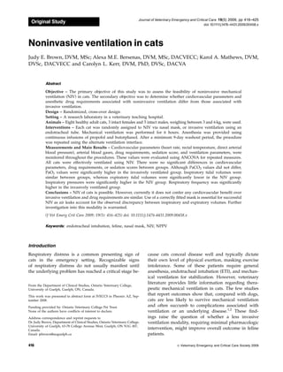
Figure 1: Application of noninvasive ventilation via nasal mask
in cat.
& Veterinary Emergency and Critical Care Society 2009, doi: 10.1111/j.1476-4431.2009.00458.x
418
J.E. Brown et al.
4. through the administration of propofol boluses
(1 mg/kg, IV) and an increase in the infusion rate by
25 mg/kg/min. The rate of butorphanol administration
was increased to a rate of 0.4 mg/kg/h after approxi-
mately 1 hour of ventilation. Once the 6-hour ventila-
tion period was complete, and spontaneous ventilation
was confirmed, mechanical ventilation was discontin-
ued and the mask or endotracheal tube was removed.
Left lateral and ventrodorsal radiographic projections
of the thorax and cranial abdomen were taken within 30
minutes of discontinuing ventilation to detect aero-
phagia, pneumothorax or other complications arising
from the ventilation. The cat was then monitored until
it was conscious, normothermic and making efforts to
ambulate.
Statistical analysis
The cross-over design allowed each animal to serve as
its own control. Physiologic data measured at each 15-
or 30-minute interval was evaluated using ANCOVA
accounting for repeated measures made over time on
the same animal. If the overall F-test was significant for
an interaction of treatment and time, paired compari-
sons were based on a multivariate t-adjustment. With
mode, time, period, and carryover as main effects,
catheter type was also included in the model as a co-
variable in the generalized linear model.n
The assumptions of normality were assessed by com-
prehensive residual analysis. A Shapiro-Wilk test in-
cluding examination of the residuals was conducted to
assess the overall normality. Where appropriate
the data were log-transformed. The P-value was set
at o0.05.
Results
The cardiovascular parameters, SA score, ventilation
parameters, arterial blood gas values, and anesthetic
drug requirements were compared between the inva-
sively ventilated group (Group I) and the NIV group
(Group NIV). No significant differences in mean heart
rate, systolic blood pressure, mean blood pressure, di-
astolic blood pressure, or temperature were found be-
tween Groups I and NIV (Table 3). There was not a
significant difference between the mean SA scores in
Group I versus Group NIV (Table 3). There were no
significant differences in propofol or butorphanol re-
quirements, evaluated on a mg/kg basis, between any
of the groups (Table 3). All cats required the maximum
butorphanol rate of 0.4 mg/kg/h.
Mean ventilatory parameters are reported in Table 4.
The respiratory frequency was divided into 2 compo-
nents: (1) the mandatory frequency, a minimum num-
ber of pressure-controlled breaths per minute delivered
by the ventilator, and (2) the spontaneous frequency,
the number of pressure-supported breaths initiated by
the subject, above and beyond the mandatory fre-
quency. Together, these values were expressed as the
total frequency. The mean total frequency and mean
mandatory frequency were both significantly higher
in Group I than in Group NIV. No significant difference
in spontaneous frequency was found between any
groups. Both the peak and mean inspiratory pressures
were significantly higher in Group NIV than in Group I.
While no significant difference in mean inspired tidal
volume was found between groups, mean expired tidal
volume was significantly lower in Group NIV than in
Group I.
Serial arterial blood gas analysis revealed signifi-
cantly higher mean PaO2, mean pH, and mean lactate
levels, and significantly lower HCO3 levels, in Group I
than in the Group NIV (Table 5). Despite the statisti-
cally significant differences in these values, all values
remained within the reference interval. There was not a
significant difference between PaCO2 or base excess
levels between groups.
Pre- and post-ventilation radiographs revealed that,
apart from the presence of air in the esophagus of cats
that had been ventilated noninvasively, no other radio-
graphic abnormalities were apparent. Gastric disten-
Table 3: Mean cardiovascular parameters, drug requirements, and sedation-anesthesia scores for invasively and noninvasively
ventilated cats
Parameter Group I (CI) Group NIV (CI) P-value
Heart rate (bpm) 141.25 (117.83–164.67) 153.18 (129.7–176.66) 0.48
Systolic BP (mm Hg) 117.38 (107.04–127.73) 126.44 (116.6–136.29) 0.16
Diastolic BP (mm Hg) 68.34 (58.35–78.35) 78.93 (68.93–88.94) 0.13
Mean BP (mm Hg) 84.77 (74.81–97.19) 94.44 (83.50–106.98) 0.23
Temperature (1C) 37.66 (37.34–37.98) 37.72 (37.42–38.03) 0.74
Total propofol (mg/kg) 42.69 (37.46–47.94) 41.89 (36.65–47.13) 0.82
Total butorphanol (mg/kg) 2.14 (2.02–2.27) 2.22 (2.10–2.35) 0.36
Sedation-Anesthesia Score 5.7 (5.55–5.91) 5.6 (5.44–5.78) 0.31
I, invasively ventilated; NIV, noninvasively ventilated, CI, confidence interval; BP, blood pressure.
& Veterinary Emergency and Critical Care Society 2009, doi: 10.1111/j.1476-4431.2009.00458.x 419
Noninvasive Ventilation in Cats
5. sion was not evident in any of the subjects. The average
decrease in packed cell volume over the course of the 2
ventilation episodes was 6% (range: 2–11%).
Of the 16 arterial catheter placements, 6 (38%) were
dorsopedal, 7 (44%) were coccygeal, and 3 (19%) were
femoral. Because of the potential impact of catheter lo-
cation on the study results, catheter location was in-
cluded as a covariable in the statistical analysis of the
data. There were significant differences in body tem-
perature, total and mandatory respiratory frequency,
peak inspiratory pressure, expiratory tidal volume, and
lactate between catheter locations. Body temperature
was lower in cats requiring femoral arterial catheters.
The differences in the other parameters were not
clinically significant as they remained within reference
intervals.
Application of NIV required continuous monitoring
and involved more intensive patient care than was an-
ticipated. Because small movements such as a shift in
head position or licking of the lips resulted in mask
displacement and air leaks, the cats required constant
monitoring to ensure appropriate mask position.
Also, active humidification of the inspired air caused
condensation accumulation within the mask, necessi-
tating intermittent (every 1–2 h) water removal. Com-
plications, necessitating discontinuation of NIV, were
encountered in 2 cats. Cat 5 became markedly hyper-
capneic (PaCO2 71.8 mm Hg) approximately 90 minutes
into the NIVepisode. At that time, the PaO2 was 101mm
Hg and had decreased from 122 mm Hg measured
30 minutes prior. The episode of hypercapnia and
relative hypoxia (expected PaO2 with FiO2 of 35% is
140–175 mm Hg) was associated with paradoxical
breathing that was asynchronous with the ventilator.
Adjustments in ventilator settings and mask position
failed to ameliorate the elevated PaCO2 and mechanical
ventilation and propofol infusion were discontinued.
Thoracic radiographs revealed a radiopaque area in the
region of the right middle lung lobe and a cardiac shift
to the right side. These changes were consistent with
atelectasis. Initially, the cat had been positioned in ster-
nal recumbency with both hind limbs to the right. Two
hours after discontinuation of NIV, the hypercapnia
had resolved. With the cat in sternal recumbency and
the hindlimbs out to either side, NIV was reinstituted
and continued for the full 6-hour study period without
further adverse events.
Cat 4 also became hypercapnic but had normal ox-
ygen tension (PaCO2 51.7 mm Hg, PaO2 162 mm Hg)
after 45 minutes of NIV and breathing efforts were
asynchronous with the ventilator. The cat had been in
full sternal recumbancy. NIV was discontinued for 30
Table 5: Mean arterial blood gas values for invasively and noninvasively ventilated cats
Parameter Group I (CI) Group NIV (CI) P-value
pH 7.35 (7.33–7.37) 7.32 (7.3–7.34)n
o0.001
PaCO2 (mm Hg) 40.32 (38.57–42.07) 42.2547 (40.51–44.0) 0.15
PaO2 (mm Hg) 172.16 (168.73–175.59) 165.58 (162.29–168.86)n
o0.001
HCO3 (mmol/L) 20.79 (19.84–21.73) 21.67 (20.74–22.59)n
0.01
Lactate (mmol/L) 0.6 (0.47–0.73) 0.51 (0.38–0.65)n
0.02
BE 3.71 ( 4.90– 2.53) 3.46 ( 4.61–2.30) 0.58
I, invasively ventilated; NIV, noninvasively ventilated, CI, confidence interval; BE, base excess.
n
Statistically significant.
Table 4: Mean ventilatory parameters for invasively and noninvasively ventilated cats
Parameter Group I (CI) Group NIV (CI) P-value
Total frequency 19.25 (16.30–22.73) 16.82 (14.28–19.82)n
0.007
Mandatory frequency 18.91 (14.41–24.82) 12.08 (9.20–15.87)n
0.03
Spontaneous frequency 6.04 (3.61–10.12) 4.67 (2.95–7.41) 0.48
Mean pressure (cm H2O) 6.31 (5.88–6.75) 7.53 (7.09–7.97)n
o0.001
Peak pressure (cm H2O) 10.94 (9.61–12.27) 14.73 (13.4–16.07)n
o0.001
Tidal volume (mL) 46.62 (35.4–57.8) 38.55 (27.3–49.8) 0.32
Tidal volume (mL/kg) 11.05 (8.39–13.70) 9.14 (6.47–11.8) –
Expiratory tidal volume (mL) 37.03 (29.3–44.7) 9.61 (1.9–17.3)n
o0.001
Expiratory tidal volume (mL/kg) 8.77 (6.94–10.59) 2.28 (0.45–4.10) –
I, invasively ventilated; NIV, noninvasively ventilated, CI, confidence interval.
n
Stastically significant.
& Veterinary Emergency and Critical Care Society 2009, doi: 10.1111/j.1476-4431.2009.00458.x
420
J.E. Brown et al.
6. minutes and resumed once PaCO2 had normalized.
NIV was administered for the full 6-hour study period
without any further complications.
Discussion
This study describes the application of NIV in cats in an
effort to determine whether the benefits of NIV ob-
served in humans extend to this species. The positive
impact of NIV on patient outcomes in the human pop-
ulation has been attributed to a reduction in complica-
tions that arise from 3 factors: (a) the process of
intubation, anesthesia, and mechanical ventilation; (b)
the loss of airway defense mechanisms; and (c) post-
ventilation effects.5
These types of complications also
arise in cats. Airway trauma associated with ETI of cats
has been reported in the veterinary literature.17–21
In
general practice, approximately 10% of all anesthesia-
related complications in cats have been attributed
to intubation.17
Post-ventilation complications such as
tracheal stenosis have also been reported in cats.21
The use of a mask rather than an endotracheal tube
eliminates these risks. Additionally, due to its invasive
nature, placement of an endotracheal tube requires the
administration of anesthetic drugs. Most available
injectable anesthetic agents cause some form of hemo-
dynamic compromise in cats. These effects, com-
pounded by the administration of positive-pressure
ventilation, can depress the cardiovascular status of the
patient. While almost all invasively ventilated human
patients are sedated or anesthetized,22
only a minority
of patients managed with NIV require any form of
sedation.23
These findings suggest that NIV may im-
prove cardiovascular parameters by virtue of requiring
less pharmacological intervention to maintain interface
tolerance.
The loss of airway defense mechanisms associated
with ETI predisposes patients to the development of
ventilator-associated pneumonia. Ventilator-associated
pneumonia is the most common ICU-acquired infection
in the human population24
and occurs in at least 11–
15% of mechanically ventilated veterinary patients.1,2,25
ETI impairs the cough reflex, interferes with mucocili-
ary clearance, and damages the tracheal epithelium
causing a breach in natural barriers to bacterial coloni-
zation of the airway. The endotracheal tube also serves
as a direct conduit through which bacteria migrate into
the lower airways26,27
and acts as a reservoir for bac-
terial accumulation.28,29
Elimination of the endotracheal
tube through the use of NIV has been shown to mark-
edly reduce the incidence of nosocomial pneumonia in
humans30
and holds promise as a means of decreasing
ventilation-associated morbidity in the veterinary pop-
ulation as well.
The study reported here demonstrates that NIV in
cats is possible, but cannot yet be considered feasible, as
it requires further refinement before being a practical
treatment option in a clinical setting. Contrary to the
initial hypothesis, the noninvasively ventilated cats did
not demonstrate significant improvements in cardio-
vascular parameters over the invasively ventilated cats.
Both groups maintained normal blood pressure and
lactate, consistent with cardiovascular stability. Also
contrary to expectation, sedation scores and anesthetic
drug requirements were similar in both groups.
Failure to identify a difference in anesthetic drug re-
quirements between groups highlights an important
distinction between the use of NIV in humans and an-
imals. Calm tolerance of a noninvasive interface is es-
sential for the success of NIV. While this state is readily
achieved in many human patients with verbal encour-
agement or mild sedation, the cats in this study required
considerably more chemical restraint to ensure compli-
ance. In order to attain a level of consciousness that
permitted tolerance of the mask, and prevented move-
ment causing gas leaks around the mask, these cats
were maintained in a state between deep sedation and
light anesthesia. In humans, this degree of altered con-
sciousness, and the consequent inability to protect the
airway from aspiration of gastric contents, is a contra-
indication to NIV according to the International
Consensus on Noninvasive Positive-Pressure Ventila-
tion in Acute Respiratory Failure.31
Based on these
guidelines, the findings reported here call into question
the applicability of NIV to deeply sedated cats. It is
interesting to note that studies exploring the use of NIV
in human patients with altered levels of consciousness
have yielded positive results. Two studies have evalu-
ated the use of NIV in human patients with acute
exacerbations of chronic obstructive pulmonary disease,
suffering from altered mentation due to hypercapneic
encephalopathy. Both studies found that NIV was
equally successful in patients with and without altered
mentation. Neither study found a significant difference
in mortality between groups.32,33
There are obvious dis-
similarities between human chronic obstructive pulmo-
nary disease patients, whose altered neurologic states
typically improve in a matter of hours, and veterinary
patients with various diagnoses, that might require pro-
longed ventilation. Nonetheless, these studies illustrate
that NIV can be successfully administered to patients
with a level of consciousness equivalent to that which
was achieved with this study. Despite these findings,
ventilating cats with an unprotected airway remains a
serious concern. A more readily tolerated NIV interface
would permit lighter levels of sedation and enhanced
airway protection. The development of such an interface
is the logical next step in refining NIV for cats.
& Veterinary Emergency and Critical Care Society 2009, doi: 10.1111/j.1476-4431.2009.00458.x 421
Noninvasive Ventilation in Cats
7. Propofol and butorphanol were selected as sedative/
anesthetic agents in this study. Propofol was selected
because its rapid metabolism would permit accurate
titration and moment-to-moment adjustments in anes-
thetic level, facilitating frequent SA score assessments.
Its use in cats has been evaluated in several prospective
studies and found to cause only mild to moderate
hemodynamic compromise in healthy individuals.34–39
However, propofol also produces respiratory depres-
sion by inciting transient apnea and bradypnea.40,41
This effect likely resulted in lower spontaneous respi-
ratory rates in the cats studied, and made the signifi-
cant differences in respiratory rate between groups
difficult to interpret. In an effort to reduce propofol
requirements, butorphanol was selected as an adjunc-
tive sedative. It was chosen over a benzodiazepine be-
cause of its analgesic properties, and over other opioids
because its respiratory and cardiovascular depressive
effects are comparatively less profound.42
Although at-
tempts were made to reduce respiratory depression by
administering a high rate of butorphanol (0.4 mg/kg/h)
and minimizing propofol administration as much
as possible, all cats required a propofol rate of at least
25–50 mg/kg/min to maintain interface tolerance.
NIV is characterized by the nature of the patient-
ventilator interface. Although some ventilators are de-
signed exclusively for NIV administration, it can be
applied using any ICU ventilator with an NIV setting,
and using any mode of ventilation (pressure- or volume-
controlled, with spontaneous or mandatory triggering).
Pressure-controlled ventilation was selected for this
study because the degree of leak at the outset of the
study was unknown and there was concern that the
administration of a fixed tidal volume provided by
volume-controlled ventilation might have resulted in
inadequate tidal volume delivery.
The significantly higher mean mandatory respiratory
frequency found in Group I is difficult to explain. In
some instances, the cats in Group NIV developed ven-
tilator asynchrony when the mandatory frequency was
increased, whereas this trend was not noted in Group I.
This tendency may have lead to the administration of
lower mandatory frequencies in Group NIV subjects.
The peak and mean inspiratory pressures were both
significantly higher in Group NIV than in Group I. This
was likely because the leak in Group NIV resulted in
the need for higher inspiratory pressures than in Group
I to attain the same ventilation targets.
The presence of a leak was most profoundly demon-
strated by the significant difference between mean ex-
piratory tidal volumes in Group NIV (2.28 mL/kg) and
Group I (8.77 mL/kg). The mean inspiratory (delivered)
tidal volumes did not differ significantly between
groups (11.05 mL/kg in Group I; 9.14 mL/kg in Group
NIV). Thus the disparity between the exhaled volumes
implies that, in Group NIV, a substantial proportion of
the delivered volume leaked from the system before
reaching the expiratory valve, whereas the leak in
Group I was negligible. The leakage in mask-ventilated
cats most likely occurred at the level of the mask or
through the mouth. Because of the small volumes and
the absence of a measuring device at the level of the
mask, it is difficult to assess how accurately these mea-
surements reflect the actual tidal volumes that were
delivered to the cats.
The slightly, but significantly, higher mean PaO2 val-
ues found in Group I may be explained by an increased
efficiency of gas exchange in this group. The mean ar-
terial blood pH was significantly higher in Group I than
in Group NIV. Although there was not a significant
difference between PaCO2 levels between groups, al-
terations in pH reflected changes in PaCO2, suggesting
that PaO2 levels were likely responsible for the signifi-
cant differences observed in blood pH between groups.
These changes are probably due to a relatively higher
degree of atelectasis in Group NIV. This occurred be-
cause the degree of inspiratory pressure sustained in
Group I could not be maintained in Group NIV due to
the ongoing leak. Lactate and HCO3 levels were sig-
nificantly higher and lower, respectively, in Group I
than in Group NIV. However, despite the statistically
significant differences in PaO2, pH, lactate, and HCO3 ,
each value remained within the target range (see Table
5) and the differences described were not considered
clinically important.
Of the cats ventilated in this study, none showed ra-
diographic evidence of gastric insufflation or pneumo-
thorax. In humans, gas distension of the stomach is a
relatively common complication of NIV, occurring in 5–
10% of patients who receive NIV.5,10
Placement of a
nasogastric tube typically resolves the problem and it
can be prevented by maintaining peak inspiratory pres-
sure below the resting upper esophageal sphincter
pressure that is reported to be 33 129
or 25 mm Hg43
in humans. In the study reported here, all the cats had
normal lungs and peak inspiratory pressures were eas-
ily maintained below 20 cm H2O (14.7 mm Hg), sub-
stantially lower than reported normal feline lower
esophageal sphincter pressures (40 mm Hg).44
Low in-
spiratory pressures and the short duration of ventila-
tion likely prevented the development of gastric
distension, pneumothorax, and other manifestations
of barotrauma.
Two cats developed hypercapnia and asynchrous
breathing within the first 2 hours of NIV. Both events
necessitated discontinuation of NIV for a short period
of time. In 1 cat, radiographic findings were consistent
with atelectasis. Radiographs of the second cat were not
Veterinary Emergency and Critical Care Society 2009, doi: 10.1111/j.1476-4431.2009.00458.x
422
J.E. Brown et al.
8. obtained. Hypercapnia resolved rapidly in both cats
once mechanical ventilation and sedation were discon-
tinued. Following their respective episodes, each cat
was successfully ventilated noninvasively for 6 consec-
utive hours without further complications. Explana-
tions for these events are not readily apparent as
hypercapnia is an unexpected complication of a ther-
apy intended to provide ventilatory support. Four pos-
sible reasons are (a) decreased minute ventilation due
to air leakage around the mask, (b) CO2 rebreathing
secondary to increased dead space of the mask, (c)
trigger-associated asynchrony, or (d) delayed cycling.
Hypoventilation occurs due to decreased effective
alveolar minute ventilation. It is possible that the per-
sistent leak from the mouth and mask edges resulted in
impaired lung inflation, ineffective breaths, and hypo-
ventilation in the 2 cats that developed hypercapnia.
This likely contributed to the development of atelectasis
in 1 of the cats. Masks contribute a larger amount of
static volume to the ventilator circuit than do endotra-
cheal tubes. Theoretically, this increase in dead space
could cause rebreathing of CO2 that is not flushed out
of the mask. However, this possibility is unlikely for
several reasons. First, evaluation of CO2 rebreathing in
humans receiving NIV has revealed a weak correlation
between static mask volume and degree of dead space.
This is because the movement of gas through the mask
can effectively reduce dynamic dead space to less than
what it would be during spontaneous ventilation with-
out positive pressure. Maintaining positive end-expira-
tory pressure 44 cm H2O has also been shown to
reduce rebreathing in some NIV ventilators45
and the
levels of positive end expiratory pressure in this study
were maintained at 5 cm H2O in all of the subjects. Fi-
nally, had mask dead space been the problem, it would
not have resolved with repositioning and cessation of
anesthetic drugs; all cats were ultimately successfully
ventilated with the masks, despite initial failures.
Another possible explanation for the hypercapnic
episodes observed is trigger-associated asynchrony,
which arises when a large leak is erroneously detected
by the ventilator as an inspiratory effort, triggering a
breath.46
This aberrant triggering leads to the admin-
istration of breaths irrespective of the subject’s efforts,
resulting in a reduction in alveolar ventilation and pre-
cipitation of NIV failure.45,47
A related problem is de-
layed cycling, which refers to the failure of ventilator to
appropriately cycle into expiration. This can occur
when a leak prevents the ventilator from meeting its
pressure target and the expiratory trigger fails to detect
the expiratory efforts of the patient. Alternatively, de-
layed cycling occurs if the programmed inspiratory
time is inappropriately long. This leads to a reduced
expiratory time and reduced lung emptying, causing
dynamic hyperinflation. With hyperinflated lungs, the
subject is less able to effectively trigger breaths and
work of breathing is increased.46
In this study, the
baseline inspiratory time was set based on maintaining
an inspiratory to expiratory (I:E) ratio of 1:2 with a
mandatory respiratory rate of 10 breaths per minute.
These settings resulted in an inspiratory time setting of
2 seconds. During the study, the inspiratory time was
found to be excessive and may have contributed to de-
layed cycling. Overall, the extent to which leak-related
pulmonary underinflation, trigger-associated asynchro-
ny, and delayed cycling contributed to the observed
hypercapnia remains undetermined. These unan-
swered questions emphasize the need for further in-
vestigation of NIV application in cats.
This study had several limitations. First, the sedation
protocol selected reflected an attempt to allow rapid,
titratable changes in sedation level to meet the objec-
tives of the study. The agents chosen, however, are not
ideal agents for use in a clinical setting and interfere
with some aspects of ventilation assessment. To eval-
uate sedation requirements, the cats were maintained
on the lowest possible rate of propofol that allowed
tolerance of the interface. Propofol boluses were ad-
ministered if the level of sedation became excessively
light. Because of the consequent intermittent periods of
apnea, these boluses generated an inconsistent breath-
ing pattern. Thus, the respiratory rates recorded may be
more reflective of the sedation status of the animal than
the mode of ventilation. Of interest, however, was that
NIV was used successfully to ventilate cats throughout
prolonged episodes of apnea.
Secondly, butorphanol was selected as an adjuctive
sedative because of its analgesic effects and its minimal
impact on blood pressure and ventilatory drive. In ap-
plication of NIV in humans, the antitussive effects of
butorphanol would be contraindicated due to the as-
sociated impairment of airway clearance. Theoretically,
these guidelines would also apply to veterinary pa-
tients, making butorphanol an inappropriate agent to
use with NIV in a clinical setting. However, because the
cats in our study required levels of sedation that pre-
vented coughing regardless of the drugs used, the an-
titussive effects of butorphanol may prove to be a moot
point in the veterinary context. Further investigations
of sedation protocols for NIV are needed to clarify this
issue.
A third limitation of the study involved the hetero-
geneity of arterial catheter sites. When dorsopedal or
coccygeal arterial catheterization was not possible, a
femoral arterial catheter was placed using a surgical
cut-down approach. Thus, catheter location was in-
cluded as a covariable in the statistical analysis of the
data. Catheter location had a statistically significant
Veterinary Emergency and Critical Care Society 2009, doi: 10.1111/j.1476-4431.2009.00458.x 423
Noninvasive Ventilation in Cats
9. impact on temperature, respiratory frequency, peak in-
spiratory pressures, and lactate. Femoral arterial cath-
eters were associated with body temperatures below
reference intervals because placement required pro-
longed periods of isoflurane anesthesia. However, the
other values remained within reference intervals, and
the statistically significant changes in these values were
not considered to be clinically important.
This investigation is, to the authors’ knowledge, the
first randomized, controlled study to assess the thera-
peutic value of NIV in cats. NIV was administered to
8 cats for 6 consecutive hours during which time blood
gases and cardiovascular parameters were maintained
within a target physiologic range. Sedation levels
and drug requirements were similar to those of inva-
sively ventilated animals. No serious complications
were encountered. As a pilot endeavor, this study illu-
minates numerous avenues in need of further investi-
gation. These include the optimization of ventilator
settings and sedation protocols for cats receiving me-
chanical ventilation, and the development of NIV in-
terfaces that are tolerated by the patient and require
minimal sedation. NIV has contributed to a dramatic
reduction in morbidity and mortality in humans and it
is important and worthwhile to explore extending its
benefits to the veterinary patient population as well.
Acknowledgement
The authors gratefully acknowledge Amanda Hathway
and James Imada for their assistance during this study.
Footnotes
a
Torbugesic butorphanol tartrate, Wyeth Animal Health, Guelph, ON,
Canada.
b
Glycopyrrolate, Sandoz, Boucherville, QC, Canada.
c
Insyte-W, Becton Dickenson Infusion Therapy Systems Inc, Sandy, UT.
d
Propofol, Novopharm, Toronto, ON, Canada.
e
Lidodan, Odan Laboratories, Montreal, PQ.
f
Aerrane, Baxter Corporation, Mississauga, ON, Canada.
g
Plasmalyte A, Baxter Corporation.
h
Arrow Radial Arterial Catherization Set, Arrow International, Reading,
PA.
i
BabyFlow Nasal CPAP Accessory, Dräger Medical, Telfor, PA.
j
Evita 4, Dräger, Lubeck, Germany.
k
Respiratory humidifier, Fischer and Paykel Healthcare Systems,
Auckland, New Zealand.
l
TCM3, Radiometer, Copenhagen, Denmark.
m
ABL 700 Series Blood Gas Analyzer, Radiometer.
n
SAS OnlineDOC (R) 9.1.3., SAS Institue Inc, 2004, Cary, NC.
References
1. Hopper K, Haskins SC, Kass PH, et al. Indications, management
and outcome of long-term positive-pressure ventilation in dogs
and cats: 148 cases (1990–2001). J Am Vet Med Assoc 2007;
230(1):64–75.
2. Lee JA, Drobatz KJ, Koch MW, et al. Indications for and outcomes
of positive-pressure ventilation in cats: 53 cases (1993–2002). J Am
Vet Med Assoc 2005; 226(6):924–931.
3. Ambrosino N, Vagheggini G. Non-invasive positive pressure ven-
tilation in the acute care setting: where are we now? Eur Resp J
2008; 31(4):874–886.
4. Hill NS. Non-invasive positive-pressure ventilation, In: Tobin MJ.
ed. Principles and Practice of Mechanical Ventilation, 2nd edn.
Toronto: McGraw-Hill; 2006, pp. 433–471.
5. Mehta S, Hill NS. Noninvasive ventilation. Am J Crit Care Med
2001; 163(2):540–577.
6. Aghai ZH, Saslow JG, Nakhla T, et al. Synchronized nasal inter-
mittent positive pressure ventilation (SNIPPV) decreased work of
breathing (WOB) in premature infants with respiratory distress
syndrome (RDS) compared to nasal continuous positive airway
pressure (NCPAP). Pediatr Pulmonol 2006; 41(9):875–881.
7. Crane SD, Elliott MW, Gilligan P, et al. Randomised controlled
comparison of continuous positive airways pressure, bilevel non-
invasive ventilation, and standard treatment in emergency
department patients with acute cardiogenic pulmonary edema.
Emerg Med J 2004; 21(2):155–161.
8. Gramlich T. Basic concepts of non-invasive positive pressure ven-
tilation, In: Pilbeam SP, Cairo JM. eds. Mechanical Ventilation:
Physiological and Clinical Applications, 4th edn. St Louis: Mosby
Elsevier; 2006, pp. 15–30.
9. Meduri GU, Cook TR, Turner RE, et al. Noninvasive positive
pressure ventilation in status asthmaticus. Chest 1996; 110(3):767–
774.
10. Liesching T, Kwok H, Hill NS. Acute applications of non-invasive
positive pressure ventilation. Chest 2003; 124(2):699–713.
11. Sauvé V, Drobatz KJ, Shokek AB, et al. Clinical course, diagnostic
findings and necropsy diagnosis in dyspneic cats with primary
pulmonary parenchymal disease: 15 cats (1996–2002). J Vet Emerg
Crit Care 2005; 15(1):38–47.
12. Mandell DC. Respiratory distress in cats, In: King LG. ed. Text-
book of Respiratory Disease in Dogs and Cats. St Louis: Saunders;
2004, pp. 12–17.
13. Ammari A, Suri M, Milisavljevic V, et al. Variables associated with
the early failure of nasal CPAP in very low birth weight infants.
J Pediatr 2005; 147(3):341–347.
14. Santin R, Brodsky N, Bhandari V. A prospective observational
study of synchronized intermittent positive pressure ventilation
(SNIPPV) as a primary mode of ventilation in infants 428 weeks
with respiratory distress syndrome. J Perinat 2004; 24(8):487–493.
15. Carrasco G. Instruments for monitoring intensive care unit seda-
tion. Crit Care 2000; 4(4):217–225.
16. Trim CM. Monitoring the anaesthetized cat, In: Hall LW, Taylor
PM. eds. Anesthesia of the Cat. Cambridge, UK: Bailliere Tindall;
1994, pp. 194–223.
17. Dyson DH, Maxie MG, Schnurr D. Morbidity and mortality as-
sociated with anesthetic management in small animal veterinary
practice in Ontario. J Am Anim Hosp Assoc 1998; 34(4):325–335.
18. Hofmeister EH, Trim CM, Kley S, et al. Traumatic endotracheal
intubation in the cat. Vet Anaesth Analg 2007; 34(3):213–216.
19. Mitchell SL, McCarthy R, Rudloff E, et al. Tracheal rupture asso-
ciated with intubation in cats: 20 cases (1996–1998). J Am Vet Med
Assoc 2000; 216(10):1592–1595.
20. Hardie EM, Spodnick GJ, Gilson SD, et al. Tracheal rupture in cats:
16 cases (1983–1998). J Am Vet Med Assoc 1999; 214(4):508–512.
21. McMillan FD. Iatrogenic tracheal stenosis in a cat. J Am Anim
Hosp Assoc 1985; 21:747–750.
22. Samuelson KA, Larsson S, Lundberg D, et al. Intentsive care se-
dation of mechanically ventilated patients: a national Swedish
survey. Intens Crit Care Nurs 2003; 19(6):350–362.
23. Devlin JW, Nava S, Fong JJ, et al. Survey of sedation practices
during non-invasive positive-pressure ventilation to treat acute
respiratory failure. Crit Care Med 2007; 35(10):2298–2302.
24. American Thoracic Society; Infectious Diseases Society of Amer-
ica. Guidelines for management of adults with hospital-acquired,
ventilator-associated, and healthcare-associated pneumonia. Am J
Respir Crit Care Med 2005; 171(4):388–416.
Veterinary Emergency and Critical Care Society 2009, doi: 10.1111/j.1476-4431.2009.00458.x
424
J.E. Brown et al.
10. 25. King LG, Hendricks JC. Use of positive-pressure ventilation in
dogs and cats: 41 cases (1990–1992). J Am Vet Med Assoc 1994;
204(7):1045–1052.
26. Davis KA. Ventilator-associated pneumonia: a review. J Intensive
Care Med 2006; 21(4):211–226.
27. Pieracci FM, Barie PS. Strategies in the prevention and manage-
ment of ventilator-associated pneumonia. Am Surg 2007; 73(5):
419–432.
28. Adair CG, Gorman SP, Feron BM, et al. Implications of endotrac-
heal tube biofilm for ventilator-associated pneumonia. Intensive
Care Med 1999; 25(10):1072–1076.
29. Koerner RJ. Contribution of endotracheal tubes to the pathogen-
esis of ventilator-associated pneumonia. J Hosp Infect 1997; 35(2):
83–89.
30. Girou E, Brun-Buisson C, Taille S, et al. Secular trends in no-
socomial infections and mortality associated with non-invasive
ventilation in patients with exacerbation of COPD and pulmonary
edema. JAMA 2003; 290(22):2985–2991.
31. American Thoracic Society.. International consensus conferences
in intensive care medicine: non-invasive positive pressure venti-
lation in acute respiratory failure. Am J Respir Crit Care Med 2001;
163(1):283–291.
32. Scala R, Naldi M, Archinucci I, et al. Non-invasive positive
pressure ventilation in patients with acute exacerbations of
COPD and varying levels of consciousness. Chest 2005; 128(3):
1657–1666.
33. Diaz GG, Alcaraz AC, Talavera JC, et al. Non-invasive positive-
pressure ventilation to treat hypercapnic coma secondary to re-
spiratory failure. Chest 2005; 127(3):952–960.
34. Liehmann L, Mosing M, Auer U. A comparison of cardiorespira-
tory variables during isoflurane–fentanyl and propofol–fentanyl
anesthesia for surgery in injured cats. Vet Anaesth Analg 2006;
33(3):158–168.
35. Pereira GG, Larsson MH, Yamaki FL, et al. Effects of propofol on
the electrocardiogram and systolic blood pressure of healthy cats
pre-medicated with acepromazine. Vet Anaesth Analg 2004;
31(3):235–238.
36. Mendes GM, Selmi AL. Use of a combination of propofol and
fentanyl, alfentanil, or sufentanil for total intravenous anesthesia
in cats. J Am Vet Med Assoc 2003; 223(11):1608–1613.
37. Ilkiw JE, Pascoe PJ, Tripp LD. Effect of variable-dose propofol
alone and in combination with two fixed doses of ketamine for
total intravenous anesthesia in cats. Am J Vet Res 2003; 64(7):907–
912.
38. Ilkiw JE, Pascoe PJ. Cardiovascular effects of propofol alone and in
combination with ketamine for total intravenous anesthesia in
cats. Am J Vet Res 2003; 64(7):913–917.
39. Correa MA, Aguiar AJ, Teixeira Neto FJ, et al. Effects of re-
mifentanil infusion regimens on cardiovascular function and re-
sponses to noxious stimulation in propofol-anesthetized cats. Am J
Vet Res 2007; 68(9):932–940.
40. Flecknell PA. Injectable anesthetics, In: Hall LW, Taylor PM. eds.
Anesthesia of the Cat. Toronto: Bailliere Tindall; 1994, pp. 129–156.
41. Plumb DC. Propofol, In: Plumb DC. ed. Veterinary Drug Handbook,
3rd edn. Ames: Iowa State University Press; 1999, pp. 632–635.
42. Tranquilli WJ. Pharmacology of preanesthetics and anesthetic ad-
juncts, In: Thurmon JC, Tranquilli WJ, Benson GJ. eds. Lumb and
Jones’ Veterinary Anesthesia, 3rd edn. Baltimore: Williams and
Wilkins; 1996, pp. 182–209.
43. Hillberg RE, Johnson DC. Non-invasive ventilation. N Engl J Med
1997; 337(24):1746–1752.
44. LiCalzi LK, Biancani P, Behar J, et al. Effect of hemorrhagic hy-
potension and hypoventilation on lower esophageal sphincter
pressure. Ann Surg 1980; 192(1):53–57.
45. Ferguson GT, Gilmartin M. CO2 rebreathing during BiPAP ven-
tilatory assistance. Am J Resp Crit Care Med 1995; 151(4):1126–
1135.
46. Jolliet P, Tassaux D. Clinical review: patient-ventilator interaction
in chronic obstructive pulmonary disease. Crit Care 2006;
10(6):236–242.
47. Miyoshi E, Fujino Y, Uchiyama A, et al. Effects of gas leak on
triggering function, humidification, and inspiratory oxygen frac-
tion during non-invasive positive airway pressure ventilation.
Chest 2005; 128(5):3691–3698.
Veterinary Emergency and Critical Care Society 2009, doi: 10.1111/j.1476-4431.2009.00458.x 425
Noninvasive Ventilation in Cats