2016 ghassemi-nasal reconstr-threelayer
•
0 likes•103 views

Dr. Alireza Ghassemi. reconstructive surgery

Recommended
Recommended
More Related Content
What's hot
What's hot (20)
Similar to 2016 ghassemi-nasal reconstr-threelayer
Similar to 2016 ghassemi-nasal reconstr-threelayer (19)
More from Klinikum Lippe GmbH
More from Klinikum Lippe GmbH (20)
Recently uploaded
Recently uploaded (20)
2016 ghassemi-nasal reconstr-threelayer
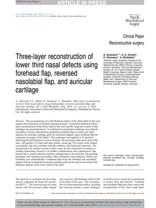
- 1. YIJOM-3515; No of Pages 5 Please cite this article in press as: Ghassemi A, et al. Three-layer reconstruction of lower third nasal defects using forehead flap, reversed nasolabial flap, and auricular cartilage, Int J Oral Maxillofac Surg (2016), http://dx.doi.org/10.1016/j.ijom.2016.08.024 Clinical Paper Reconstructive surgery Three-layer reconstruction of lower third nasal defects using forehead flap, reversed nasolabial flap, and auricular cartilage A. Ghassemi, S.S. Ahmed, H. Ghanepur, A. Modabber: Three-layer reconstruction of lower third nasal defects using forehead flap, reversed nasolabial flap, and auricular cartilage. Int. J. Oral Maxillofac. Surg. 2016; xxx: xxx–xxx. # 2016 International Association of Oral and Maxillofacial Surgeons. Published by Elsevier Ltd. All rights reserved. A. Ghassemi1,2 , S. S. Ahmed3 , H. Ghanepur4 , A. Modabber5 1 Klinikum Lippe, Academic Hospital of the University of Hannover, Detmold, Germany; 2 Medical Faculty, RWTH Aachen University, Aachen, Germany; 3 Oral and Maxillofacial Surgery, Dr. Z.A. Dental College, Aligarh Muslim University, Aligarh, India; 4 Department of Maxillofacial Surgery, Shahid Beheshti Hospital, University of Medical Science, Babol, Iran; 5 Department of Oral and Maxillofacial Surgery, RWTH Aachen University Hospital, Aachen, Germany Abstract. The reconstruction of a full-thickness defect of the distal third of the nose requires the restoration of all three anatomical layers. A practical method for three- layer reconstruction of the lower third of the nose and the long-term results of this technique are presented herein. A combined reconstruction technique was utilized, including a reverse subcutaneous pedicled nasolabial flap to restore the nasal mucosa, an auricular cartilage graft for structural support, and a forehead flap for cutaneous coverage of the defect. This technique was applied in 21 patients following the full-thickness excision of basal cell carcinoma of the lower part of the nose. All patients (12 male and nine female; mean age 59.8 years) were treated successfully and were satisfied with the aesthetic and functional outcomes. The wound had to be further revised in three cases for the correction of contour or residual deformities; however, no further complications were experienced. One patient had a wound infection and the cartilage had to be removed. The grafting procedure was repeated successfully after resolution of the infection. Donor site morbidity was unremarkable. Combined flaps from the forehead and nasolabial regions with an incorporated auricular cartilage graft can be used to reconstruct full- thickness defects of the lower third of the nose. Key words: three-layer nasal reconstruction; reversed nasolabial flap; auricular cartilage; forehead flap. Accepted for publication 23 August 2016 The nasal ala is a common site for malig- nancies, especially for basal cell carcino- ma (BCC).1,2 The local excision of a nasal tumour with the necessary safety margin may result in a full-thickness defect of the lower third of the nose.3 The reconstruc- tion of a lower nasal third defect, including the nasal tip, presents a major challenge, as all three layers need to be reconstructed to restore form and function.4 Forehead and nasolabial flaps have been used in the reconstruction of the lower nasal third, Int. J. Oral Maxillofac. Surg. 2016; xxx: xxx–xxx http://dx.doi.org/10.1016/j.ijom.2016.08.024, available online at http://www.sciencedirect.com 0901-5027/000001+05 # 2016 International Association of Oral and Maxillofacial Surgeons. Published by Elsevier Ltd. All rights reserved.
- 2. especially of the nasal ala.4–7 Other recon- structive options include the muco-peri- chondrial flap from septum, free skin graft and oral mucosa for the inner lining, com- bined with a forehead flap as skin cover- age.5–9 To restore a defect of the lower third of the nose properly, it is critical to focus on both function and aesthetics.10–12 All of the three missing anatomical layers should be replaced optimally: the thin nasal vestibule, the supportive cartilage, and the nasal skin.7 The provision of a reliable and sufficient nasal lining is con- sidered the most challenging aspect of nasal reconstruction. Inadequate recon- struction of the nasal lining is complicated by contracture of the skin cover and does not allow the simultaneous placement of a cartilage graft for skeletal framework support. The present authors have developed a method for the reconstruction of full- thickness defects of the distal third of the nose, using a combined forehead flap as skin cover, a subcutaneously pedicled reverse nasolabial flap as the nasal lining, and an auricular cartilage graft as the cartilaginous skeletal support. The techni- cal feasibility and technical performance of this alternative approach, as well as its aesthetic and functional outcomes, are discussed herein. Methods This study was approved by the necessary institutional review board and all patients signed an informed consent agreement to participate in the study. Twenty-one patients have been treated for BCC of the lower third of the nose at the study institution since March 2010 (Table 1). The surgical excision of the nasal ala included all three layers: the overlying skin coverage, the inner mucosal lining, and the interpositioning lateral crural cartilage. The patients were asked about the aesthetic outcome and any airway obstruction. Two surgeons evaluated the outcomes after more than 1 year. Surgical procedure The surgical method utilized to accom- plish the full-thickness reconstruction of the lower third nasal defects is illustrated in Fig. 1. The BCC was resected and a tumour- free margin was achieved in the first stage (Fig. 2a ). Resection of the safety margin and reconstruction of the lost part was performed in a second stage. A nasolabial flap was incised and dissected as a subcu- taneously pedicled flap, up to the ala base, as close as possible to the defect (Fig. 1 and 2b). Subcutaneous remnants of muscle are included in the flap in order to preserve a safe blood supply. The flap was reversed and its cranial border sutured to the caudal border of the remaining nasal mucosa, up to the base of the columella (Fig. 2b and c). The cutaneous coverage of the nasolabial flap was turned downward and replaced the lost nasal mucosa. After marking the size and shape of the lost skin according to the healthy side, a forehead flap was dis- sected as skin coverage (Fig. 2c and d). The distal end of the flap was carefully thinned out for easy fitting by considering the vascularization. Taking into account the shrinkage of flaps during the healing phase, a flap larger than the defect size was elevated. This should prevent pin-cushion- ing or alar displacement. Tension-free closure of the donor site was achieved in all cases. Vaseline-soaked gauze was inserted into the nasal hole to support the recon- structed ala. Antibiotics were prescribed for 5 days. After 7–10 days, the forehead flap was elevated and both flaps thinned out as far as possible. A cres- cent-shaped auricular cartilage graft was inserted for stabilization of the nasal ala. A mattress 4–0 Prolene suture, one laterally in the base of the ala and one into the tip region, secured the lateral and medial edges of the cartilage graft (Fig. 2d). After 2 weeks, the pedicle of the forehead flap was transected and carefully adapted. The final adaptation and debulking of the flaps, as required, was performed after 3–5 weeks (Fig. 2e). Figure 3 shows the outcome of the reconstruction after 18 months. Results The technique described was applied in 21 patients (12 men and nine women); their mean age was 59.8 years (range 43–84 2 Ghassemi et al. YIJOM-3515; No of Pages 5 Please cite this article in press as: Ghassemi A, et al. Three-layer reconstruction of lower third nasal defects using forehead flap, reversed nasolabial flap, and auricular cartilage, Int J Oral Maxillofac Surg (2016), http://dx.doi.org/10.1016/j.ijom.2016.08.024 Table 1. Patient characteristics. Sex Age range, years Defect location Total Right ala Left ala Female 53–76 6 3 9 Male 43–84 7 5 12 Total 13 8 21 Fig. 1. Artistic illustration of the technique used. (a) Incision of the cranially pedicled nasolabial flap. (b) The cranial edge of the reversed nasolabial flap is sutured with the caudal edge of the preserved nasal mucosa. An auricular cartilage graft is placed on the subcutaneous surface of the nasolabial flap in the second step. (c) Dissection and adaptation of a paramedian forehead flap as skin coverage.
- 3. years). The complete procedure could be performed under local anaesthesia in sev- en patients. Although the defect size was variable following excision of the tumour with a safety margin, it included the nasal ala in all patients. The flap had to be revised in three cases, and this resolved without any remarkable consequences. Antibiotics were continued for up to 10 days. The auricular cartilage was completely removed in one case, due to an infection of the surgical site. The area was grafted with new auricular cartilage after complete resolution of the wound infection. There was no case of flap ne- crosis or loss. All individuals, with the exception of two patients, were satisfied with the aesthetic and functional outcomes (Table 2). However, the two examiners who were involved in the surgery and post-surgical follow-up, found the ala still to be too thick and requiring improvement when compared with the healthy side. All patients reported some discomfort due to obstruction, but none of the patients com- plained of subjective nasal obstruction after 1 year. Discussion A reversed nasolabial flap was utilized to replace the inner mucosal nasal lining, a forehead flap for the overlying skin cov- erage, and an auricular cartilage graft as skeletal support. Malignancies such as BCC are the most common tumour of the skin and are most often found on the nasal ala.1,2 Excision of the tumour of the affected nasal region should include an adequate safety margin to reduce the rate of recurrence.3 This can subsequently lead to a large full-thickness defect. The surgi- cal choice of the preferred donor site will differ based on the location, size, tissues involved, medical comorbidities, and the desired outcome, taking into consideration airway patency, the aesthetic outcome, and the preferred technique or expertise of the surgeon.10,12 The nose consists of complex contours with alternating shadows and highlights, which make its reconstruction challeng- ing.10–12 To achieve a better aesthetic outcome, the surgeon should consider all of these aspects in the reconstruction plan, since any small deformity or residual de- fect will be easily visible. The principles of nasal subunits in facial aesthetics should be respected as much as possible to improve the aesthetic and functional Reconstruction of lower third nasal defects 3 YIJOM-3515; No of Pages 5 Please cite this article in press as: Ghassemi A, et al. Three-layer reconstruction of lower third nasal defects using forehead flap, reversed nasolabial flap, and auricular cartilage, Int J Oral Maxillofac Surg (2016), http://dx.doi.org/10.1016/j.ijom.2016.08.024 Fig. 2. (a) Full-thickness excision of a basal cell carcinoma of the left lower third part of the nose in a 60-year-old man. (b) The nasolabial flap is incised according to the defect size. The cranial edge of the flap is dissected and sutured to the caudal edge of the preserved nasal mucosa using 4–0 Vicryl. (c) The nasolabial flap is sutured and fitted into the defect completely to replace the lost nasal mucosa. The forehead flap is marked accordingly. (d) The forehead flap is elevated and adjusted to the defect as skin coverage. (e) Cartilage graft seen between the two flaps during the debulking procedure.
- 4. outcomes, although strict adherence to these principles is not mandatory.13–16 If a full-thickness defect is present, all three missing anatomical layers have to be replaced accordingly. This includes the thin, well-vascularized inner mucosal lin- ing, the skeletal support to avoid nasal collapse and airway obstruction, and skin coverage with similar colour and tex- ture.6,7,11,12 To date, many local and distant skin flaps have been suggested for skin cover- age.4–9,12,17–23 The reconstruction of the nasal mucosa is considered to be the most challenging aspect of the full-thickness nasal reconstruction. Numerous techni- ques have been described for this pur- pose.5,7–9,17–21 The pedicled forehead flap is one of the best options to replace a larger skin area and has become the gold standard for nasal reconstruction includ- ing the tip.11 It offers a large skin area with secured vascularization, along with excel- lent skin colour and texture. Turnover forehead flaps for internal lining, com- bined with the composite crus of the helix graft for external lining and mechanical support to the ala, has been described for partial lateral defects.19,21 However, it is difficult to fold a thick flap such as a forehead flap to shape the ala. Further- more, kinking of the flap at the hinge between the overlying skin cover and the inner lining may compromise the blood supply distally, especially if the distal portion is thinned to prevent airway obstruction. In addition, harvesting the tissue required from a single donor site can result in a large defect and make primary tension-free closure difficult. Lo- cal flaps of the nasal muco-perichondrium from the remaining septum or nasal side- wall can provide an excellent local tissue match with a good source of vasculariza- tion, which may support healing of the cartilage graft.9 Nevertheless, elevation of the muco-perichondrial flap results in ex- posure of the septum. Furthermore, this may be absent or impossible for larger defects. Alternative options for surgical reconstruction include full-thickness skin grafts, the turn-over island nasal skin flap, the transverse orbicularis oris myocuta- neous flap, the upper lip flap, the free mucosa graft, and the grafted or prefabri- cated forehead flap, up to microvascular free tissue transfer.7–9,14,17,18,20,21 The nasolabial flap has been used pre- viously for different parts of the nose, especially the ala.6,21–23 However, it does not offer sufficient tissue for the full-thick- ness reconstruction of the lower third of the nose. Free tissue transfer has been used in conjunction with pre-lamination and pre-fabrication for nasal reconstruc- tion.17,18 These procedures require a lon- ger operating time, multiple surgical steps, a good medical condition, and the avail- ability of the surgical expertise and infra- structure. Additionally, these procedures result in significant morbidity and suffer from different qualities in, for example, bulkiness, colour, and texture. The healing of a free skin graft is not always reliable; there may be tissue shrinkage, the recon- structed site may not be as supportive as a cartilage graft, and there are differences in skin colour and texture . The present authors have developed a method combining a reversed nasolabial flap to reconstruct the inner lining, a pedicled forehead flap as skin cover, and a crescent-shaped auricular cartilage graft from the conchal bowl as skeletal support. This was inserted in a second step after 7–10 days, to avoid shrinkage and the pin-cushioning effect of the lower border of the flaps. In this step, the forehead flap was partially separated 4 Ghassemi et al. YIJOM-3515; No of Pages 5 Please cite this article in press as: Ghassemi A, et al. Three-layer reconstruction of lower third nasal defects using forehead flap, reversed nasolabial flap, and auricular cartilage, Int J Oral Maxillofac Surg (2016), http://dx.doi.org/10.1016/j.ijom.2016.08.024 Fig. 3. Result seen after 18 months: (a) frontal view; (b) profile view. Table 2. Scoring of the result by the two examiners and the patient. Evaluation/examiner Unacceptable Acceptable Good Excellent Examiner A 0 14 7 0 Examiner B 0 12 9 0 Patient 2 14 5 0
- 5. from the nasolabial flap along the caudal border for thinning and incorporating the cartilage graft. During the flap pedi- cle separation phase after 2 weeks, fur- ther refinement of the ala was performed. The colour and texture of the forehead skin matched the original and the remaining nasal skin perfectly (Fig. 2e and f). This method offers a good alternative for reconstruction, especially in patients with subtotal nasal defects and an absence of septal cartilage. It can be used to form the inner lining from the base of the ala to the columellar foot, considering that the columella may lack vascularization if the flap is supplied by random pattern. The combined method is relatively easy to perform and offers low surgical mor- bidity. Furthermore, the defect of each donor site is reduced in size, which allows easier and tension-free wound closure. Although the thickness of the nasolabial flap as an inner lining is aesthetically problematic, attention to the proper utili- zation of the supportive cartilage graft and ancillary debulking procedures will ulti- mately improve the functional and aes- thetic outcomes. In the present case series, the aesthetic and functional out- comes were favourable and none of the patients complained of alar collapse. However, the two examiners considered the residual thickness of the ala still to be too great, which was difficult to improve (Table 2). A simple, multi-stage reconstructive procedure for full-thickness dorsal nasal defects is presented. It consists of a naso- labial skin flap as a reversed flap for nasal lining, a forehead flap as skin cover, and an auricular cartilage graft for skeletal support. This is a safe technique that can be applied successfully for the resto- ration of the lower third of the nose. The flaps originate from two different donor sites and allow tension-free primary clo- sure of the donor sites. However, the technique suffers from possible graft loss, requires multiple steps, and the bulky reconstructed ala can suffer from subopti- mal aesthetics. Funding None. Competing interests None. Ethical approval Not applicable. Patient consent Patient consent was obtained. Acknowledgement. We would like to ex- press our sincere appreciation to Mr Wolfgang Graulich from the Institute for Anatomy of RWTH Aachen for his valuable contribution of the artistic illus- trations. References 1. Janjua OS, Qureshi SM. Basal cell carcino- ma of the head and neck region: an analysis of 171 cases. J Skin Cancer 2012;2012: 943472. 2. Diepgen TL, Mahler V. The epidemiology of skin cancer. Br J Dermatol 2002;146:1–6. 3. Wieckiewicz W, Bieniek A, Wieckiewicz M, Sroczyk L. Interdisciplinary treatment of BCC located on the nose—review of litera- ture. Adv Clin Exp Med 2013;22:289–93. 4. Mazzola RF, Marcus S. History of total nasal reconstruction with particular emphasis on the folded forehead flap technique. Plast Reconstr Surg 1983;72:408–14. 5. Spear Hatoko M, Tada H, Shirai T. Useful- ness of hard palate mucosa graft as nasal lining in alar reconstruction. Plast Reconstr Surg 1995;95:390–5. 6. Millard DR. Reconstructive rhinoplasty for the lower two-thirds of the nose. Plast Reconstr Surg 1976;57:722–8. 7. Burget GC, Menick FJ. Nasal support and lining: the marriage of beauty and blood supply. Plast Reconstr Surg 1989;84:189– 202. 8. Mavili ME, Murat P, Gursu KG. Upper lip flap for reconstruction of a full-thickness ala nasi defect. Plast Reconstr Surg 1994;94: 1064–8. 9. Aneeshkumar MK, Chueng K, Hart R, Trites J, Taylor M. Pivoted composite nasal septal flap for reconstruction of the nose. Eur Arch Otorhinolaryngol 2013;270:2445–50. 10. Barton Jr FE. Aesthetic aspects of nasal reconstruction. Clin Plast Surg 1988;15: 155–66. 11. Menick FJ. Aesthetic refinements in use of forehead for nasal reconstruction. The para- median forehead flap. Clin Plast Surg 1990;17:607–22. 12. Menick FJ. Nasal reconstruction. Plast Reconstr Surg 2010;125:138e–50e. 13. Gonzales-Ulloa M. Selective regional plastic restoration by means of esthetic unities. Rev Bras Cir 1957;33:527–33. 14. Ulug TB, Kuran I. Nasal reconstruction based on subunit principle combined with turn-over island nasal skin flap for nasal lining restora- tion. Head Neck Surg 2008;61:521–6. 15. Burget GC, Menick FJ. The subunit principle in nasal reconstruction. Plast Reconstr Surg 1985;76:239–47. 16. Rohrich RJ, Griffin JR, Ansari M, Beran SJ, Potter JK. Nasal reconstruction—beyond aes- thetic subunits: a 15-year review of 1334 cases. Plast Reconstr Surg 2004;114:1405–16. 17. Moore EJ, Storme SA, Kasperbauer JL, Sherris DA, Manning LA. Vascularized ra- dial forearm free tissue transfer for lining in nasal reconstruction. Laryngoscope 2003;113: 2078–85. 18. Upton J, Ferraro N, Healy G, Khouri R, Merrell C. The use of prefabricated flaps for lining of the oral and nasal cavities. Plast Reconstr Surg 1994;94:573–9. 19. Parkhouse N, Evans D. Reconstruction of the ala of the nose using a composite free flap from the pinna. Br J Plast Surg 1985;38:306–13. 20. Menick FJ. A new modified method for nasal lining: the Menick technique for folded lin- ing. J Surg Oncol 2006;94:509–14. 21. Spear SL, Kroll SS, Romm S. A new twist to the nasolabial flap for reconstruction of lat- eral alar defects. Plast Reconstr Surg 1987;79:915–20. 22. Iwao F. Alar reconstruction with subcutane- ous pedicled nasolabial flap: difficulties, considerations, and conclusions for this pro- cedure. Dermatol Surg 2005;31:1351–4. 23. Arden RL, Miguel GS. The subcutaneous melolabial island flap for nasal alar recon- struction: a clinical review with nuances in technique. Laryngoscope 2012;122:1685–9. Address: Syed S. Ahmed Department of Oral and Maxillofacial Surgery Dr. Z.A. Dental College Aligarh Muslim University Aligarh 202002 India Tel: +91 9411981399 Fax: +91 571 2721184 E-mail: drssahmed@msn.com Reconstruction of lower third nasal defects 5 YIJOM-3515; No of Pages 5 Please cite this article in press as: Ghassemi A, et al. Three-layer reconstruction of lower third nasal defects using forehead flap, reversed nasolabial flap, and auricular cartilage, Int J Oral Maxillofac Surg (2016), http://dx.doi.org/10.1016/j.ijom.2016.08.024