Interpreting diagnostic tests for sars co v-2 - Dr. Freddy Flores Malpartida
•
0 likes•46 views

Sobre COVID 19

Recommended
Recommended
More Related Content
What's hot
What's hot (20)
Similar to Interpreting diagnostic tests for sars co v-2 - Dr. Freddy Flores Malpartida
Similar to Interpreting diagnostic tests for sars co v-2 - Dr. Freddy Flores Malpartida (20)
More from Freddy Flores Malpartida
More from Freddy Flores Malpartida (20)
Recently uploaded
Recently uploaded (20)
Interpreting diagnostic tests for sars co v-2 - Dr. Freddy Flores Malpartida
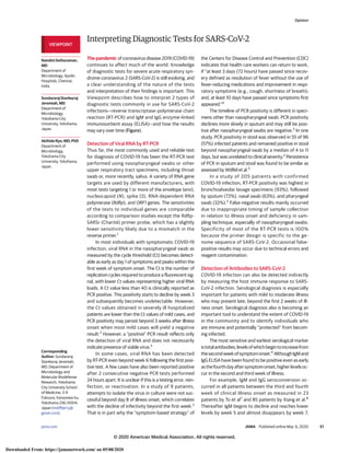
- 1. Interpreting Diagnostic Tests for SARS-CoV-2 The pandemic of coronavirus disease 2019 (COVID-19) continues to affect much of the world. Knowledge of diagnostic tests for severe acute respiratory syn- drome coronavirus 2 (SARS-CoV-2) is still evolving, and a clear understanding of the nature of the tests and interpretation of their findings is important. This Viewpoint describes how to interpret 2 types of diagnostic tests commonly in use for SARS-CoV-2 infections—reverse transcriptase–polymerase chain reaction (RT-PCR) and IgM and IgG enzyme-linked immunosorbent assay (ELISA)—and how the results may vary over time (Figure). Detection of Viral RNA by RT-PCR Thus far, the most commonly used and reliable test for diagnosis of COVID-19 has been the RT-PCR test performed using nasopharyngeal swabs or other upper respiratory tract specimens, including throat swab or, more recently, saliva. A variety of RNA gene targets are used by different manufacturers, with most tests targeting 1 or more of the envelope (env), nucleocapsid (N), spike (S), RNA-dependent RNA polymerase (RdRp), and ORF1 genes. The sensitivities of the tests to individual genes are comparable according to comparison studies except the RdRp- SARSr (Charité) primer probe, which has a slightly lower sensitivity likely due to a mismatch in the reverse primer.1 In most individuals with symptomatic COVID-19 infection, viral RNA in the nasopharyngeal swab as measured by the cycle threshold (Ct) becomes detect- able as early as day 1 of symptoms and peaks within the first week of symptom onset. The Ct is the number of replication cycles required to produce a fluorescent sig- nal, with lower Ct values representing higher viral RNA loads. A Ct value less than 40 is clinically reported as PCR positive. This positivity starts to decline by week 3 and subsequently becomes undetectable. However, the Ct values obtained in severely ill hospitalized patients are lower than the Ct values of mild cases, and PCR positivity may persist beyond 3 weeks after illness onset when most mild cases will yield a negative result.2 However, a “positive” PCR result reflects only the detection of viral RNA and does not necessarily indicate presence of viable virus.3 In some cases, viral RNA has been detected by RT-PCR even beyond week 6 following the first posi- tive test. A few cases have also been reported positive after 2 consecutive negative PCR tests performed 24 hours apart. It is unclear if this is a testing error, rein- fection, or reactivation. In a study of 9 patients, attempts to isolate the virus in culture were not suc- cessful beyond day 8 of illness onset, which correlates with the decline of infectivity beyond the first week.3 That is in part why the “symptom-based strategy” of the Centers for Disease Control and Prevention (CDC) indicates that health care workers can return to work, if “at least 3 days (72 hours) have passed since recov- ery defined as resolution of fever without the use of fever-reducing medications and improvement in respi- ratory symptoms (e.g., cough, shortness of breath); and, at least 10 days have passed since symptoms first appeared.”4 The timeline of PCR positivity is different in speci- mens other than nasopharyngeal swab. PCR positivity declines more slowly in sputum and may still be posi- tive after nasopharyngeal swabs are negative.3 In one study, PCR positivity in stool was observed in 55 of 96 (57%) infected patients and remained positive in stool beyond nasopharyngeal swab by a median of 4 to 11 days, but was unrelated to clinical severity.2 Persistence of PCR in sputum and stool was found to be similar as assessed by Wölfel et al.3 In a study of 205 patients with confirmed COVID-19 infection, RT-PCR positivity was highest in bronchoalveolar lavage specimens (93%), followed by sputum (72%), nasal swab (63%), and pharyngeal swab (32%).5 False-negative results mainly occurred due to inappropriate timing of sample collection in relation to illness onset and deficiency in sam- pling technique, especially of nasopharyngeal swabs. Specificity of most of the RT-PCR tests is 100% because the primer design is specific to the ge- nome sequence of SARS-CoV-2. Occasional false- positive results may occur due to technical errors and reagent contamination. Detection of Antibodies to SARS-CoV-2 COVID-19 infection can also be detected indirectly by measuring the host immune response to SARS- CoV-2 infection. Serological diagnosis is especially important for patients with mild to moderate illness who may present late, beyond the first 2 weeks of ill- ness onset. Serological diagnosis also is becoming an important tool to understand the extent of COVID-19 in the community and to identify individuals who are immune and potentially “protected” from becom- ing infected. The most sensitive and earliest serological marker istotalantibodies,levelsofwhichbegintoincreasefrom thesecondweekofsymptomonset.6 AlthoughIgMand IgG ELISA have been found to be positive even as early asthefourthdayaftersymptomonset,higherlevelsoc- cur in the second and third week of illness. For example, IgM and IgG seroconversion oc- curred in all patients between the third and fourth week of clinical illness onset as measured in 23 patients by To et al7 and 85 patients by Xiang et al.8 Thereafter IgM begins to decline and reaches lower levels by week 5 and almost disappears by week 7, VIEWPOINT Nandini Sethuraman, MD Department of Microbiology, Apollo Hospitals, Chennai, India. Sundararaj Stanleyraj Jeremiah, MD Department of Microbiology, Yokohama City University, Yokohama, Japan. Akihide Ryo, MD, PhD Department of Microbiology, Yokohama City University, Yokohama, Japan. Corresponding Author: Sundararaj Stanleyraj Jeremiah, MD, Department of Microbiology and Molecular Biodefense Research, Yokohama City University School of Medicine, 3-9 Fukuura, Kanazawa-ku, Yokohama 236-0004, Japan (rediffjerry@ gmail.com). Opinion jama.com (Reprinted) JAMA Published online May 6, 2020 E1 © 2020 American Medical Association. All rights reserved.© 2020 American Medical Association. All rights reserved. Downloaded From: https://jamanetwork.com/ on 05/08/2020
- 2. whereas IgG persists beyond 7 weeks.9 In a study of 140 pa- tients, combined sensitivity of PCR and IgM ELISA directed at nucleocapsid (NC) antigen was 98.6% vs 51.9% with a single PCR test. During the first 5.5 days, quantitative PCR had a higher posi- tivity rate than IgM, whereas IgM ELISA had a higher positivity rate after day 5.5 of illness.10 ELISA-based IgM and IgG antibody tests have greater than 95% specificity for diagnosis of COVID-19. Testing of paired serum samples with the initial PCR and the second 2 weeks later can fur- ther increase diagnostic accuracy. Typically, the majority of anti- bodies are produced against the most abundant protein of the virus, which is the NC. Therefore, tests that detect antibodies to NC would be the most sensitive. However, the receptor-binding domain of S (RBD-S) protein is the host attachment protein, and antibodies to RBD-S would be more specific and are expected to be neutralizing. Therefore, using one or both antigens for detecting IgG and IgM would result in high sensitivity.7 Antibodies may, however, have cross-reactivity with SARS-CoV and possibly other coronaviruses. Rapid point-of-care tests for detection of antibodies have been widely developed and marketed and are of variable quality. Many manufacturers do not reveal the nature of antigens used. These tests are purely qualitative in nature and can only indicate the presence or absence of SARS-CoV-2 antibodies. The presence of neutralizing antibodies can only be confirmed by a plaque reduction neutralization test. However, high titers of IgG antibod- ies detected by ELISA have been shown to positively correlate with neutralizing antibodies.7 The long-term persistence and duration of protection conferred by the neutralizing antibodies remains unknown. Conclusions Using available evidence, a clinically useful timeline of diag- nostic markers for detection of COVID-19 has been devised (Figure). Most of the available data are for adult populations who are not immunocompromised. The time course of PCR positivity and seroconversion may vary in children and other groups, including the large population of asymptomatic individuals who go undiagnosed without active surveillance. Many questions remain, particularly how long potential immunity lasts in indi- viduals, both asymptomatic and symptomatic, who are infected with SARS-CoV-2. Figure. Estimated Variation Over Time in Diagnostic Tests for Detection of SARS-CoV-2 Infection Relative to Symptom Onset Week −2 Week −1 Week 1 Week 2 Week 3 Week 4 Week 5 Week 6 Symptom onset After symptom onsetBefore symptom onset Antibody detection PCR - Likely positiveDetection unlikelya PCR - Likely negativeb SARS-CoV-2 exposure Increasingprobabilityofdetection Nasopharyngeal swab PCR Stool PCRVirus isolation from respiratory tract Bronchoalveolar lavage/sputum PCR IgM antibody IgG antibody Estimated time intervals and rates of viral detection are based on data from several published reports. Because of variability in values among studies, estimated time intervals should be considered approximations and the probability of detection of SARS-CoV-2 infection is presented qualitatively. SARS-CoV-2 indicates severe acute respiratory syndrome coronavirus 2; PCR, polymerase chain reaction. a Detection only occurs if patients are followed up proactively from the time of exposure. b More likely to register a negative than a positive result by PCR of a nasopharyngeal swab. Opinion Viewpoint E2 JAMA Published online May 6, 2020 (Reprinted) jama.com © 2020 American Medical Association. All rights reserved. Downloaded From: https://jamanetwork.com/ on 05/08/2020
- 3. ARTICLE INFORMATION Published Online: May 6, 2020. doi:10.1001/jama.2020.8259 Conflict of Interest Disclosures: None reported. REFERENCES 1. Nalla AK, Casto AM, Huang MW, et al. Comparative performance of SARS-CoV-2 detection assays using seven different primer/probe sets and one assay kit. J Clin Microbiol. 2020;JCM.00557-20. Published online April 8, 2020. doi:10.1128/JCM. 00557-20 2. Zheng S, Fan J, Yu F, et al. Viral load dynamics and disease severity in patients infected with SARS-CoV-2 in Zhejiang province, China, January-March 2020: retrospective cohort study. BMJ. 2020;369:m1443. Published online April 21, 2020. doi:10.1136/bmj.m1443 3. Wölfel R, Corman VM, Guggemos W, et al. Virological assessment of hospitalized patients with COVID-2019. Nature. 2020. Published online April 1, 2020. doi:10.1038/s41586-020-2196-x 4. CDC. Return-to-work criteria for healthcare workers. Updated April 30, 2020. Accessed May 3, 2020. https://www.cdc.gov/coronavirus/2019- ncov/hcp/return-to-work.html 5. Wang W, Xu Y, Gao R, et al. Detection of SARS-CoV-2 in different types of clinical specimens. JAMA. 2020. Published online March 11, 2020. doi:10. 1001/jama.2020.3786 6. Lou B, Li T, Zheng S, et al Serology characteristics of SARS-CoV-2 infection since the exposure and post symptoms onset. medRxiv. Preprint posted March 27, 2020. doi:10.1101/2020. 03.23.20041707 7. To KK-W, Tsang OT-Y, Leung W-S, et al. Temporal profiles of viral load in posterior oropharyngeal saliva samples and serum antibody responses during infection by SARS-CoV-2: an observational cohort study. Lancet Infect Dis. 2020;20(5):565-574. doi:10.1016/S1473-3099(20)30196-1 8. Xiang F, Wang X, He X, et al. Antibody detection and dynamic characteristics in patients with COVID-19. Clin Infect Dis. 2020;ciaa461. Published online April 19, 2020. doi:10.1093/cid/ciaa461 9. Xiao AT, Gao C, Zhang S. Profile of specific antibodies to SARS-CoV-2: the first report. J Infect. 2020;S0163-4453(20)30138-9. Published online March 21, 2020. doi:10.1016/j.jinf.2020.03.012 10. Guo L, Ren L, Yang S, et al. Profiling early humoral response to diagnose novel coronavirus disease (COVID-19). Clin Infect Dis. 2020;ciaa310. Published online March 21, 2020. doi:10.1093/cid/ ciaa310 Viewpoint Opinion jama.com (Reprinted) JAMA Published online May 6, 2020 E3 © 2020 American Medical Association. All rights reserved. Downloaded From: https://jamanetwork.com/ on 05/08/2020