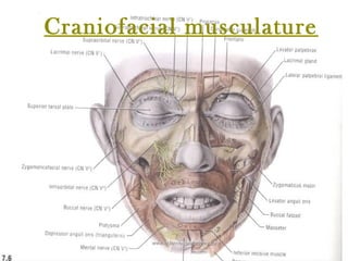
Cranio facial musculature
•
0 likes•419 views

The Indian Dental Academy is the Leader in continuing dental education , training dentists in all aspects of dentistry and offering a wide range of dental certified courses in different formats.for more details please visit www.indiandentalacademy.com

Recommended
Recommended
More Related Content
What's hot
What's hot (20)
Similar to Cranio facial musculature
Similar to Cranio facial musculature (20)
More from Indian dental academy
More from Indian dental academy (20)
Recently uploaded
Recently uploaded (20)
Cranio facial musculature
- 2. Contents:-Contents:- introductionintroduction Development of the muscles.Development of the muscles. classification of craniofacial musculature.classification of craniofacial musculature. the buccinator mechanism.the buccinator mechanism. the orbicularis oris.the orbicularis oris. the muscles of mastication.the muscles of mastication. accessory muscles of masticationaccessory muscles of mastication The tongue.The tongue. www.indiandentalacademy.comwww.indiandentalacademy.com
- 3. Contents:-Contents:- The temporo mandibular jointThe temporo mandibular joint Physiology of muscles.Physiology of muscles. Reflex of musclesReflex of muscles myotactic reflex.myotactic reflex. Methods to study muscles.Methods to study muscles. EmgEmg Is it the one of them?Is it the one of them? The equilibrium theoryThe equilibrium theorywww.indiandentalacademy.comwww.indiandentalacademy.com
- 4. Contents:-Contents:- Muscle and growth.Muscle and growth. Muscle adaptation in malocclusionMuscle adaptation in malocclusion Soft tissue environment of patients withSoft tissue environment of patients with malocclusion.malocclusion. Muscular adaptation after orthognathic surgeryMuscular adaptation after orthognathic surgery Effects of orthodontic treatment on theEffects of orthodontic treatment on the neuromuscular functions.neuromuscular functions. Muscles and its treatment modalities.Muscles and its treatment modalities. ConclusionConclusion www.indiandentalacademy.comwww.indiandentalacademy.com
- 5. Introduction:-Introduction:- A Man to increase the strength ofA Man to increase the strength of skeleton has been blessed by 639skeleton has been blessed by 639 muscles which includes 6 million ofmuscles which includes 6 million of muscle fibers.muscle fibers. Each fibers has 1000 fibrilsEach fibers has 1000 fibrils Thus there is 6000 billion muscle fiberThus there is 6000 billion muscle fiber working at one time.working at one time. www.indiandentalacademy.comwww.indiandentalacademy.com
- 6. In orthodontics and in real life weIn orthodontics and in real life we judge / diagnose a individual when wejudge / diagnose a individual when we look at him.look at him. But here when we look at patient weBut here when we look at patient we look at him in repose/rest and we try tolook at him in repose/rest and we try to see the dentofacial complex and theirsee the dentofacial complex and their intimate relationship to each otherintimate relationship to each other www.indiandentalacademy.comwww.indiandentalacademy.com
- 7. This is just the static analysis but importantThis is just the static analysis but important is the dynamic appreciation on how theyis the dynamic appreciation on how they function , as they function, how they affectfunction , as they function, how they affect the growth and the relationships of parts ,the growth and the relationships of parts , these constitutes the stomatoganthicthese constitutes the stomatoganthic system.system. Here musculature plays a very importantHere musculature plays a very important part.part. www.indiandentalacademy.comwww.indiandentalacademy.com
- 8. We orthodontist try to achieve theWe orthodontist try to achieve the perfect equilibrium between the part ofperfect equilibrium between the part of stomatoganthic system which includesstomatoganthic system which includes the muscle.the muscle. so it is of definite importance that weso it is of definite importance that we should know in and out about them.should know in and out about them. www.indiandentalacademy.comwww.indiandentalacademy.com
- 9. Development of muscle.Development of muscle. During the 3rd week of I.U life the embryo undergoes gastrulation toDuring the 3rd week of I.U life the embryo undergoes gastrulation to form a trilaminar disk i.e.form a trilaminar disk i.e. the cells from epiblast migrate to primitive streak and to primitivethe cells from epiblast migrate to primitive streak and to primitive node detach from the epiblast and they invaginated the hypoblast tonode detach from the epiblast and they invaginated the hypoblast to displace it and form the three layers.displace it and form the three layers. Three germ layers derived from the epiblastThree germ layers derived from the epiblast endodermendoderm mesodermmesoderm ectodermectoderm Three germ layers derived from the cells epiblast www.indiandentalacademy.comwww.indiandentalacademy.com
- 10. The mesodermal layer is further dividedThe mesodermal layer is further divided into:-into:- headhead paraxialparaxial intermediateintermediate laterallateral The mesoderm on either side of the notochordalThe mesoderm on either side of the notochordal process thickens to form longitudinal columns ofprocess thickens to form longitudinal columns of tissue called the paraxial mesoderm.tissue called the paraxial mesoderm. These segments into paired blocks of tissueThese segments into paired blocks of tissue called the somites. Of these the cranial ones arecalled the somites. Of these the cranial ones are called as somitomeres.called as somitomeres. www.indiandentalacademy.comwww.indiandentalacademy.com
- 11. There are seven of somitomeres approx. InThere are seven of somitomeres approx. In register with that of the pharyngeal arches.register with that of the pharyngeal arches. The skeletal muscles of the head and neckThe skeletal muscles of the head and neck develop from this somitomeres and the mostdevelop from this somitomeres and the most cranial somites.cranial somites. The pharyngeal arches develop during theThe pharyngeal arches develop during the 4th wk. of the I.U life.4th wk. of the I.U life. They significantly contribute to theThey significantly contribute to the development of head, face neck and nasaldevelopment of head, face neck and nasal cavity, mouth and to some extent the larynxcavity, mouth and to some extent the larynx and the pharynx.and the pharynx. www.indiandentalacademy.comwww.indiandentalacademy.com
- 12. The branchial arches have incomplete cleftsThe branchial arches have incomplete clefts between the arches, the external ectodermalbetween the arches, the external ectodermal branchial grooves and internal endodermalbranchial grooves and internal endodermal pharyngeal pouches.pharyngeal pouches. The branchial groove of the first branchial archThe branchial groove of the first branchial arch persists as external acoustic meatus which ispersists as external acoustic meatus which is covered by the tympanic membrane.covered by the tympanic membrane. All the grooves disappear but they remain asAll the grooves disappear but they remain as the tympanic cavity, auditory tube, tonsil,the tympanic cavity, auditory tube, tonsil, thymus, parathyroid, and the thyroid gland.thymus, parathyroid, and the thyroid gland. www.indiandentalacademy.comwww.indiandentalacademy.com
- 14. Cartilage components derived fromCartilage components derived from pharyngeal arches are:-pharyngeal arches are:- Arch I [mandibular arch]:-Arch I [mandibular arch]:- this is major contributor tothis is major contributor to the development of the face.the development of the face. The cartilage of the first arch is called as theThe cartilage of the first arch is called as the meckelsmeckels cartilagecartilage.. The dorsal end of the meckels cartilage becomes ossifiedThe dorsal end of the meckels cartilage becomes ossified to form 2 bones of the middle ear ossicle :-to form 2 bones of the middle ear ossicle :- 1.1. malleusmalleus 2.2. IncusIncus The middle portion of the meckels cartilage regresses, butThe middle portion of the meckels cartilage regresses, but its perichondrium forms the sphenomandibular ligament.its perichondrium forms the sphenomandibular ligament. www.indiandentalacademy.comwww.indiandentalacademy.com
- 15. The ventral part of the meckels cartilageThe ventral part of the meckels cartilage forms a horse shaped structure in the shapeforms a horse shaped structure in the shape of the future mandible.of the future mandible. The mesenchymal tissue lateral to theThe mesenchymal tissue lateral to the cartilage undergoes intra membranouscartilage undergoes intra membranous ossification to produce the mandible as theossification to produce the mandible as the original meckels cartilage disappears.original meckels cartilage disappears. www.indiandentalacademy.comwww.indiandentalacademy.com
- 16. Arch II [hyoid arch]:-Arch II [hyoid arch]:- this cartilage isthis cartilage is called as thecalled as the reicherts cartilagereicherts cartilage It dorsal end becomes ossified to produceIt dorsal end becomes ossified to produce the other middle ear ossicle, the stapes, andthe other middle ear ossicle, the stapes, and the styloid process of the temporal bone.the styloid process of the temporal bone. A portion of the perichondrium of theA portion of the perichondrium of the cartilage forms the stylohyoid ligament.cartilage forms the stylohyoid ligament. This also contributes in the development ofThis also contributes in the development of the hyoid bone specially the lesser cornu,the hyoid bone specially the lesser cornu, and the superior portion of the body.and the superior portion of the body. www.indiandentalacademy.comwww.indiandentalacademy.com
- 17. The cartilage of the 3rd arch gives rise toThe cartilage of the 3rd arch gives rise to the greater cornu and the inferior part of thethe greater cornu and the inferior part of the body of the hyoid bone.body of the hyoid bone. The cartilage of the 4th and the 6th archesThe cartilage of the 4th and the 6th arches fuses together to form the laryngealfuses together to form the laryngeal cartilage, including the thyroid, cricoids, andcartilage, including the thyroid, cricoids, and arytenoids cartilage except the epiglottis.arytenoids cartilage except the epiglottis. www.indiandentalacademy.comwww.indiandentalacademy.com
- 18. Muscle components :-Muscle components :- They are derived from the cranialThey are derived from the cranial somites and the cells that migrate atsomites and the cells that migrate at this region from the somitomeres.this region from the somitomeres. Arch I:Arch I: muscles derived are: themuscles derived are: the muscles of mastication, anterior bellymuscles of mastication, anterior belly of digastric, mylohyoid, tensor veliof digastric, mylohyoid, tensor veli palatine, tensor tympani.palatine, tensor tympani. www.indiandentalacademy.comwww.indiandentalacademy.com
- 20. Arch II: the muscles of facial expression.Arch II: the muscles of facial expression. These are relatively thinner and haveThese are relatively thinner and have their origin and insertion in the skin andtheir origin and insertion in the skin and are present throughout the face and theare present throughout the face and the neck.neck. Arch III: it gives rise to stylopharyngeousArch III: it gives rise to stylopharyngeous muscle.muscle. Arch IV and arch V: - muscles of theArch IV and arch V: - muscles of the pharynx and the larynx. Arch IV give risepharynx and the larynx. Arch IV give rise to cricothyroid and arch V to intrinsicto cricothyroid and arch V to intrinsic muscles of larynx.muscles of larynx.www.indiandentalacademy.comwww.indiandentalacademy.com
- 21. Classification ofClassification of muscles:-muscles:- they can be primarily classified as:-they can be primarily classified as:- 1.1. facial musclesfacial muscles 2.2. Muscles of mastication.Muscles of mastication. The facial muscles: they are related to theThe facial muscles: they are related to the orbital margins plus the eyelids, theorbital margins plus the eyelids, the external nose & nostrils, the lips, cheeks,external nose & nostrils, the lips, cheeks, mouth, the pinna, scalp and the cervicalmouth, the pinna, scalp and the cervical skin.skin. These are also called as muscles of facialThese are also called as muscles of facial expression as that is their function.expression as that is their function. www.indiandentalacademy.comwww.indiandentalacademy.com
- 22. Muscles of mastication: they are chieflyMuscles of mastication: they are chiefly concerned with the movements of theconcerned with the movements of the TMJTMJ The division reflects the differentThe division reflects the different embryonic origins and innervations of theembryonic origins and innervations of the two groups.two groups. But all the functions such as mastication,But all the functions such as mastication, deglutition, respiration, swallowing,deglutition, respiration, swallowing, speech, communicative and emotionalspeech, communicative and emotional expression, ocular, nasal and aural actionexpression, ocular, nasal and aural action are the effect of close cooperation of twoare the effect of close cooperation of two groups.groups. www.indiandentalacademy.comwww.indiandentalacademy.com
- 23. According to moyers:-According to moyers:- 1.1. facial musclesfacial muscles 2.2. jaw musclesjaw muscles 3.3. Portal muscles.Portal muscles. www.indiandentalacademy.comwww.indiandentalacademy.com
- 24. Characteristics of the facialCharacteristics of the facial muscles:-muscles:- The primary function is expression of the emotions.The primary function is expression of the emotions. The facial muscles are capable of performing 7000The facial muscles are capable of performing 7000 expressions according to Coleman.expressions according to Coleman. They are also responsible for the maintenance ofThey are also responsible for the maintenance of the posture of the facial structures.the posture of the facial structures. Paresis of the orbicularis oculi leads to theParesis of the orbicularis oculi leads to the drooping of the lower eyelid.drooping of the lower eyelid. Paralysis of the orbicularis oris will lead to angularParalysis of the orbicularis oris will lead to angular cheliosis and the drooling.cheliosis and the drooling. www.indiandentalacademy.comwww.indiandentalacademy.com
- 25. The facial muscle also contributes toThe facial muscle also contributes to stabilization of the mandible duringstabilization of the mandible during the infantile swallowing and chewingthe infantile swallowing and chewing and swallowing in the occlusallyand swallowing in the occlusally compromised adults.compromised adults. It is also important for the visual andIt is also important for the visual and the spoken communications.the spoken communications. www.indiandentalacademy.comwww.indiandentalacademy.com
- 26. Characteristics of the jawCharacteristics of the jaw muscles:-muscles:- The mandible being maintained against theThe mandible being maintained against the gravity by the stretch reflex of the elevators.gravity by the stretch reflex of the elevators. EMG studies have shown of [postural position]EMG studies have shown of [postural position] that the inframandibular groups of muscles arethat the inframandibular groups of muscles are more active than the levator.more active than the levator. Mandibular movement assisted by the levatorMandibular movement assisted by the levator and the depressors can’t be considered just theand the depressors can’t be considered just the interplay between these 2 but is very muchinterplay between these 2 but is very much thought as the intricate muscular web where thethought as the intricate muscular web where the teeth and the joints acts as the stops.teeth and the joints acts as the stops. www.indiandentalacademy.comwww.indiandentalacademy.com
- 27. The head posture also affects the posture of theThe head posture also affects the posture of the mandible for e.g. when there is extension of themandible for e.g. when there is extension of the head there is increase in the freeway space andhead there is increase in the freeway space and when there is flexion there is decrease in thewhen there is flexion there is decrease in the freeway space.freeway space. Changes in the head posture also results changesChanges in the head posture also results changes in the anteroposterior positioning of the posture ofin the anteroposterior positioning of the posture of the mandible.the mandible. One of the most important factors is the posture ofOne of the most important factors is the posture of the mandible affecting the development of thethe mandible affecting the development of the jaws.jaws. E.g. during the mouth breathing there is effect onE.g. during the mouth breathing there is effect on the growth of both maxilla as well as the mandiblethe growth of both maxilla as well as the mandible due to alteration in position of the mandible, hyoiddue to alteration in position of the mandible, hyoid and the tongue.and the tongue. www.indiandentalacademy.comwww.indiandentalacademy.com
- 28. But in case where there is Sunday biteBut in case where there is Sunday bite there is no resultant correction probablythere is no resultant correction probably because the dorsal position of thebecause the dorsal position of the mandible during the functional activitiesmandible during the functional activities cancels the biologic signals to the jointcancels the biologic signals to the joint structures.structures. Thus functional appliances work the bestThus functional appliances work the best as they are worn for the most hours of theas they are worn for the most hours of the day .day . www.indiandentalacademy.comwww.indiandentalacademy.com
- 29. Characteristics of the portalCharacteristics of the portal muscles:-muscles:- Portal muscle is the word coined byPortal muscle is the word coined by Bosma to denote the upper alimentaryBosma to denote the upper alimentary tract and the respiratory tract.tract and the respiratory tract. These muscles serve some functions ofThese muscles serve some functions of our interest such as posture, respiration,our interest such as posture, respiration, feeding.feeding. www.indiandentalacademy.comwww.indiandentalacademy.com
- 30. The muscles include the muscles ofThe muscles include the muscles of the tongue [both the intrinsic and thethe tongue [both the intrinsic and the extrinsic] the soft palate, theextrinsic] the soft palate, the pharyngeal pillars, the pharynx proper,pharyngeal pillars, the pharynx proper, and the larynx.and the larynx. www.indiandentalacademy.comwww.indiandentalacademy.com
- 31. According to grays:-According to grays:- they are broadly classifies as:they are broadly classifies as: 1.1. epicranial musclesepicranial muscles 2.2. circumorbital musclescircumorbital muscles 3.3. the nasal musculaturethe nasal musculature 4.4. the buccolabial musculaturethe buccolabial musculature www.indiandentalacademy.comwww.indiandentalacademy.com
- 32. The epicranial musculatureThe epicranial musculature The epicranius consists of two mainThe epicranius consists of two main partsparts 1.1. occipitofrontalisoccipitofrontalis 2.2. Temporoparietalis.Temporoparietalis. www.indiandentalacademy.comwww.indiandentalacademy.com
- 34. The scalp:-The scalp:- The scalp essentially consists of fiveThe scalp essentially consists of five layers i.e.layers i.e. 1.1. superficial fasciasuperficial fascia 2.2. connective tissueconnective tissue 3.3. epicranial aponeurosisepicranial aponeurosis 4.4. loose aerolar tissueloose aerolar tissue 5.5. pericraniumpericranium www.indiandentalacademy.comwww.indiandentalacademy.com
- 35. The superficial fascia in the scalp is firm andThe superficial fascia in the scalp is firm and fibro-adipose adherent to the skin and thefibro-adipose adherent to the skin and the underlying epicranius and the aponeurosis,underlying epicranius and the aponeurosis, the galea aponeurotica [epicranialthe galea aponeurotica [epicranial aponeurosis.]aponeurosis.] It is continuous with the superficial fascia ofIt is continuous with the superficial fascia of the back of the neck; laterally, it is prolongedthe back of the neck; laterally, it is prolonged to the temporal region where it losses itsto the temporal region where it losses its texture.texture. www.indiandentalacademy.comwww.indiandentalacademy.com
- 36. The occipito frontalisThe occipito frontalis It is a broad, musculofibrous layer,It is a broad, musculofibrous layer, covers the dome of the skull, from thecovers the dome of the skull, from the nuchal lines to the eyebrows.nuchal lines to the eyebrows. It consists of 4 parts---- 2 occipital andIt consists of 4 parts---- 2 occipital and 2 frontal connected by the epicranial2 frontal connected by the epicranial aponeurosisaponeurosis www.indiandentalacademy.comwww.indiandentalacademy.com
- 37. Occipital part: - each of them are thin,Occipital part: - each of them are thin, quadrilateral arises by tendinous fibersquadrilateral arises by tendinous fibers from the lat. 2/3rd of the highest nuchalfrom the lat. 2/3rd of the highest nuchal lines of the occipital bone and the mastoidlines of the occipital bone and the mastoid part of the temporal bone. It ends in thepart of the temporal bone. It ends in the epicranial aponeurosis.epicranial aponeurosis. Frontal part: - they are thin, quadrilateral,Frontal part: - they are thin, quadrilateral, and adherent to superficial fascia. It isand adherent to superficial fascia. It is broader than the occipital part and itsbroader than the occipital part and its fibers are longer and paler.fibers are longer and paler. www.indiandentalacademy.comwww.indiandentalacademy.com
- 38. It has got no bony attachments. Its medial fibers areIt has got no bony attachments. Its medial fibers are contiguous with those of the procerus; its intermediatecontiguous with those of the procerus; its intermediate fibers blend with the corrugator supercilli and thefibers blend with the corrugator supercilli and the orbicularis oculi; its lateral fibers are also blended withorbicularis oculi; its lateral fibers are also blended with the latter muscle over the zygomatic process of thethe latter muscle over the zygomatic process of the frontal bone.frontal bone. From these attachments the fibers are directed upwardsFrom these attachments the fibers are directed upwards to join the epicranial aponeurosis in front of the coronalto join the epicranial aponeurosis in front of the coronal suture. The medial margins of the frontal slips are joinedsuture. The medial margins of the frontal slips are joined together for some distance above the root of the nose;together for some distance above the root of the nose; but between the occipital bellies there is considerable,but between the occipital bellies there is considerable, but variable interval occupied by the epicranialbut variable interval occupied by the epicranial aponeurosis.aponeurosis. www.indiandentalacademy.comwww.indiandentalacademy.com
- 39. Galea aponeurotica:-Galea aponeurotica:- It covers the upper part of the cranium andIt covers the upper part of the cranium and along with the epicranius it forms thealong with the epicranius it forms the continuous fibromuscular sheet extendingcontinuous fibromuscular sheet extending from the occipital to the eyebrows. Behind, infrom the occipital to the eyebrows. Behind, in the interval between the occipital parts of thethe interval between the occipital parts of the occipitofrontalis, it is attached on theoccipitofrontalis, it is attached on the external protuberance or highest nuchalexternal protuberance or highest nuchal lines of the occipital bone.lines of the occipital bone. In front it splits to enclose the frontal partsIn front it splits to enclose the frontal parts and sends a short narrow prolongationand sends a short narrow prolongation between them.between them. www.indiandentalacademy.comwww.indiandentalacademy.com
- 40. It is united to the skin by firm, fibrousIt is united to the skin by firm, fibrous superficial fascia; it is connected to thesuperficial fascia; it is connected to the pericranium by loose aerolar tissuepericranium by loose aerolar tissue which allows its free movement. Thewhich allows its free movement. The latter carrying it with the skin of the scalplatter carrying it with the skin of the scalp Nerve supply: - the occipital part isNerve supply: - the occipital part is supplied by the posterior auricularsupplied by the posterior auricular branch and the frontal part by thebranch and the frontal part by the temporal branches of the facial nerve.temporal branches of the facial nerve. www.indiandentalacademy.comwww.indiandentalacademy.com
- 41. Action:- the occipital slips draw theAction:- the occipital slips draw the scalp downward , the frontal slipsscalp downward , the frontal slips acting from above raise the eyebrowsacting from above raise the eyebrows and the skin of the root of the nose ;and the skin of the root of the nose ; acting from below they draw the scalpacting from below they draw the scalp forwards; throwing the integument offorwards; throwing the integument of the forehead into transverse wrinkles.the forehead into transverse wrinkles. They act in tandem in expressionThey act in tandem in expression like surprise, horror or fright etc.like surprise, horror or fright etc. www.indiandentalacademy.comwww.indiandentalacademy.com
- 42. The fourth layer of the scalp:The fourth layer of the scalp: it is made up of loose aerolar tissue. itit is made up of loose aerolar tissue. it extends anteriorly to the eyelids; andextends anteriorly to the eyelids; and posteriorly to the highest nuchal linesposteriorly to the highest nuchal lines and on each side to the superiorand on each side to the superior temporal lines.temporal lines. The fifth layer is called as pericranium:-itThe fifth layer is called as pericranium:-it is loosely attached to the surface of theis loosely attached to the surface of the bones, but is firmly adherent to theirbones, but is firmly adherent to their sutures where the sutural ligaments bindsutures where the sutural ligaments bind the pericranium to the endocranium.the pericranium to the endocranium. www.indiandentalacademy.comwww.indiandentalacademy.com
- 43. Temporoparietalis:-Temporoparietalis:- It is variably developed sheet of muscle thatIt is variably developed sheet of muscle that lies between the frontal part of the occipitolies between the frontal part of the occipito frontalis and the ant. And sup. Auricularfrontalis and the ant. And sup. Auricular muscles.muscles. A thin muscular slip, the transverse nuchae,A thin muscular slip, the transverse nuchae, is present in about 25 percent of the people;is present in about 25 percent of the people; it arises from the external occipitalit arises from the external occipital protuberance or from the superior nuchalprotuberance or from the superior nuchal lines, present either sup. Or deep tolines, present either sup. Or deep to trapezius; it is frequently inserted with thetrapezius; it is frequently inserted with the auricularis posterior, but may join the post.auricularis posterior, but may join the post. Edge of the sterocleidomastoid.Edge of the sterocleidomastoid. www.indiandentalacademy.comwww.indiandentalacademy.com
- 44. Circumorbital andCircumorbital and palpeberal musculature.palpeberal musculature. The muscle that come under thisThe muscle that come under this heading are:-heading are:- 1.1. orbicularis oculiorbicularis oculi 2.2. corrugator supercillicorrugator supercilli 3.3. levator palpebrae superiorislevator palpebrae superioris www.indiandentalacademy.comwww.indiandentalacademy.com
- 45. The orbicularis oculi:-The orbicularis oculi:- it is a broad, flat, elliptical muscle thatit is a broad, flat, elliptical muscle that occupies the eyelids, surrounds theoccupies the eyelids, surrounds the circumference of the orbit and spreadscircumference of the orbit and spreads on the temporal region and the cheek.on the temporal region and the cheek. it consists of the orbital , palpebraeit consists of the orbital , palpebrae and lacrimal parts.and lacrimal parts. www.indiandentalacademy.comwww.indiandentalacademy.com
- 46. The orbital partThe orbital part it is reddish and thicker than the palpeberalit is reddish and thicker than the palpeberal fasiculifasiculi Origin:-arises from the nasal part of the frontalOrigin:-arises from the nasal part of the frontal bone, from the frontal process of the maxilla andbone, from the frontal process of the maxilla and from the medial palpeberal ligament, whichfrom the medial palpeberal ligament, which interrupts the bony attachment.interrupts the bony attachment. It fibers form the complete ellipses without theIt fibers form the complete ellipses without the interruption on the lateral side, the upper onesinterruption on the lateral side, the upper ones blending with the frontal part of theblending with the frontal part of the occipitofrontalis and the corrugator.occipitofrontalis and the corrugator.www.indiandentalacademy.comwww.indiandentalacademy.com
- 47. Insertion:-some of the fibers areInsertion:-some of the fibers are inserted into the skin and theinserted into the skin and the subcutaneous tissue of the eyebrow.subcutaneous tissue of the eyebrow. They constitute the depressorThey constitute the depressor supercilli.supercilli. www.indiandentalacademy.comwww.indiandentalacademy.com
- 48. The palpeberal partThe palpeberal part - it is thin and pale. It arises from the medial- it is thin and pale. It arises from the medial palpeberal ligament chiefly from its superficialpalpeberal ligament chiefly from its superficial but also from its deep parts, though not frombut also from its deep parts, though not from the lower margin, it arises also from the bonethe lower margin, it arises also from the bone immd. Above and below the ligament.immd. Above and below the ligament. The fibers sweep across the eyelids in front ofThe fibers sweep across the eyelids in front of the orbital septum and at the lateralthe orbital septum and at the lateral commissure and interlace to form the lateralcommissure and interlace to form the lateral palpeberal raphe. A small group of the finepalpeberal raphe. A small group of the fine fibers lies close to the margin of each eyelid,fibers lies close to the margin of each eyelid, behind the eyelashes; it is named as the ciliarybehind the eyelashes; it is named as the ciliary bundle.bundle. www.indiandentalacademy.comwww.indiandentalacademy.com
- 49. The lacrimal part: -The lacrimal part: - it lies behind the lacrimal sac but separatedit lies behind the lacrimal sac but separated from it by the lacrimal fascia.from it by the lacrimal fascia. It is attached to lacrimal fascia, to the upperIt is attached to lacrimal fascia, to the upper part of the crest of the lacrimal bone, andpart of the crest of the lacrimal bone, and adjacent part of the lateral part of the lacrimaladjacent part of the lateral part of the lacrimal bone.bone. Passing laterally behind the lacrimal sac thePassing laterally behind the lacrimal sac the muscle divides into upper and lower slips; somemuscle divides into upper and lower slips; some fibers are inserted into the tarsi of the eyelidsfibers are inserted into the tarsi of the eyelids close to the lacrimal canaliculi, but mostclose to the lacrimal canaliculi, but most continue across in front of the tarsi andcontinue across in front of the tarsi and interlace in the lateral palpeberal raphe.interlace in the lateral palpeberal raphe. www.indiandentalacademy.comwww.indiandentalacademy.com
- 50. The medial palpeberal ligament:- it isThe medial palpeberal ligament:- it is about 4mm in the length and 2mm in theabout 4mm in the length and 2mm in the breadth, is attached to the frontalbreadth, is attached to the frontal process of the maxilla in front of theprocess of the maxilla in front of the nasolacrimal groove. Crossing thenasolacrimal groove. Crossing the lacrimal sac it divides into 2 parts i.e.lacrimal sac it divides into 2 parts i.e. upper and lower parts each one attachedupper and lower parts each one attached to the medial end of the correspondingto the medial end of the corresponding tarsus.tarsus. It is separated from the lacrimal sacIt is separated from the lacrimal sac by the fascia.by the fascia. www.indiandentalacademy.comwww.indiandentalacademy.com
- 51. Nerve supply: - temporal and zygomaticNerve supply: - temporal and zygomatic branch of the facial nerve.branch of the facial nerve. Actions: - the orbicularis oculi: - it is theActions: - the orbicularis oculi: - it is the sphincter muscle of the eyelids.sphincter muscle of the eyelids. The palpeberal portionThe palpeberal portion acts underacts under voluntary control closing the lids gently asvoluntary control closing the lids gently as in sleep or blinking; the orbital portion isin sleep or blinking; the orbital portion is more frequently under voluntary controlmore frequently under voluntary control www.indiandentalacademy.comwww.indiandentalacademy.com
- 52. During the eye closure there is lowering ofDuring the eye closure there is lowering of the upper as well as elevation of the lowerthe upper as well as elevation of the lower eyelid. thus palpeberal part has depressoreyelid. thus palpeberal part has depressor and elevator fasicles.when the entireand elevator fasicles.when the entire muscle contracts than the skin of themuscle contracts than the skin of the forehead , temple and cheek is drawnforehead , temple and cheek is drawn towards the medial angle of the orbit, andtowards the medial angle of the orbit, and the eyelids are not only firmly closed butthe eyelids are not only firmly closed but they are moved in toto medially. The skin isthey are moved in toto medially. The skin is thrown in the folds on the lateral angle ofthrown in the folds on the lateral angle of the eyelids due to this action which arethe eyelids due to this action which are called as crow’s feet.called as crow’s feet. www.indiandentalacademy.comwww.indiandentalacademy.com
- 53. The lacrimal part of the muscle draws the eyelidsThe lacrimal part of the muscle draws the eyelids and the lacrimal papillae medially, and exertsand the lacrimal papillae medially, and exerts traction on the lacrimal fascia and it dilates thetraction on the lacrimal fascia and it dilates the lacrimal sac.lacrimal sac. Thus the muscle has important action in tearThus the muscle has important action in tear transport.transport. The muscle is also an important element in facialThe muscle is also an important element in facial expression and the ocular reflexes.expression and the ocular reflexes. Partial closure of the palpeberal fissure togetherPartial closure of the palpeberal fissure together with bunching and the protrusion of the eyebrowswith bunching and the protrusion of the eyebrows diminish the entry of the light.diminish the entry of the light. www.indiandentalacademy.comwww.indiandentalacademy.com
- 54. These action of the upper orbital fibersThese action of the upper orbital fibers and their peripheral extension causeand their peripheral extension cause vertical furrowing above the bridge of thevertical furrowing above the bridge of the nose. This is called as blink reflex and itsnose. This is called as blink reflex and its protective value is obvious.protective value is obvious. www.indiandentalacademy.comwww.indiandentalacademy.com
- 55. The corrugatorThe corrugator supercilli:supercilli: it is a small pyramidal muscle, at theit is a small pyramidal muscle, at the medial end of the eyebrows, deep to themedial end of the eyebrows, deep to the frontal part of the occipitofrontalis and thefrontal part of the occipitofrontalis and the orbicularis oculi .orbicularis oculi . From the medial end of the superciliaryFrom the medial end of the superciliary arch its fibers pass slightly laterally andarch its fibers pass slightly laterally and slightly upwards to the deep surface ofslightly upwards to the deep surface of the skin above the middle of thethe skin above the middle of the supraorbital margin.supraorbital margin.www.indiandentalacademy.comwww.indiandentalacademy.com
- 57. Nerve supply: - the temporal branches ofNerve supply: - the temporal branches of the facial nerve.the facial nerve. Action: - draws the eyebrows mediallyAction: - draws the eyebrows medially and downwardsand downwards Together with the orbicularis oculiTogether with the orbicularis oculi causing vertical wrinkles of the forehead.causing vertical wrinkles of the forehead. It assists in drawing the eyebrowsIt assists in drawing the eyebrows downwards in the bright sunlight and isdownwards in the bright sunlight and is also involved in frowningalso involved in frowning www.indiandentalacademy.comwww.indiandentalacademy.com
- 58. The nasal musculature:The nasal musculature: This group comprises of threeThis group comprises of three muscles:-muscles:- 1.1. the procerusthe procerus 2.2. the nasalisthe nasalis 3.3. The depressor septi.The depressor septi. www.indiandentalacademy.comwww.indiandentalacademy.com
- 59. The procerus: -The procerus: - it is a small pyramidal slip continuous withit is a small pyramidal slip continuous with the medial side of the frontal part of thethe medial side of the frontal part of the occipitofrontalis.occipitofrontalis. Origin:-It arises from the fascia coveringOrigin:-It arises from the fascia covering the lower part of the nasal bone and thethe lower part of the nasal bone and the upper part of the lateral nasal cartilage.upper part of the lateral nasal cartilage. Insertion:-it is inserted into the skin overInsertion:-it is inserted into the skin over the lower part of the forehead betweenthe lower part of the forehead between the eyebrows.the eyebrows. www.indiandentalacademy.comwww.indiandentalacademy.com
- 60. Action: - it draws down the medial angleAction: - it draws down the medial angle of the eyebrow and incidentally producesof the eyebrow and incidentally produces wrinkles over the bridge of the nose.wrinkles over the bridge of the nose. It is active in frowning and concentration.It is active in frowning and concentration. It also aids in reducing the glare of theIt also aids in reducing the glare of the sunlight.sunlight. www.indiandentalacademy.comwww.indiandentalacademy.com
- 61. The nasalis: -The nasalis: - it consists of transverse and alar partsit consists of transverse and alar parts which may be continuous at the origin.which may be continuous at the origin. The transverse part:-[compressor naris] –The transverse part:-[compressor naris] – it arises from the maxilla just lateral to theit arises from the maxilla just lateral to the nasal notch; its fibers proceed upwardsnasal notch; its fibers proceed upwards and medially and expand into a thinand medially and expand into a thin aponeurosis, which is continuous on theaponeurosis, which is continuous on the bridge of the nose with that of the musclebridge of the nose with that of the muscle of the opposite side, and with theof the opposite side, and with the aponeurosis of the procerus.aponeurosis of the procerus.www.indiandentalacademy.comwww.indiandentalacademy.com
- 62. The alar part [dilator naris]—it arises from theThe alar part [dilator naris]—it arises from the maxilla, below and medial to the transverse part. Itmaxilla, below and medial to the transverse part. It is attached to the cartilaginous ala nasi.is attached to the cartilaginous ala nasi. Actions: - the transverse part: - it compresses theActions: - the transverse part: - it compresses the nasal aperture at the junction of the vestibule withnasal aperture at the junction of the vestibule with the nasal cavity.the nasal cavity. The alar part draws the ala downwards andThe alar part draws the ala downwards and laterally and so assists in widening the ant. Nasallaterally and so assists in widening the ant. Nasal aperture.aperture. These actions are visible in deep respiration,These actions are visible in deep respiration, especially in its inspiratory phase, and they alsoespecially in its inspiratory phase, and they also accompany certain emotional states.accompany certain emotional states. www.indiandentalacademy.comwww.indiandentalacademy.com
- 64. The depressor septi: -The depressor septi: - often regarded as the part of theoften regarded as the part of the dilator nasi; is attached to maxilladilator nasi; is attached to maxilla above the central incisor roots.above the central incisor roots. Present immediately deep to thePresent immediately deep to the mucous membrane of the upper lip.mucous membrane of the upper lip. www.indiandentalacademy.comwww.indiandentalacademy.com
- 65. Action: - it assists the alar part of theAction: - it assists the alar part of the nasalis in widening the nasal aperturenasalis in widening the nasal aperture while deep inspiration.while deep inspiration. Nerve supply: - All the nasal musculatureNerve supply: - All the nasal musculature supplied by superior buccal branches ofsupplied by superior buccal branches of the facial nerve.the facial nerve. www.indiandentalacademy.comwww.indiandentalacademy.com
- 66. buccolabial musculature:-buccolabial musculature:- These are the muscle slips which control theThese are the muscle slips which control the shape of buccal orifice and the posture of theshape of buccal orifice and the posture of the lipslips They include: the retractors and elevators ofThey include: the retractors and elevators of the upper lip viz:-the upper lip viz:- 1.1. levator labii superioris alaeque nasilevator labii superioris alaeque nasi 2.2. levator labii superiorislevator labii superioris 3.3. the zygomaticus majorthe zygomaticus major 4.4. the zygomaticus minorthe zygomaticus minor 5.5. risoriusrisorius 6.6. levator anguli orislevator anguli oriswww.indiandentalacademy.comwww.indiandentalacademy.com
- 68. the depressor and retractors of thethe depressor and retractors of the lower lip viz:-lower lip viz:- 1.1. depressor labii inferiorisdepressor labii inferioris 2.2. depressor anguli orisdepressor anguli oris 3.3. mentalismentalis www.indiandentalacademy.comwww.indiandentalacademy.com
- 69. The levator labii superioris alaequeThe levator labii superioris alaeque nasinasi Origin:-it arises from the upper part of theOrigin:-it arises from the upper part of the frontal process of the maxilla and, passingfrontal process of the maxilla and, passing obliquely downwards and laterally, dividesobliquely downwards and laterally, divides into medial and lateral slips.into medial and lateral slips. Insertion:-The medial slips is inserted intoInsertion:-The medial slips is inserted into greater alar cartilage and skin of the ala ofgreater alar cartilage and skin of the ala of the nose.the nose. www.indiandentalacademy.comwww.indiandentalacademy.com
- 70. the lateral slip is prolonged into thethe lateral slip is prolonged into the lateral part of the upper lip, and blendslateral part of the upper lip, and blends with the levator labii superioris andwith the levator labii superioris and orbicularis orisorbicularis oris Action: - the lateral slip raises and evertsAction: - the lateral slip raises and everts the lipthe lip The medial slip acts as dilator of theThe medial slip acts as dilator of the nostril.nostril. Nerve supply: - it is supplied by theNerve supply: - it is supplied by the buccal branches of the facial nerve.buccal branches of the facial nerve. www.indiandentalacademy.comwww.indiandentalacademy.com
- 71. The levator labii superioris: -The levator labii superioris: - Origin:-it starts immediately above theOrigin:-it starts immediately above the infra-orbital margin at the lower margininfra-orbital margin at the lower margin of the orbital opening.of the orbital opening. It arises from the maxilla and theIt arises from the maxilla and the zygomatic bone.zygomatic bone. www.indiandentalacademy.comwww.indiandentalacademy.com
- 72. Insertion:-Its fibers converge into the muscularInsertion:-Its fibers converge into the muscular substance of the upper lip between the lateral slipsubstance of the upper lip between the lateral slip of the levator labii superioris alaeque nasi andof the levator labii superioris alaeque nasi and levator anguli oris.levator anguli oris. Action: - it raises and everts the upper lip.Action: - it raises and everts the upper lip. Along with the zygomaticus major it forms theAlong with the zygomaticus major it forms the nasolabial furrow, from the side of the nose to thenasolabial furrow, from the side of the nose to the upper lip.upper lip. The furrow deepens while expressing sadnessThe furrow deepens while expressing sadness and seriousness.and seriousness. Nerve supply: - it is supplied by the buccalNerve supply: - it is supplied by the buccal branches of the facial nerve.branches of the facial nerve. www.indiandentalacademy.comwww.indiandentalacademy.com
- 73. The zygomaticus minor: -The zygomaticus minor: - Origin:-arises from the lateral surface of theOrigin:-arises from the lateral surface of the zygomatic bone immediately behind thezygomatic bone immediately behind the zygomaticomaxillary suture.zygomaticomaxillary suture. Insertion:-it passes downward and mediallyInsertion:-it passes downward and medially into the muscular substance of the upper lip.into the muscular substance of the upper lip. It is separated from the levator labiiIt is separated from the levator labii superioris by a short interval.superioris by a short interval. www.indiandentalacademy.comwww.indiandentalacademy.com
- 74. Action:-it elevates the upper lip and alsoAction:-it elevates the upper lip and also produces the nasolabial furrow.produces the nasolabial furrow. Nerve supply: - it is supplied by theNerve supply: - it is supplied by the buccal branches of the facial nerve.buccal branches of the facial nerve. When the levator labii superioris alaequeWhen the levator labii superioris alaeque nasi, the levator labii superioris and thenasi, the levator labii superioris and the zygomaticus minor are in action togetherzygomaticus minor are in action together they express contempt and disdain.they express contempt and disdain. www.indiandentalacademy.comwww.indiandentalacademy.com
- 75. The levator anguli oris: -The levator anguli oris: - Origin: it arises from the canine fossa,Origin: it arises from the canine fossa, just below the infra –orbital margin.just below the infra –orbital margin. Insertion:-it is inserted into the angle ofInsertion:-it is inserted into the angle of the mouth, intermingling with fibers of thethe mouth, intermingling with fibers of the zygomaticus major, depressor anguli oriszygomaticus major, depressor anguli oris and orbicularis oris...and orbicularis oris... Between the levator anguli oris and theBetween the levator anguli oris and the levator labii superioris are the infra orbitallevator labii superioris are the infra orbital vessels and nerves.vessels and nerves. www.indiandentalacademy.comwww.indiandentalacademy.com
- 76. Action: - it raises the angle of theAction: - it raises the angle of the mouthmouth It is instrumental in producing theIt is instrumental in producing the nasolabial furrow.nasolabial furrow. Nerve supply: - it is supplied by theNerve supply: - it is supplied by the buccal branches of the facial nerve.buccal branches of the facial nerve. www.indiandentalacademy.comwww.indiandentalacademy.com
- 77. The zygomaticus major: -The zygomaticus major: - Origin:-extends from the zygomatic boneOrigin:-extends from the zygomatic bone in front of the zygomaticotemporal suture.in front of the zygomaticotemporal suture. Insertion:-to the angle of the mouth,Insertion:-to the angle of the mouth, where it blends with the fibers of thewhere it blends with the fibers of the levator anguli oris, orbicularis oris and thelevator anguli oris, orbicularis oris and the depressor anguli oris.depressor anguli oris. www.indiandentalacademy.comwww.indiandentalacademy.com
- 78. Actions: - it draws the angle of the mouthActions: - it draws the angle of the mouth upward & laterally as in laughing.upward & laterally as in laughing. Nerve supply: - it is supplied by theNerve supply: - it is supplied by the buccal branches of the facial nerve.buccal branches of the facial nerve. The zygomaticus major and minor and theThe zygomaticus major and minor and the levator labii superioris are sometimeslevator labii superioris are sometimes enclosed by thin sheet of muscle calledenclosed by thin sheet of muscle called as musculus malaris and are continuousas musculus malaris and are continuous with the orbicularis oculi. [Lightoller 1925]with the orbicularis oculi. [Lightoller 1925] www.indiandentalacademy.comwww.indiandentalacademy.com
- 79. The depressor labii inferioris:The depressor labii inferioris: -- Origin:-it is quadrilateral in shape andOrigin:-it is quadrilateral in shape and arises from the oblique line of thearises from the oblique line of the mandible between the mental foramenmandible between the mental foramen and the symphysis menti. At its origin it isand the symphysis menti. At its origin it is continuous with the platysma.continuous with the platysma. Insertion:-it passes upwards and mediallyInsertion:-it passes upwards and medially into the skin of the lower lip, blending withinto the skin of the lower lip, blending with its fellow and orbicularis oris.its fellow and orbicularis oris. www.indiandentalacademy.comwww.indiandentalacademy.com
- 80. Action - it draws the lower lip downwardAction - it draws the lower lip downward and a little laterally in masticatoryand a little laterally in masticatory activityactivity It contributes to expression of irony.It contributes to expression of irony. Nerve supply: - it receives supply fromNerve supply: - it receives supply from the mandibular marginal branch of thethe mandibular marginal branch of the facial nerve.facial nerve. www.indiandentalacademy.comwww.indiandentalacademy.com
- 81. The depressor anguli oris: -The depressor anguli oris: - Origin:-arise from the oblique line of theOrigin:-arise from the oblique line of the mandible, below and lateral to themandible, below and lateral to the depressor labii inferioris.depressor labii inferioris. Insertion:-it converges into the narrowInsertion:-it converges into the narrow fasciculus blending with the other musclesfasciculus blending with the other muscles at the angle of the mouth.at the angle of the mouth. It is continuous with the platysma at itsIt is continuous with the platysma at its origin and at its insertion with theorigin and at its insertion with the orbicularis oris and risorius;orbicularis oris and risorius; www.indiandentalacademy.comwww.indiandentalacademy.com
- 82. Some of the fibers are directly continuousSome of the fibers are directly continuous with that of the levator anguli oris, andwith that of the levator anguli oris, and others accidentally cross to the other sideothers accidentally cross to the other side these are called as the transversus menti.these are called as the transversus menti. Action: - draws the angle of the mouthAction: - draws the angle of the mouth downward and laterally while opening ofdownward and laterally while opening of the mouth and during expression of thethe mouth and during expression of the sadness.sadness. Nerve supply: - it receives supply fromNerve supply: - it receives supply from the mandibular marginal branch of thethe mandibular marginal branch of the facial nerve.facial nerve. www.indiandentalacademy.comwww.indiandentalacademy.com
- 83. The mentalis:The mentalis: Origin:-it is a conical fasciculus at theOrigin:-it is a conical fasciculus at the side of the frenulum of the lower lip. Itside of the frenulum of the lower lip. It arises from the incisive fossa of thearises from the incisive fossa of the mandible.mandible. Insertion:-it descends to be attachedInsertion:-it descends to be attached to the skin of the chin.to the skin of the chin. www.indiandentalacademy.comwww.indiandentalacademy.com
- 84. Action:- it raises and protrude the lower lipAction:- it raises and protrude the lower lip and at same time wrinkles the skin of theand at same time wrinkles the skin of the chin.chin. It helps in drinking and in expressingIt helps in drinking and in expressing disdain and doubt.disdain and doubt. There is continuous activity in the muscleThere is continuous activity in the muscle also during the sleep according to EMGalso during the sleep according to EMG studies.studies. Nerve supply: - it receives supply fromNerve supply: - it receives supply from the mandibular marginal branch of thethe mandibular marginal branch of the facial nerve.facial nerve. www.indiandentalacademy.comwww.indiandentalacademy.com
- 85. The buccinator:The buccinator: it is thin quadrilateral muscle occupyingit is thin quadrilateral muscle occupying the interval between the maxilla and thethe interval between the maxilla and the mandible, in the cheek.mandible, in the cheek. It is attached to the outer surfaces of theIt is attached to the outer surfaces of the alveolar processes of the maxilla and thealveolar processes of the maxilla and the mandible, opposite to molar region andmandible, opposite to molar region and behind, the anterior border of thebehind, the anterior border of the pterygomandibular raphe, whichpterygomandibular raphe, which separates it from the superior constrictorseparates it from the superior constrictor of the pharynx.of the pharynx. www.indiandentalacademy.comwww.indiandentalacademy.com
- 87. Between the maxillary tuberosity and theBetween the maxillary tuberosity and the upper end of the raphe a few fibers ariseupper end of the raphe a few fibers arise from the tendinous band which bridge thefrom the tendinous band which bridge the gap between the maxilla and the pterygoidgap between the maxilla and the pterygoid hamulus.hamulus. The tendon of the tensor veli palatini on itsThe tendon of the tensor veli palatini on its way to the soft plate pierces the pharyngealway to the soft plate pierces the pharyngeal wall in the small gap which lies behind thiswall in the small gap which lies behind this tendinous band.tendinous band. www.indiandentalacademy.comwww.indiandentalacademy.com
- 88. The fibers of the buccinator convergeThe fibers of the buccinator converge towards the angle of the mouth , wheretowards the angle of the mouth , where the central fibers intersect each other,the central fibers intersect each other, those from below being continuous withthose from below being continuous with the upper segment of the orbicularis oris,the upper segment of the orbicularis oris, and those from above with the lowerand those from above with the lower segment of orbicularis oris.segment of orbicularis oris. The lowest and the highest fibers areThe lowest and the highest fibers are continuous forward into the correspondingcontinuous forward into the corresponding lip without decussation.lip without decussation. www.indiandentalacademy.comwww.indiandentalacademy.com
- 89. Relations:-Relations:- it is covered by the buccopharyngealit is covered by the buccopharyngeal fascia and lies in the same plane asfascia and lies in the same plane as that of the superior constrictor.that of the superior constrictor. Superiorly, posteriorly a large mass ofSuperiorly, posteriorly a large mass of fat separates it from the ramus of thefat separates it from the ramus of the mandible, masseter, and small portionmandible, masseter, and small portion of the temporalis.this is called as theof the temporalis.this is called as the suctorial pad.suctorial pad. www.indiandentalacademy.comwww.indiandentalacademy.com
- 91. Anteriorly, superficial surface of the muscleAnteriorly, superficial surface of the muscle is related to the zygomaticus majoris related to the zygomaticus major risorius, levator and depressor anguli oris,risorius, levator and depressor anguli oris, the parotid duct which pierces it oppositethe parotid duct which pierces it opposite to the 3rd molar tooth. the facial artery andto the 3rd molar tooth. the facial artery and facial vein crosses it; the facial nerve andfacial vein crosses it; the facial nerve and the buccal nerves also cross it .the buccal nerves also cross it . The deep surface is related to the buccalThe deep surface is related to the buccal glands and the mucous membrane of theglands and the mucous membrane of the mouth.mouth. www.indiandentalacademy.comwww.indiandentalacademy.com
- 92. Nerve supply: - supplied by the lowerNerve supply: - supplied by the lower buccal branch of the facial nervebuccal branch of the facial nerve Action: - it compresses the cheek againstAction: - it compresses the cheek against the teeth so helps in mastication as thethe teeth so helps in mastication as the food is passed between them.food is passed between them. It helps in blowing, hence the nameIt helps in blowing, hence the name buccinator= the trumpeter.buccinator= the trumpeter. www.indiandentalacademy.comwww.indiandentalacademy.com
- 93. The buccinator mechanismThe buccinator mechanism There is a strong interdependence ofThere is a strong interdependence of muscles and bone and the major factormuscles and bone and the major factor in this environmental balance is thein this environmental balance is the musculature. They are the potent forcemusculature. They are the potent force whether in active state or at rest.whether in active state or at rest. The teeth and the supporting structureThe teeth and the supporting structure are under constant pressure from theare under constant pressure from the contiguous musculaturecontiguous musculature www.indiandentalacademy.comwww.indiandentalacademy.com
- 95. The integrity of the dental arch and itsThe integrity of the dental arch and its relation with the same arch and therelation with the same arch and the opposing arch is maintained by theopposing arch is maintained by the morphogenetic pattern, which is modified bymorphogenetic pattern, which is modified by the stabilizing and active functional force ofthe stabilizing and active functional force of musclesmuscles Environmental factors are the contactEnvironmental factors are the contact relations and resistance afforded by therelations and resistance afforded by the buttressing effect of contiguous teeth,buttressing effect of contiguous teeth, occlusal interdigitation and the bone buildingocclusal interdigitation and the bone building – resorption balance maintained in the– resorption balance maintained in the periodontal membrane.periodontal membrane.www.indiandentalacademy.comwww.indiandentalacademy.com
- 96. Thus stability is dependent on the 1.Thus stability is dependent on the 1. Genetic. 2. Environmental 3. EpigeneticGenetic. 2. Environmental 3. Epigenetic factors 4. Morphologic factors 5.factors 4. Morphologic factors 5. Physiologic.Physiologic. Acc. To Winders the tongue exerts two toAcc. To Winders the tongue exerts two to three times more pressure on the dentitionthree times more pressure on the dentition than the lips and the cheeks but the netthan the lips and the cheeks but the net effect is maintained as the tonal contraction,effect is maintained as the tonal contraction, peripheral fiber recruitment of the buccal &peripheral fiber recruitment of the buccal & labial muscles and the atm. Pressure teamlabial muscles and the atm. Pressure team up to offset the momentarily greaterup to offset the momentarily greater functional force of the tongue.functional force of the tongue. www.indiandentalacademy.comwww.indiandentalacademy.com
- 98. Acc. to Lear and Moorress the enigmaAcc. to Lear and Moorress the enigma between the dental arch and the musclebetween the dental arch and the muscle function remains as there are limitations suchfunction remains as there are limitations such as measuring equipment; hydraulic nature ofas measuring equipment; hydraulic nature of response, size and sample and even theresponse, size and sample and even the geometry of the dental arch which do notgeometry of the dental arch which do not permit definitive form- function conclusions.permit definitive form- function conclusions. www.indiandentalacademy.comwww.indiandentalacademy.com
- 99. The buccinator mechanism.The buccinator mechanism. The orbicularis oris muscle decussating fibers joins theThe orbicularis oris muscle decussating fibers joins the right and left fibers in the lips. The buccinator mech.right and left fibers in the lips. The buccinator mech. Runs laterally and posteriorly around the corner of theRuns laterally and posteriorly around the corner of the mouth, joining other fibers of the buccinator musclemouth, joining other fibers of the buccinator muscle which insert into the pterygomandibular raphe just behindwhich insert into the pterygomandibular raphe just behind the dentition. Here it intermingles with the fibers of thethe dentition. Here it intermingles with the fibers of the sup. Constrictor muscle and continues posteriorly andsup. Constrictor muscle and continues posteriorly and medially to anchor at the origin of the superior constrictormedially to anchor at the origin of the superior constrictor muscles, the pharyngeal tubercle of the occipital bone.muscles, the pharyngeal tubercle of the occipital bone. The tongue pressure opposes the buccinator mech.The tongue pressure opposes the buccinator mech. www.indiandentalacademy.comwww.indiandentalacademy.com
- 100. The pterygomandibular raphe:The pterygomandibular raphe: -- It is the interlacing of the tendinous fibersIt is the interlacing of the tendinous fibers stretched from the hamulus of the medialstretched from the hamulus of the medial pterygoid plate to the posterior end of thepterygoid plate to the posterior end of the mylohyoid line of the mandible.mylohyoid line of the mandible. Medially it is covered by the mucousMedially it is covered by the mucous membrane of the mouth.membrane of the mouth. www.indiandentalacademy.comwww.indiandentalacademy.com
- 101. Laterally, it is separated from theLaterally, it is separated from the ramus of the mandible by quantity oframus of the mandible by quantity of fat.fat. Posteriorly, it gives attachment to thePosteriorly, it gives attachment to the superior constrictor of the pharynx.superior constrictor of the pharynx. Anteriorly to the part of theAnteriorly to the part of the buccinator.buccinator. www.indiandentalacademy.comwww.indiandentalacademy.com
- 102. The orbicularis oris: -The orbicularis oris: - Is made of the several strata whichIs made of the several strata which surround the orifice of the mouth butsurround the orifice of the mouth but have different directions. It consists partlyhave different directions. It consists partly of the fibers derived from the other facialof the fibers derived from the other facial muscles which pass into the lips, partly ofmuscles which pass into the lips, partly of fibers proper to them.fibers proper to them. Of the former there is no. of them derivedOf the former there is no. of them derived from buccinator, and from the deeperfrom buccinator, and from the deeper stratum.stratum. www.indiandentalacademy.comwww.indiandentalacademy.com
- 104. Some of the buccinator fibers viz: thoseSome of the buccinator fibers viz: those near the middle of the muscle- decussate atnear the middle of the muscle- decussate at the angle of the mouth; the uppermost andthe angle of the mouth; the uppermost and the lowermost fibers pass across the lipsthe lowermost fibers pass across the lips from side to side without decussation.from side to side without decussation. Superficial to this is the second strata,Superficial to this is the second strata, formed by the levator and the depressorformed by the levator and the depressor anguli oris, which cross each other at theanguli oris, which cross each other at the angle of the mouth; the fibers from theangle of the mouth; the fibers from the levator pass to the lower lip and that fromlevator pass to the lower lip and that from the depressor into the upper lip. Alongthe depressor into the upper lip. Along which they run to reach the skin at thewhich they run to reach the skin at the anterior median line.anterior median line. www.indiandentalacademy.comwww.indiandentalacademy.com
- 105. Fibers are also derived from the levator labiiFibers are also derived from the levator labii superioris, zygomaticus major and minor, andsuperioris, zygomaticus major and minor, and the depressor labii inferioris; these interminglethe depressor labii inferioris; these intermingle with the transverse fibers described above, andwith the transverse fibers described above, and have principally an oblique direction.have principally an oblique direction. Thus some eight or nine muscle thus convergeThus some eight or nine muscle thus converge at the angle of the mouth and interlace here at aat the angle of the mouth and interlace here at a palpable nodular mass ,palpable nodular mass , The modiulous.The modiulous. this canthis can be fixed in a given position by the combinedbe fixed in a given position by the combined action of the depressor anguli oris , zygomaticusaction of the depressor anguli oris , zygomaticus major ,levator anguli oris.. This thus serves to fixmajor ,levator anguli oris.. This thus serves to fix the attachments of the orbicularis oris and thethe attachments of the orbicularis oris and the buccinator.buccinator. www.indiandentalacademy.comwww.indiandentalacademy.com
- 106. Within the lips the fibers of the orbicularisWithin the lips the fibers of the orbicularis oris are divisible into two fasiculi, theoris are divisible into two fasiculi, the marginal and the peripheral.marginal and the peripheral. These combine to form the labial bandsThese combine to form the labial bands that are traceable to the modiulousthat are traceable to the modiulous [lightoller 1925, burkitt and lightoller in[lightoller 1925, burkitt and lightoller in 1926, 1927]1926, 1927] The fibers of the lip are in obliqueThe fibers of the lip are in oblique direction, and pass from the deep surfacedirection, and pass from the deep surface of the skin to the mucous membrane, thruof the skin to the mucous membrane, thru the thickness of the lip.the thickness of the lip. www.indiandentalacademy.comwww.indiandentalacademy.com
- 107. Finally there are fibers of the muscle bandsFinally there are fibers of the muscle bands that are attached to the maxilla above andthat are attached to the maxilla above and mandible below.mandible below. In the upper lip these constitute the incisiveIn the upper lip these constitute the incisive labii superioris which arises from thelabii superioris which arises from the alveolar border of the maxilla, opposite toalveolar border of the maxilla, opposite to the lateral incisor tooth, and arching laterallythe lateral incisor tooth, and arching laterally which is continuous with the other muscleswhich is continuous with the other muscles at angle of the mouth.at angle of the mouth. www.indiandentalacademy.comwww.indiandentalacademy.com
- 108. The additional fibers of the lower lipThe additional fibers of the lower lip constitute a slip of incisive labii inferioris, onconstitute a slip of incisive labii inferioris, on each side; the slips arise from the mandible;each side; the slips arise from the mandible; lateral to the mentalis, and mingles with thelateral to the mentalis, and mingles with the other muscles at the angle of the mouth.other muscles at the angle of the mouth. In a study on children on fetal lips [14-25In a study on children on fetal lips [14-25 weeks] Latham and Deaton in 1976weeks] Latham and Deaton in 1976 conclude that orbicular oris fibers interlaceconclude that orbicular oris fibers interlace and cross the midline to their cutaneousand cross the midline to their cutaneous insertions, thus creating the ridges of theinsertions, thus creating the ridges of the philtrum of the upper lip.philtrum of the upper lip. www.indiandentalacademy.comwww.indiandentalacademy.com
- 109. Nerve supply: - it is supplied by the lowerNerve supply: - it is supplied by the lower buccal and the mandibular marginal branchbuccal and the mandibular marginal branch of the facial nerve.of the facial nerve. Actions: - its ordinary action is to effectActions: - its ordinary action is to effect direct closure of the lips, by its deep anddirect closure of the lips, by its deep and oblique fibers it compresses the lips againstoblique fibers it compresses the lips against the teeth.the teeth. The superficial part, consisting principally ofThe superficial part, consisting principally of the decussating fibers , brings the lipsthe decussating fibers , brings the lips together and protrudes them. Thetogether and protrudes them. The orbicularis oris and other muscles of the lipsorbicularis oris and other muscles of the lips play an important part in articulation, as wellplay an important part in articulation, as well as in mastication. [Duckworth 1947]as in mastication. [Duckworth 1947] www.indiandentalacademy.comwww.indiandentalacademy.com
- 110. The risorius: -The risorius: - Origin:-arises from the parotid fasciaOrigin:-arises from the parotid fascia Insertion:-is inserted into the skin at theInsertion:-is inserted into the skin at the angle of the mouth.angle of the mouth. It is a narrow bundle of fibers, broad at itsIt is a narrow bundle of fibers, broad at its origin.origin. It may vary much in its size and form; likeIt may vary much in its size and form; like may arise from the zygomatic arch,may arise from the zygomatic arch, external ear or the fascia over theexternal ear or the fascia over the mastoid process.mastoid process. www.indiandentalacademy.comwww.indiandentalacademy.com
- 111. Nerve supply: - is supplied by the buccalNerve supply: - is supplied by the buccal branches of the facial nerve.branches of the facial nerve. Action: - it retracts the angle of the mouthAction: - it retracts the angle of the mouth and produces the sardonic expression.and produces the sardonic expression. Facial muscles also play an important roleFacial muscles also play an important role in the speech and feeding and drinking.in the speech and feeding and drinking. Their importance in mastication hasTheir importance in mastication has always been a topic of EMG study.always been a topic of EMG study. www.indiandentalacademy.comwww.indiandentalacademy.com
- 115. These muscles immediately areThese muscles immediately are concerned with the movements of theconcerned with the movements of the mandible [and speech],mandible [and speech], These muscles are viz:-These muscles are viz:- 1.1. MasseterMasseter 2.2. TemporalisTemporalis 3.3. Pterygoid muscles.Pterygoid muscles. www.indiandentalacademy.comwww.indiandentalacademy.com
- 117. The masseter: -The masseter: - A strong layer of fascia derived from the deep cervicalA strong layer of fascia derived from the deep cervical fascia is named the parotid fascia; it covers thefascia is named the parotid fascia; it covers the masseter and is firmly connected with it. It is attached tomasseter and is firmly connected with it. It is attached to the lower border of the zygomatic arch and invests thethe lower border of the zygomatic arch and invests the parotid glandparotid gland.. www.indiandentalacademy.comwww.indiandentalacademy.com
- 118. It is quadrilateral in shape and consists of threeIt is quadrilateral in shape and consists of three superimposed layers blending anteriorly.superimposed layers blending anteriorly. The superficial layer, the largest arises by a thickThe superficial layer, the largest arises by a thick aponeurosis from the zygomatic process of theaponeurosis from the zygomatic process of the maxilla and from the anterior 2/3rds of the lowermaxilla and from the anterior 2/3rds of the lower border of the zygomatic arch.border of the zygomatic arch. Its fibers pass downwards and backwards, to beIts fibers pass downwards and backwards, to be inserted into the angle and lower half of the lateralinserted into the angle and lower half of the lateral surface of the ramus of the mandible.surface of the ramus of the mandible. Intramuscular septa in this region are responsibleIntramuscular septa in this region are responsible for the ridge on the bone.for the ridge on the bone. www.indiandentalacademy.comwww.indiandentalacademy.com
- 119. The middle layer: - it arises from the deepThe middle layer: - it arises from the deep surface of the anterior 2/3rd and thesurface of the anterior 2/3rd and the posterior 1/3posterior 1/3rdrd of the lower borderof the lower border zygomatic arch.zygomatic arch. It is inserted in the middle of the ramus ofIt is inserted in the middle of the ramus of the mandible.the mandible. The deep layer: - it arises from the deepThe deep layer: - it arises from the deep surface of the zygomatic arch and issurface of the zygomatic arch and is inserted into the upper part of the ramus ofinserted into the upper part of the ramus of the mandible and the coronoid process.the mandible and the coronoid process. www.indiandentalacademy.comwww.indiandentalacademy.com
- 120. The middle and the deep layers constituteThe middle and the deep layers constitute to form a cruciate muscle. [Where theto form a cruciate muscle. [Where the fasiculi run in 2-3 directions]fasiculi run in 2-3 directions] As it is close to the skin it can be palpatedAs it is close to the skin it can be palpated when it is thrown into contractionwhen it is thrown into contraction vigorously as in clenching of the teeth.vigorously as in clenching of the teeth. Acc. To mcconnaill 1975 the mostAcc. To mcconnaill 1975 the most superficial fibers are continuous thru theirsuperficial fibers are continuous thru their attachment at the lower border of theattachment at the lower border of the mandible, into the attachment of the medialmandible, into the attachment of the medial pterygoid muscle.pterygoid muscle. www.indiandentalacademy.comwww.indiandentalacademy.com
- 121. Nerve supply: - is supplied by the branchNerve supply: - is supplied by the branch of the anterior trunk of the mandibularof the anterior trunk of the mandibular nervenerve Actions: - it elevates the mandible toActions: - it elevates the mandible to occlude the teeth in mastication. Itsocclude the teeth in mastication. Its activity in the resting position of theactivity in the resting position of the mandible is minimal.mandible is minimal. In clenching of the teeth.In clenching of the teeth. It has little effect in side to sideIt has little effect in side to side movements, protraction and the retractionmovements, protraction and the retraction of the mandible.of the mandible. www.indiandentalacademy.comwww.indiandentalacademy.com
- 122. The temporal fascia:-The temporal fascia:- It covers the temporalis muscle. It is a strong,It covers the temporalis muscle. It is a strong, fibrous investment covered, laterally, by thefibrous investment covered, laterally, by the auricularis anterior and superior, the galeaauricularis anterior and superior, the galea aponeurotica and part of the orbicularisaponeurotica and part of the orbicularis oculi.The superficial temporal vessels and theoculi.The superficial temporal vessels and the auriculotemporal nerve ascend over it.auriculotemporal nerve ascend over it. Above it is a single layer attached to the wholeAbove it is a single layer attached to the whole of the sup. Temporal line and below it is twoof the sup. Temporal line and below it is two layers one attached to the lateral and the otherlayers one attached to the lateral and the other to the medial margin of the upper zygomaticto the medial margin of the upper zygomatic arch.arch. www.indiandentalacademy.comwww.indiandentalacademy.com
- 123. A small quantity of fat, the zygomaticA small quantity of fat, the zygomatic branch of the superficial temporalbranch of the superficial temporal artery, the zygomatico temporalartery, the zygomatico temporal branch of the maxillary nerve liebranch of the maxillary nerve lie between these layers.between these layers. The deep surface of the fascia affordsThe deep surface of the fascia affords attachment to the superficial fibers ofattachment to the superficial fibers of the temporalis.the temporalis. www.indiandentalacademy.comwww.indiandentalacademy.com
- 125. The temporalis: -The temporalis: - Origin:-It is a fan- shaped muscle. It arises fromOrigin:-It is a fan- shaped muscle. It arises from the whole of the temporal fossa [except the partthe whole of the temporal fossa [except the part formed by the zygomatic arch] and the deepformed by the zygomatic arch] and the deep surface of the temporal fascia.surface of the temporal fascia. www.indiandentalacademy.comwww.indiandentalacademy.com
- 126. Insertion:-Its fibers converge and descend into aInsertion:-Its fibers converge and descend into a tendon which passes thru the gap between thetendon which passes thru the gap between the zygomatic arch and the side of the skull, to bezygomatic arch and the side of the skull, to be attached to the medial surface, apex, anteriorattached to the medial surface, apex, anterior and posterior borders of the coronoid process,and posterior borders of the coronoid process, and the anterior border of the ramus of theand the anterior border of the ramus of the mandible nearly as far as the last molar tooth.mandible nearly as far as the last molar tooth. Nerve supply: - supplied by the deep temporalNerve supply: - supplied by the deep temporal branch of the ant. Trunk of the mandibularbranch of the ant. Trunk of the mandibular nerve.nerve. Actions: - it elevates the mandible i.e. closes theActions: - it elevates the mandible i.e. closes the mouth and approximates the teeth.mouth and approximates the teeth. It is also contributor to the side to side grindingIt is also contributor to the side to side grinding movements.movements. www.indiandentalacademy.comwww.indiandentalacademy.com
- 127. About its action on the elevation of theAbout its action on the elevation of the mandible there are lots of studies thatmandible there are lots of studies that states the temporalis is active in thestates the temporalis is active in the forcible elevation but not involved inforcible elevation but not involved in the slow elevation without occlusion.the slow elevation without occlusion. [Vitti and basmajian 1977][Vitti and basmajian 1977] It’s not easy to palpate but theIt’s not easy to palpate but the contraction of the temporalis musclecontraction of the temporalis muscle can be felt.can be felt. www.indiandentalacademy.comwww.indiandentalacademy.com
- 128. The lateral pterygoid: -The lateral pterygoid: - It is a short and thick muscle with twoIt is a short and thick muscle with two parts of the head:-parts of the head:- www.indiandentalacademy.comwww.indiandentalacademy.com
- 130. 1.1. Upper from the infra temporal surfaceUpper from the infra temporal surface and from the infra temporal crest of theand from the infra temporal crest of the greater wing of sphenoid bone,greater wing of sphenoid bone, 2.2. Lower head from the lateral surface ofLower head from the lateral surface of the lateral pterygoid plate.the lateral pterygoid plate. Insertion:-its fibers pass backwards andInsertion:-its fibers pass backwards and laterally to be inserted in the depressionlaterally to be inserted in the depression on the front of the neck of the mandible,on the front of the neck of the mandible, and into the articular capsule and diskand into the articular capsule and disk of the TMJ.of the TMJ. www.indiandentalacademy.comwww.indiandentalacademy.com
- 131. In the 3rd month of the I.U life the lateralIn the 3rd month of the I.U life the lateral pterygoid muscle is inserted in thepterygoid muscle is inserted in the mesenchyme condensed around the dev.mesenchyme condensed around the dev. Condyle of the mandible but a part of itsCondyle of the mandible but a part of its tendon sweeps backwards above thetendon sweeps backwards above the condyle and gains into the portion of thecondyle and gains into the portion of the meckels cartilage which later forms themeckels cartilage which later forms the head of the malleus.head of the malleus. Then gets inserted into the articular disk ofThen gets inserted into the articular disk of the TMJ and the attachment with thethe TMJ and the attachment with the malleus does not persist.malleus does not persist. www.indiandentalacademy.comwww.indiandentalacademy.com
- 132. Nerve supply: - it is supplied by the br.Nerve supply: - it is supplied by the br. from the anterior trunk of the mandibularfrom the anterior trunk of the mandibular nerve.nerve. Actions: - it assists in opening the mouthActions: - it assists in opening the mouth by pulling forward the condylar process ofby pulling forward the condylar process of the mandible and the articular disk whilethe mandible and the articular disk while the head of the condyle rotates on thethe head of the condyle rotates on the articular disk. [Posselt 1952]articular disk. [Posselt 1952] www.indiandentalacademy.comwww.indiandentalacademy.com
- 133. In the closure the backward gliding of theIn the closure the backward gliding of the articular disk and the condyle of thearticular disk and the condyle of the mandible is controlled by the slowmandible is controlled by the slow elongation of the lateral pterygoid, whileelongation of the lateral pterygoid, while the masseter and the temporalis restorethe masseter and the temporalis restore the jaw to the occlusal positionthe jaw to the occlusal position Along with the medial pterygoid of theAlong with the medial pterygoid of the same side the lateral pterygoid advancessame side the lateral pterygoid advances the condyle of that side so that the jawthe condyle of that side so that the jaw rotates about the vertical axis.rotates about the vertical axis. www.indiandentalacademy.comwww.indiandentalacademy.com
- 134. When the medial and the lateral pterygoid actWhen the medial and the lateral pterygoid act together than the mandible protrudes so that thetogether than the mandible protrudes so that the lower incisors project in front of the upper andlower incisors project in front of the upper and they also produce the side to side movement asthey also produce the side to side movement as in chewing when the two muscles contractin chewing when the two muscles contract alternatetly.alternatetly. According to [McNamara 1972—EMG study]According to [McNamara 1972—EMG study] both the heads of the lateral pterygoid haveboth the heads of the lateral pterygoid have diff. actions i.e. the upper head is being involveddiff. actions i.e. the upper head is being involved in the chewing and the lower head is inin the chewing and the lower head is in protrusion.protrusion. www.indiandentalacademy.comwww.indiandentalacademy.com
- 135. The medial pterygoid: -The medial pterygoid: - It is quadrilateral in shape and consists of twoIt is quadrilateral in shape and consists of two headsheads Deep head:-attaches to the medial surface ofDeep head:-attaches to the medial surface of the lateral pterygoid plate and the groovedthe lateral pterygoid plate and the grooved surface of the pyramidal process of thesurface of the pyramidal process of the palatine bonepalatine bone superficial head:- from the lateral surfaces ofsuperficial head:- from the lateral surfaces of the pyramidal processes of the palatine bonethe pyramidal processes of the palatine bone and the tuberosity of the maxilla.and the tuberosity of the maxilla. www.indiandentalacademy.comwww.indiandentalacademy.com
- 137. Insertion:-its fibers pass downwards andInsertion:-its fibers pass downwards and backwards and are attached to a strongbackwards and are attached to a strong tendinous lamina, to the posterior-inferiortendinous lamina, to the posterior-inferior part of the medial surface of the ramus andpart of the medial surface of the ramus and the angle of the mandible, as high as thethe angle of the mandible, as high as the mandibular foramen and nearly as forwardmandibular foramen and nearly as forward as the mylohyoid groove.as the mylohyoid groove. www.indiandentalacademy.comwww.indiandentalacademy.com
- 138. Nerve supply:-it is supplied by theNerve supply:-it is supplied by the branch of the mandibular nerve.branch of the mandibular nerve. Acc. to a study done by SchumacherAcc. to a study done by Schumacher et.Al on the ramifications of theet.Al on the ramifications of the muscular nerve of the masticatorymuscular nerve of the masticatory muscles a very similar mode ofmuscles a very similar mode of branching in all these muscles isbranching in all these muscles is observed.observed. www.indiandentalacademy.comwww.indiandentalacademy.com
- 139. Actions: - it assists in elevating theActions: - it assists in elevating the mandible. Acting with lateral pterygoid itmandible. Acting with lateral pterygoid it protrudes the mandible.protrudes the mandible. When the 2 pterygoid of one side are inWhen the 2 pterygoid of one side are in action the corresponding side is swungaction the corresponding side is swung forward and to the opposite side with slightforward and to the opposite side with slight degree of rotation.degree of rotation. www.indiandentalacademy.comwww.indiandentalacademy.com
- 141. Palpation of muscles:-Palpation of muscles:- The muscles of mastication are palpated forThe muscles of mastication are palpated for tenderness or pain during the screeningtenderness or pain during the screening examination.examination. It is accomplished mainly by the palmar surfaceIt is accomplished mainly by the palmar surface of the middle finger, with the index finger and theof the middle finger, with the index finger and the forefinger testing the adjacent areas.forefinger testing the adjacent areas. Soft but firm pressure is applied to theSoft but firm pressure is applied to the designated muscles, the fingers compresses thedesignated muscles, the fingers compresses the adjacent tissues in a small circular motion.adjacent tissues in a small circular motion. www.indiandentalacademy.comwww.indiandentalacademy.com
- 143. A single firm thrust of 1 or 2 seconds isA single firm thrust of 1 or 2 seconds is usually better than many light thrusts.usually better than many light thrusts. During that the patient is asked for theDuring that the patient is asked for the symptoms.symptoms. The temporalis: -The temporalis: - it has three functionalit has three functional areas and each is independently palpated.areas and each is independently palpated. Anterior region: - palpated above theAnterior region: - palpated above the zygomatic arch and anterior to the TMJ.zygomatic arch and anterior to the TMJ. Middle region: - directly above TMJ andMiddle region: - directly above TMJ and superior to the zygomatic arch.superior to the zygomatic arch. www.indiandentalacademy.comwww.indiandentalacademy.com
- 144. Posterior region: - palpated above and behindPosterior region: - palpated above and behind the ear. Otherwise, the patient is asked to clenchthe ear. Otherwise, the patient is asked to clench the teeth so that the temporalis contracts andthe teeth so that the temporalis contracts and this is felt with hands placed on the abovethis is felt with hands placed on the above specified locations.specified locations. The masseter musclesThe masseter muscles are palpated bilaterally atare palpated bilaterally at their superior and inferior attachments.their superior and inferior attachments. The fingers are placed on the zygomatic archesThe fingers are placed on the zygomatic arches and then dropped down slightly just anterior toand then dropped down slightly just anterior to the tmj for palpating superior part.the tmj for palpating superior part. Secondly, the fingers are placed on the inferiorSecondly, the fingers are placed on the inferior border of the rami to palpate inferior attachment.border of the rami to palpate inferior attachment. www.indiandentalacademy.comwww.indiandentalacademy.com
- 145. Medial pterygoid: -Medial pterygoid: - palpated at thepalpated at the intersection of medial surfaces ofintersection of medial surfaces of mandibular angles. Finger tips are placedmandibular angles. Finger tips are placed on the inferior border of the mandible aton the inferior border of the mandible at the angles and are rolled medially andthe angles and are rolled medially and superiorly. Ask the patient to clench thesuperiorly. Ask the patient to clench the teeth if it is difficult to locate the muscle.teeth if it is difficult to locate the muscle. www.indiandentalacademy.comwww.indiandentalacademy.com
- 146. The lateral pterygoid palpation is difficult.The lateral pterygoid palpation is difficult. It is accomplished by placing the forefingerIt is accomplished by placing the forefinger behind the maxillary tuberosity, right abovebehind the maxillary tuberosity, right above the occlusal plane, with the palmar surfacethe occlusal plane, with the palmar surface of the finger directed medially toward theof the finger directed medially toward the pterygoid hamulus.pterygoid hamulus. If there is tenderness in the superior head ofIf there is tenderness in the superior head of the lateral pterygoid muscle than it indicatesthe lateral pterygoid muscle than it indicates abnormal functional loading of the joint.abnormal functional loading of the joint. www.indiandentalacademy.comwww.indiandentalacademy.com
- 147. The finding are classified into fourThe finding are classified into four categories.-categories.- 1. Zero: - no tenderness or pain is reported1. Zero: - no tenderness or pain is reported by the patient,by the patient, 2. One: - patient’s response is recorded.2. One: - patient’s response is recorded. Here the palpations cause discomfort.Here the palpations cause discomfort. 3. Two:-there is definite discomfort or pain.3. Two:-there is definite discomfort or pain. 4. Three: - patient shows evasive action4. Three: - patient shows evasive action or verbally expresses desire not toor verbally expresses desire not to palpate.palpate. www.indiandentalacademy.comwww.indiandentalacademy.com
- 148. The accessory muscles ofThe accessory muscles of facial expression:-facial expression:- The superficial and the lateralThe superficial and the lateral cervical muscles:-cervical muscles:- These include viz:-These include viz:- 1.1. The platysmaThe platysma 2.2. The trapeziusThe trapezius 3.3. The sterocleidomastoid musclesThe sterocleidomastoid muscles www.indiandentalacademy.comwww.indiandentalacademy.com
- 149. The platysma: -The platysma: - Origin:-it is a broad sheet arising form theOrigin:-it is a broad sheet arising form the fascia covering the upper part of the pectoralisfascia covering the upper part of the pectoralis major and the deltoid.major and the deltoid. Insertion:-it is inserted at the lower border of theInsertion:-it is inserted at the lower border of the body of the mandible and the posterior fibersbody of the mandible and the posterior fibers cross the mandible and the lower anterior partcross the mandible and the lower anterior part of masseter to be attached to the skin and theof masseter to be attached to the skin and the subcutaneous tissue of the lower part of thesubcutaneous tissue of the lower part of the face. And many fibers blending at the angle andface. And many fibers blending at the angle and the lower part of the mouth.the lower part of the mouth.www.indiandentalacademy.comwww.indiandentalacademy.com
- 150. Action: - when the entire platysma is inAction: - when the entire platysma is in action it diminishes the concavityaction it diminishes the concavity between the jaw and the side of thebetween the jaw and the side of the neck.neck. www.indiandentalacademy.comwww.indiandentalacademy.com
- 151. Its anterior portion i.e. the thickest partIts anterior portion i.e. the thickest part helps in depressing the mandiblehelps in depressing the mandible It also serves to draw down the lower lipIt also serves to draw down the lower lip and the angle of the mouth as inand the angle of the mouth as in expression of horror or surprise.expression of horror or surprise. The trapezius and the sterocleidomastoidThe trapezius and the sterocleidomastoid maintain the posture of the head.maintain the posture of the head. www.indiandentalacademy.comwww.indiandentalacademy.com
- 152. The accessory muscles.The accessory muscles. These are the muscle’s which whenThese are the muscle’s which when involved assist the main group ofinvolved assist the main group of muscles in the function ofmuscles in the function of stomatoganthic function:stomatoganthic function: 1.1. The suprahyoid musclesThe suprahyoid muscles 2.2. The infra hyoid musclesThe infra hyoid muscles www.indiandentalacademy.comwww.indiandentalacademy.com
- 153. The suprahyoid muscles: -The suprahyoid muscles: - 1.1. The digastric .The digastric . 2.2. The stylohyoid.The stylohyoid. 3.3. The mylohyoid .The mylohyoid . 4.4. The geniohyoid.The geniohyoid. www.indiandentalacademy.comwww.indiandentalacademy.com
- 154. The digastric: -The digastric: - It has two bellies that are united by anIt has two bellies that are united by an intermediate tendon. Hence the name.intermediate tendon. Hence the name. The posterior belly: - it s longer than theThe posterior belly: - it s longer than the anterior and is attached to the mastoidanterior and is attached to the mastoid notch of the temporal bone and passesnotch of the temporal bone and passes downwards and forwards.downwards and forwards. www.indiandentalacademy.comwww.indiandentalacademy.com
- 155. The anterior belly: - it is attached to theThe anterior belly: - it is attached to the digastric fossa on the base of the mandibledigastric fossa on the base of the mandible close to the median plane and passesclose to the median plane and passes downward and backwards.downward and backwards. www.indiandentalacademy.comwww.indiandentalacademy.com
- 156. Actions: - it depresses the mandible and canActions: - it depresses the mandible and can elevate the hyoid bone.elevate the hyoid bone. On EMG studies it has been confirmed thatOn EMG studies it has been confirmed that they always act together and 2ndary to thethey always act together and 2ndary to the lateral pterygoid for the depression thelateral pterygoid for the depression the mandible and are required only during themandible and are required only during the maximal depression. [Moyers 1950]maximal depression. [Moyers 1950] The posterior belly is active during theThe posterior belly is active during the swallowing and the chewing.swallowing and the chewing. www.indiandentalacademy.comwww.indiandentalacademy.com
- 157. The tongue:-The tongue:- The tongue begins itsThe tongue begins its activities even beforeactivities even before the birth when itthe birth when it function thefunction the swallowing ofswallowing of amniotic fluid. It isamniotic fluid. It is one of the best dev.one of the best dev. Muscle / structure inMuscle / structure in the human body atthe human body at birth.birth. www.indiandentalacademy.comwww.indiandentalacademy.com