More Related Content
Similar to Diag pré natal de ventrículo único
Similar to Diag pré natal de ventrículo único (20)
Diag pré natal de ventrículo único
- 1. Ultrasound Obstet Gynecol 2015; 45: 657–663
Published online 23 April 2015 in Wiley Online Library (wileyonlinelibrary.com). DOI: 10.1002/uog.14634
Perinatal outcome after prenatal diagnosis of single-ventricle
cardiac defects
R. S. BEROUKHIM*†, K. GAUVREAU†, O. J. BENAVIDEZ*†, C. W. BAIRD‡, T. LAFRANCHI† and
W. TWORETZKY†
*Department of Pediatric/Congenital Cardiology, Massachusetts General Hospital for Children, Boston, MA, USA; †Department of
Cardiology, Boston Children’s Hospital, Boston, MA, USA; ‡Department of Cardiac Surgery, Boston Children’s Hospital, Boston, MA, USA
KEYWORDS: congenital heart disease; fetal; hypoplastic left heart syndrome; outcomes; postnatal; prenatal; termination of
pregnancy; tricuspid atresia
ABSTRACT
Objectives To investigate the perinatal outcome of cases
with a prenatal diagnosis of single-ventricle cardiac
defects, single ventricle being defined as a dominant right
ventricle (RV) or left ventricle (LV), in which biventricular
circulation was not possible.
Methods We reviewed patients with a prenatal diagnosis
of single-ventricle cardiac defects, made at one insti-
tution between 1995 and 2008. Cases diagnosed with
double-inlet LV, tricuspid atresia, pulmonary atresia with
intact ventricular septum and severe RV hypoplasia and
those with hypoplastic left heart syndrome (HLHS) were
included in the study population. Patients with HLHS
were identified prenatally as being standard risk or high
risk (HLHS with highly restrictive or intact atrial septum,
mitral stenosis with aortic atresia and/or LV coronary
artery sinusoids). Patients with an address over 200 miles
from the hospital, diagnosed with heterotaxy syndrome
or referred for fetal intervention, were excluded.
Results We identified 312 cases of single-ventricle cardiac
defect (208 dominant RV; 104 dominant LV) that
were diagnosed prenatally. Most (96%) patients with
a dominant RV had HLHS. Among the total 312 cases
there were 98 (31%) elective terminations of pregnancy
(TOP), 12 (4%) cases of spontaneous fetal demise, 12
(4%) cases lost to prenatal follow-up and 190 (61%)
live births. Among the 199 patients that underwent fetal
echocardiography before 24 weeks’ gestation, there were
97 (49%) cases of elective TOP. There was no difference
in prenatal outcome between those with a dominant RV
and those with a dominant LV (P = 0.98). Of the 190
live births, five received comfort care. With an average
of 7 years’ follow-up (to obtain data on the Fontan
procedure), transplantation-free survival was lower in
Correspondence to: Dr R. Beroukhim, Department of Pediatric Cardiology, Massachusetts General Hospital for Children, 175 Cambridge
Street, Boston, MA 02114, USA (e-mail: rsberoukhim@partners.org)
Accepted: 7 July 2014
those with a dominant RV than in those with a dominant
LV (standard-risk HLHS odds ratio (OR), 3.0 (P = 0.01);
high-risk HLHS OR, 8.8 (P < 0.001)).
Conclusions The prenatal outcome of cases with
single-ventricle cardiac defects was similar between those
with a dominant RV and those with a dominant LV, how-
ever postnatal intermediate-term survival favored those
with a dominant LV. High-risk HLHS identified prena-
tally was associated with the lowest transplantation-free
survival. Copyright © 2014 ISUOG. Published by John
Wiley & Sons Ltd.
INTRODUCTION
Patients with single-ventricle cardiac defects, broadly
defined for the purposes of our study as those in whom
biventricular circulation is not possible, are at risk of
long-term morbidity, including heart failure, neurological
injury, multisystem organ failure and early death. Since
the introduction of surgical palliation in the late 1970s and
early 1980s, postnatal outcomes into early adulthood have
been studied in detail. The 10-year survival rate of infants
with a morphological single left ventricle (LV) (tricuspid
atresia and double-inlet left ventricle) is estimated to
be between 70% and 80% based on series published
previously1,2. The outcomes of hypoplastic left heart
syndrome (HLHS) are not as well characterized because
recent improvements in palliative surgical techniques and
postoperative care have resulted in improved short-term
survival3
.
Various studies have focused on the postnatal survival
of infants born with single-ventricle cardiac defects1,4,5
.
However, with the ubiquitous use of prenatal ultrasound
and, increasingly, fetal echocardiography, cardiac defects
can be diagnosed from as early as the second trimester
Copyright © 2014 ISUOG. Published by John Wiley & Sons Ltd. ORIGINAL PAPER
- 2. 658 Beroukhim et al.
of pregnancy. Knowledge of perinatal survival of cases
with single-ventricle cardiac defects from the time of the
initial diagnosis can inform patients and providers who
are faced with such diagnoses during pregnancy. This
allows for decision-making regarding the pregnancy and
for planning of delivery and treatment at a high-quality
regional center. Furthermore, studies have shown a benefit
to postnatal survival either when serious cardiac defects
are diagnosed prenatally or when neonates are delivered
near a cardiac surgical center6,7
.
The primary aim of this study was to determine
the prenatal and postnatal outcomes after the prenatal
diagnosis of single-ventricle cardiac defects and, most
importantly, to assess differences between patients
with a dominant right ventricle (RV) and those with
a dominant LV. We included cases with elective
termination of pregnancy (TOP), intrauterine fetal demise,
non-intervention after birth and survival after surgical
palliation by the Fontan procedure. The secondary aim
was to determine prenatally identified risk factors for
prenatal and postnatal death.
METHODS
We included patients with a prenatal diagnosis of
dominant RV or dominant LV cardiac defects, diagnosed
between 1995 and 2008 at Boston Children’s Hospital,
Boston, MA, USA. These dates were chosen in order
to allow for sufficient follow-up on Fontan completion in
2012. For this study, we included fetuses with unequivocal
single-ventricle cardiac defects. For dominant LV, we
included double-inlet LV, tricuspid atresia and pulmonary
atresia with intact ventricular septum and severe RV
hypoplasia. Fetuses with dominant RV had either no
apparent LV or an LV that was too small to support
circulation. If the fetal cardiologist was equivocal about
single or biventricular circulation in the report, the patient
was excluded. We also excluded patients with heterotaxy
syndrome, an address over 200 miles from the hospital or
those referred for fetal intervention.
Maternal and pregnancy data for prenatal variables
included maternal age at diagnosis, gravidity, parity,
incidence of twin gestations and gestational age at first
fetal echocardiography. Fetal data obtained included the
cardiac diagnosis, prenatal diagnosis of genetic syndrome
or an additional non-cardiac malformation, presence
of hydrops, ventricular dysfunction or severe valve
regurgitation and presence of high-risk HLHS phenotype
(HLHS with highly restrictive or intact atrial septum,
mitral stenosis and aortic atresia or LV coronary artery
sinusoids).
Among the liveborn infants that received surgical
palliation, we collected the following data for post-
natal variables: gestational age at birth, birth weight
and cardiac diagnosis. The accuracy of the diagnosis
of ventricular morphology (dominant RV or dominant
LV) by fetal echocardiography was confirmed by exam-
ination of the postnatal echocardiogram and operative
reports. Surgical data obtained included the age and
type of first-stage palliative surgical procedure under-
taken (Stage-1 Norwood or other first-stage procedure
such as systemic-to-pulmonary artery shunt, pulmonary
artery band or bidirectional superior cavopulmonary
anastomosis). Data regarding subsequent surgery such as
cavopulmonary anastomosis and the final-stage Fontan
procedure (complete cavopulmonary anastomosis) or
heart transplantation were also collected. In addition,
the study cohort was divided into two groups according
to the period in which a prenatal diagnosis was made
(1995–2002 or 2003–2008).
We examined the following prenatal and postnatal
outcomes: elective TOP, spontaneous fetal demise,
survival to live birth, death after comfort care, timing
of postnatal death, need for heart transplantation and
survival after surgical palliation. Patients who underwent
a single fetal examination with no further data were
considered lost to fetal follow-up. Patients who underwent
initial surgical palliation without sufficient follow-up
demonstrating completion of the Fontan procedure,
transplant or death were considered lost to postnatal
follow-up. Patients who survived the Fontan procedure
but were subsequently lost to follow-up were taken into
account and ‘censored’ in the survival analysis.
Statistical analysis
Categorical variables were summarized as n (%) and
compared among subgroups of patients using Fisher’s
exact test. Continuous variables were summarized as
median (range) and compared using the Wilcoxon
signed-rank test. In prenatal and postnatal groups, logistic
regression analysis was used to estimate the odds ratios
(OR) for transplantation or death that are associated
with patient and pregnancy characteristics. Kaplan–Meier
survival analysis was used to estimate the time from birth
to death or transplant, and survival times were compared
across diagnostic groups using the log-rank test.
Approval was obtained from The Scientific Review
Committee of the Department of Cardiology, the Boston
Children’s Hospital Committee on Clinical Investigation
and the Brigham and Women’s Hospital Institutional
Review Board.
RESULTS
Between 1995 and 2008, there were 312 patients with
a prenatal diagnosis of a single-ventricle cardiac defect;
208 (67%) had a dominant RV and 104 (33%) had a
dominant LV, diagnosed at a mean gestational age of 24
(range, 18–41) weeks. Most (96%) fetuses with dominant
RV had HLHS, whereas the majority (73%) of fetuses
with dominant LV had tricuspid atresia or double-inlet
left ventricle (Table 1). Twenty-four percent of patients
with dominant RV had a high-risk HLHS phenotype
(defined as HLHS variants with highly restrictive or intact
atrial septum, mitral stenosis with aortic atresia and/or
LV coronary artery sinusoids). The mean maternal age at
prenatal diagnosis was 30 (range, 15–45) years, with a
Copyright © 2014 ISUOG. Published by John Wiley & Sons Ltd. Ultrasound Obstet Gynecol 2015; 45: 657–663.
- 3. Fetal single-ventricle outcomes 659
Table 1 Characteristics of 312 fetuses diagnosed prenatally with
single-ventricle cardiac defects
Characteristic Value
Dominant left ventricle 104 (33)
Tricuspid atresia 46/104 (44)
Double-inlet left ventricle 30/104 (29)
Pulmonary atresia with intact
ventricular septum
18/104 (17)
Single left ventricle 5/104 (5)
Left dominant atrioventricular canal
defect
3/104 (3)
TGA with straddling tricuspid valve 2/104 (2)
Dominant right ventricle 208 (67)
Standard-risk HLHS 150/208 (72)
High-risk HLHS* 50/208 (24)
Right dominant atrioventricular canal 8/208 (4)
Twin gestation 29 (9)
Hydrops 6 (2)
Prenatal genetic diagnosis and/or other
major malformation
26 (8)
GA at first fetal echocardiogram (weeks) 21 (15–41)
GA at delivery (weeks) 38 (31–41)
Birth weight (kg) 3.1 (1.4–4.5)
Age of survivors at follow-up (years) 7.2 (1.9–15.1)
Data are given as n (%) or median (range). *HLHS with highly
restrictive or intact atrial septum, mitral stenosis with aortic atresia
and/or coronary artery sinusoids. GA, gestational age; HLHS,
hypoplastic left heart syndrome; TGA, transposition of the great
arteries.
mean of two (range, 1–9) previous pregnancies reported
and one (range, 0–6) living child.
Among the entire cohort (n = 312), 190 (61%) fetuses
were liveborn. TOP was chosen in 98 (31%) cases, with
similar rates of TOP between those with single-RV and
single-LV cardiac defects (Table 2). Twelve (4%) patients
had spontaneous fetal demise between 18 and 32 weeks’
gestation, with similar rates in both RV (n = 8 (4%))
and LV (n = 4 (4%)) groups. Among the 12 patients lost
to fetal follow-up, two had a severe brain malformation
including anencephaly, two had trisomy 18 and one was
hydropic.
Of the 199 fetuses in which the diagnosis was made
before 24 weeks’ gestation, TOP was chosen in 97 (49%)
cases, nine (5%) had spontaneous fetal demise, nine (5%)
were lost to follow-up and 84 (42%) were liveborn.
Within this subgroup there was no difference in prenatal
outcome for fetuses with dominant RV or dominant LV,
nor was there a difference in prenatal outcome between
high-risk and standard-risk single-RV patients (P = 0.98).
Of the 190 liveborn infants, the parents elected for com-
fort care in five cases, two died awaiting surgery and 183
(96%) underwent surgery (Table 3). Postnatal interven-
tions depended on the ventricular morphology and presur-
gical anatomy. Patients with HLHS underwent a Stage-1
Norwood operation, with either RV-to-pulmonary artery
conduit (Sano shunt) or Blalock–Taussig shunt (n = 125)
or hybrid palliation (n = 4, all of whom died). Patients
with dominant single-LV defects underwent right mod-
ified Blalock–Taussig shunt (n = 39), pulmonary artery
banding (n = 7), bidirectional Glenn shunt (n = 7) or RV
outflow tract stent (n = 1) as their initial intervention.
None of the patients was listed for a heart transplant as
their primary treatment. Of the liveborn patients, none
had an incorrect fetal diagnosis of dominant RV or dom-
inant LV.
Of the 183 patients who underwent postnatal inter-
vention, eight (4%) had a subsequent heart transplant for
failed surgical palliation, four (2%) were lost to follow-up
prior to completion of the Fontan procedure and 129
(70%) completed the Fontan procedure and survived free
of transplantation at the most recent follow-up. There was
a higher rate of transplantation-free survival in those with
a dominant LV than in those with a dominant RV (86%
vs 58%; P < 0.001) with an average follow-up of 7 years
(Table 3). There was a temporal increase in the number
of patients diagnosed prenatally after 1998 (Figure 1).
On logistic regression analysis, there were no risk
factors for TOP, including no statistically significant
differences in dominant ventricular morphology, twin
gestation, fetal genetic diagnosis or major malformation,
hydrops, severe ventricular dysfunction or valve regur-
gitation, distance from the hospital, maternal factors or
period of study (Table 4). However, of the five patients
with hydrops and follow-up data, none survived to the
first palliative surgery (Table 5). Details of the patients
with a prenatal diagnosis of genetic or non-cardiac mal-
formation are provided in Table 6.
Patients with a dominant RV (the majority had HLHS)
had a lower survival rate than did those with a dominant
LV, and prenatally identified high-risk HLHS phenotype
was associated with lower postnatal transplantation-free
survival compared to those with standard-risk HLHS
(Table 7 and Figure 2). In pregnancies in which the
diagnosis was made before 24 weeks’ gestation compared
to those diagnosed after, there was no difference in the
incidence of twin gestation, fetal genetic diagnosis or
major malformation, maternal factors, birth weight or
gestational age at delivery, and there was no difference
in transplantation-free survival rates between the two
surgical periods.
DISCUSSION
We performed this study to determine the perinatal
outcome of cases with single-ventricle cardiac defects
diagnosed prenatally and to ascertain whether there is
a difference in perinatal outcome between those with
dominant RV and those with dominant LV defects.
Because the majority of patients with dominant RV had
HLHS, our results became more broadly a comparison
between HLHS and a variety of diagnoses with dominant
LV morphology. We found that liveborn patients with
dominant LV had better transplantation-free survival than
did those with HLHS. In contrast, there was no difference
in the rate of elective TOP in those diagnosed before 24
weeks’ gestation among the two groups.
In general, it is not known what the prenatal
detection rates for congenital heart defects in the
broader community are, though studies have shown that
Copyright © 2014 ISUOG. Published by John Wiley & Sons Ltd. Ultrasound Obstet Gynecol 2015; 45: 657–663.
- 4. 660 Beroukhim et al.
Table 2 Prenatal outcome of patients diagnosed with single-ventricle cardiac defects before and after 24 weeks’ gestation, according to
presence of dominant left (LV) or right (RV) ventricle
Prenatal outcome
All cases
(n = 312)
Dominant LV
(n = 104)
Dominant RV
(n = 208)
Termination of pregnancy 98 (31) 32 (31) 66 (32)
In-utero demise 12 (4) 4 (4) 8 (4)
Lost to follow-up after echo before 24 weeks’ gestation 9 (3) 3 (3) 6 (3)
Lost to follow-up after echo after 24 weeks’ gestation 3 (1) — 3 (1)
Live birth 190 (61) 65 (63) 125 (60)
Data are given as n (%). P = 0.91 for comparison of prenatal outcome between dominant LV vs dominant RV patients. echo,
echocardiography.
Table 3 Postnatal outcome of all liveborn patients with single-ventricle cardiac defects according to presence of dominant left (LV) or right
(RV) ventricle
Postnatal outcome
All cases
(n = 190)
Dominant LV
(n = 65)
Dominant RV
(n = 125) P†
Comfort care 5 (3) 1 (2) 4 (3) < 0.001
Heart transplant for failed palliation 8 (4) 1 (2) 7 (6) < 0.001
Death or transplant after intent to treat 52 (27) 8 (12) 44 (35) < 0.001
Transplantation-free survival after
Fontan procedure
129 (68) 56 (86) 73 (58) < 0.001
Lost to postnatal follow-up before
Fontan procedure
4 (2) — 4 (3) < 0.001
Age of transplantation-free survivors at
follow-up (years)*
7.3 (1.9–15.1) 7.0 (2.6–15.1) 7.4 (1.9–14.8) 0.68
Data are given as n (%) or median (range). *n = 129: excluding those lost to follow-up. †Postnatal outcome compared between those with
dominant LV and dominant RV.
35
30
25
20
Numberofpatients
15
10
5
0
2008
2007
2006
2005
2004
2003
2002
2001
2000
1999
1998
1997
1996
1995
Year of study
Figure 1 Prenatal and postnatal outcomes of all fetuses diagnosed
prenatally with single-ventricle cardiac defects between 1995 and
2008, according to year of diagnosis. Outcomes comprise: (1) lost
to fetal or postnatal follow-up ( ); (2) elective termination of
pregnancy ( ); (3) intrauterine fetal demise ( ); (4) comfort
care ( ); (5) intent to treat, transplantation after failed surgical
palliation ( ); (6) intent to treat, survival after Fontan procedure
( ); (7) intent to treat, died before or after surgical palliation ( ).
There were no differences in outcome between the two study
periods of 1995–2002 and 2003–2008.
single-ventricle cardiac defects have the highest rates
of prenatal detection8
. Our patient cohort probably
reflects a biased sample of patients from the New
England region, USA, of which many were diagnosed
at an outside center and referred for either surgery or
additional prenatal consultation. An unknown number
Table 4 Logistic regression analysis to determine risk factors for
prenatal death, including intrauterine fetal demise (n = 12) and
termination of pregnancy (n = 98), from patient and pregnancy
characteristics of cases diagnosed prenatally with single-ventricle
cardiac defects (n = 312)
Variable Odds ratio (95% CI) P
Diagnosis
Dominant LV 1.0 —
High-risk HLHS* 1.06 (0.53–2.15) 0.87
Standard-risk HLHS 1.04 (0.62–1.74) 0.89
Twin gestation 0.68 (0.29–1.58) 0.37
Fetal genetic diagnosis or major
malformation
1.39 (0.61–3.13) 0.43
Hydrops/severe dysfunction/severe
valve regurgitation
1.87 (0.46–7.62) 0.38
Distance from hospital† 1.09 (1.01–1.17) 0.04
Maternal age‡ 1.04 (1.00–1.08) 0.05
Gravidity 1.07 (0.91–1.26) 0.41
Parity 0.86 (0.69–1.08) 0.20
Year of first fetal echo before 2003 1.46 (0.92–2.34) 0.11
Hydrops 1.86 (0.37–9.37) 0.45
*HLHS with highly restrictive or intact atrial septum, mitral
stenosis with aortic atresia and/or coronary artery sinusoids.
†Distance > 10 miles was considered significant. ‡Difference in
maternal age > 1 year was considered significant. echo,
echocardiogram; HLHS, hypoplastic left heart syndrome; LV, left
ventricle.
of patients were diagnosed at an outside center and
may have chosen TOP. Thus, it is likely that the
actual rate of TOP for single-ventricle cardiac defects
among pregnancies diagnosed before 24 weeks’ gestation
Copyright © 2014 ISUOG. Published by John Wiley & Sons Ltd. Ultrasound Obstet Gynecol 2015; 45: 657–663.
- 5. Fetal single-ventricle outcomes 661
Table 5 Outcome of six fetuses with hydrops among 312 fetuses
diagnosed prenatally with single-ventricle cardiac defects
Diagnosis
Extracardiac
abnormality Outcome
Tricuspid atresia TOP
Standard-risk HLHS Turner syndrome IUFD
Standard-risk HLHS Turner syndrome IUFD
High-risk HLHS Lost to prenatal
follow-up
Standard-risk HLHS Live birth,
comfort care
High-risk HLHS Hydronephrosis Live birth, died
before surgery
HLHS, hypoplastic left heart syndrome; IUFD, intrauterine fetal
demise; TOP, termination of pregnancy.
is higher than the 48% reported in our study. Our
rate of TOP is similar to that published previously9,
though higher than that reported by the Children’s
Hospital of Philadelphia, USA (11.7% of all patients
with HLHS)10
. The rate of spontaneous fetal demise
was low and there were no statistically significant risk
factors for prenatal death including dominant ventricular
morphology (RV vs LV), twin gestation, the presence of
other fetal anomalies or genetic diagnosis, the presence
of hydrops and period of diagnosis. We believe that the
data in the study accurately reflect postnatal outcomes in
our region, given the relatively low number of patients
that were lost to follow-up and that the majority of
patients with single-ventricle type defects are managed at
our center.
The factors that guided parental decision-making, aside
from the presence of congenital heart disease, remain
largely unknown given the retrospective nature of this
study. Defining the type of dominant ventricle anatomy
(RV or LV) is a critical part of the fetal echocardiogram
and influences prenatal counseling on surgical options.
However, our data suggest that a distinction between the
type of dominant ventricle anatomy did not influence the
rate of TOP, nor did the presence of a high-risk HLHS
phenotype.
To our knowledge, this is the first study demonstrating
decreased transplantation-free survival in patients with
prenatally diagnosed HLHS vs dominant LV. There
are several theoretical explanations for our findings,
including differences in complexity of the initial surgical
palliation, differences in RV vs LV myocardium and
systemic atrioventricular valves and the timing of the first
operation. Whereas patients with HLHS require aortic
arch reconstruction at the time of the Stage-1 Norwood
operation, many patients with dominant LV have a
well-developed aortic arch, thus reducing the complexity
of their first operation. This in part explains why HLHS
patients had lower survival than dominant-LV patients.
However, we included all-cause mortality in our data
including comfort care, death before Stage-1 palliation
Table 6 Cardiac diagnosis and outcome of 26 patients diagnosed prenatally with non-cardiac anomaly among 312 fetuses diagnosed
prenatally with single-ventricle cardiac defects
Non-cardiac anomaly Cardiac diagnosis Outcome
Left lung hypoplasia/agenesis Left dominant AVC TOP
Left congenital diaphragmatic hernia HLHS TOP
Renal, liver and genital anomalies HLHS TOP
Congenital diaphragmatic hernia HLHS TOP
Trisomy 9, Dandy–Walker malformation HLHS TOP
46,XY/45,XO mosaicism HLHS TOP
Trisomy 18 HLHS TOP
Cleft lip and severe brain malformation HLHS Fetal LTFU
Congenital pulmonary adenomatoid malformation HLHS Fetal LTFU
Club foot HLHS Fetal LTFU
Anencephaly HLHS Fetal LTFU
Probable trisomy 18 (club foot, clenched hands, brain cysts) Right dominant AVC Fetal LTFU
Hydrocephalus, cleft palate, omphalocele, hydronephrosis, clubbed feet, extra digits HLHS IUFD
Turner syndrome, hydrops HLHS IUFD
Turner syndrome, hydrops HLHS IUFD
Trisomy 13, Arnold–Chiari malformation HLHS IUFD
Omphalocele, cleft lip, duodenal atresia Tricuspid atresia Comfort care
Trisomy 18 HLHS Postnatal LTFU
Posterior urethral valves with severe hydronephrosis HLHS Postnatal death
Cleft lip HLHS Postnatal death
Turner syndrome HLHS Postnatal death
Omphalocele, pentalogy of Cantrell Tricuspid atresia Survived to Fontan
Possible trisomy 18 (clenched hands, IUGR, micrognathia)* HLHS Survived to Fontan
Severe IUGR, Dandy–Walker malformation, agenesis of right kidney HLHS Survived to Fontan
Omphalocele Tricuspid atresia Survived to Fontan
Neurofibromatosis Type 1 Single left ventricle Survived to Fontan
*Patient diagnosed postnatally with mosaicism for chromosome 17p11.2 duplication. AVC, atrioventricular canal defect; HLHS,
hypoplastic left heart syndrome; IUFD, intrauterine fetal demise; IUGR, intrauterine growth restriction; LTFU, lost to follow-up; TOP,
termination of pregnancy.
Copyright © 2014 ISUOG. Published by John Wiley & Sons Ltd. Ultrasound Obstet Gynecol 2015; 45: 657–663.
- 6. 662 Beroukhim et al.
Table 7 Logistic regression analysis to determine risk factors for
postnatal death or transplantation, including comfort care (n = 5),
heart transplant (n = 8) and death after intent to treat (n = 44),
from patient and pregnancy characteristics of all live births
(n = 190) among 312 fetuses diagnosed prenatally with
single-ventricle cardiac defects
Variable Odds ratio (95% CI) P
Diagnosis
Dominant LV 1.0 —
High-risk HLHS* 8.81 (3.18–24.5) < 0.001
Standard-risk HLHS 2.97 (1.30–6.76) 0.01
Twin gestation 1.65 (0.63–4.28) 0.31
Fetal genetic diagnosis or
major malformation
1.93 (0.50–7.48) 0.34
Distance from hospital† 0.93 (0.85–1.01) 0.08
Maternal age at prenatal
diagnosis‡
0.96 (0.91–1.01) 0.15
Gravidity 0.82 (0.64–1.05) 0.12
Parity 0.90 (0.68–1.21) 0.50
Year of first fetal echo before
2003
0.92 (0.49–1.74) 0.80
Hydrops (2 live births, none
survived)
— —
Congenital heart surgery
performed before 2003
1.12 (0.60–2.08) 0.73
Gestational age at delivery 0.91 (0.75–1.12) 0.39
Birth weight 0.90 (0.51–1.59) 0.71
*HLHS with highly restrictive or intact atrial septum, mitral
stenosis with aortic atresia and/or coronary artery sinusoids.
†Distance > 10 miles was considered significant. ‡Difference in
age > 1 year was considered significant. echo, echocardiogram;
HLHS, hypoplastic left heart syndrome; LV, left ventricle.
and surgical and interstage mortality. Of the 44 patients
with dominant RV who eventually died after surgical
palliation, over 50% survived for more than 60 days after
surgery. Thus the difference in survival is not entirely
a reflection of surgical mortality, but may be related
to other factors as well. Studies have suggested that
patients with single RV have reduced systolic and diastolic
function compared to those with single LV11
. Also, the
tricuspid valve is thought to be less competent than
the mitral valve as a systemic atrioventricular valve12
.
RV dominance in single-ventricle cardiac disease has
been shown to be a risk factor for death prior to the
bidirectional cavopulmonary anastomosis13. While RV
morphology is thought by some to be a risk factor
for early failure of the completed Fontan procedure12,14,
survival after the Fontan procedure was not influenced by
ventricular morphology in other studies15,16. However,
a major limitation of these previous studies is that any
differences in survival prior to completion of the Fontan
procedure were not captured, thus rendering them less
useful for prenatal counseling.
Patients with prenatally identified high-risk HLHS
had a worse postnatal outcome than did standard-risk
HLHS and single-LV patients. Those included in the
high-risk group had mitral stenosis and aortic atresia,
coronary artery sinusoids and/or an intact or highly
restrictive atrial septum. All these factors have been
identified previously as risk factors for a worse postnatal
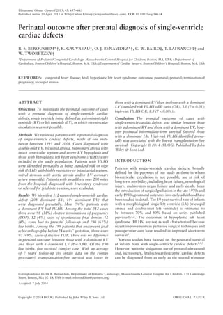
100
80
60
40
Freedomfromdeathortransplant(%)
20
0
0
64
92
29
48
58
11
25
32
6
10
6
3
2 4 6 8
Age (years)
10 12 14 16
Figure 2 Kaplan–Meier survival plot showing difference in
postnatal transplantation-free survival after Fontan procedure of all
live births with prenatal diagnosis of dominant LV ( , n = 65)
and dominant RV categorized into high-risk ( , n = 30) or
standard-risk ( , n = 95) hypoplastic left heart syndrome. Log
rank test P < 0.001. Values at bottom of plot indicate number of
patients alive and not lost to follow-up at each time point,
displayed in same order as curves.
outcome. Patients with mitral stenosis and aortic atresia
have a higher incidence of coronary artery anomalies
and LV coronary artery fistulae, which are thought to
increase the risk of myocardial ischemia during the Stage-1
operation17
. Earlier studies have also shown that patients
with severe restriction of the atrial septum have a survival
disadvantage, probably owing to pulmonary venous
hypertension, ‘arterialization’ of the pulmonary veins,
lymphangiectasia and poor pulmonary mechanics18,19
.
In the period of the Baltimore–Washington Infant Study
(c. 1981), the prevalence of tricuspid atresia and HLHS
was 0.039 and 0.267 per 1000 live births, respectively20.
With improved detection of heart defects over the last
20 years, the option of TOP has probably resulted in a
decrease in the prevalence of single-ventricle heart disease
in the liveborn population21. This has implications for
healthcare costs and finance in the present era.
Our study has a number of limitations. The study group
was a preselected population of patients with a wide
variety of single-ventricle cardiac defects, excluding those
with borderline hypoplasia of the RV or LV, heterotaxy
syndrome and prenatal intervention candidates. Thus our
findings are not applicable universally to all patients
who are not biventricular candidates. There were other
limitations, given the retrospective nature of the study.
Many patients were referred to our center after a prenatal
diagnosis at an outside facility, and the timing of our first
fetal echocardiogram may have been weeks to months
after the initial diagnosis. Furthermore, our reported
termination rate may be an underestimate, as there were
potentially other patients who chose TOP prior to referral.
While none of the patients with hydrops survived to
the first palliative surgery, the number of patients with
hydrops was small and their outcomes varied between
elective TOP, intrauterine fetal demise, comfort care and
Copyright © 2014 ISUOG. Published by John Wiley & Sons Ltd. Ultrasound Obstet Gynecol 2015; 45: 657–663.
- 7. Fetal single-ventricle outcomes 663
postnatal death. Thus, the study was underpowered to
determine whether hydrops was a risk factor for any one
of these outcomes. Finally, we did not assess long-term
mortality of single-ventricle cardiac disease, which may
influence prenatal decision-making.
CONCLUSIONS
In a high percentage (31%) of cases of fetal single-ventricle
cardiac defects the parents chose TOP. Whereas the
prenatal outcome of patients with a dominant RV
(HLHS) and a dominant LV were the same, there was a
considerable difference in postnatal survival between these
two groups, favoring longer intermediate-term survival in
single-LV patients. Prenatally identified high-risk HLHS
phenotype portends a worse postnatal outcome. Factors
affecting TOP remain unclear and are not associated with
ventricular morphology.
REFERENCES
1. Sittiwangkul R, Azakie A, Van Arsdell GS, Williams WG, McCrindle BW. Outcomes
of tricuspid atresia in the Fontan era. Ann Thorac Surg 2004; 77: 889–894.
2. Tham EB, Wald R, McElhinney DB, Hirji A, Goff D, Del Nido PJ, Hornberger LK,
Nield LE, Tworetzky W. Outcome of fetuses and infants with double inlet single left
ventricle. Am J Cardiol 2008; 101: 1652–1656.
3. Ohye RG, Sleeper LA, Mahony L, Newburger JW, Pearson GD, Lu M, Goldberg CS,
Tabbutt S, Frommelt PC, Ghanayem NS, Laussen PC, Rhodes JF, Lewis AB, Mital
S, Ravishankar C, Williams IA, Dunbar-Masterson C, Atz AM, Colan S, Minich
LL, Pizarro C, Kanter KR, Jaggers J, Jacobs JP, Krawczeski CD, Pike N, McCrindle
BW, Virzi L, Gaynor JW. Comparison of shunt types in the Norwood procedure for
single-ventricle lesions. New Engl J Med 2010; 362: 1980–1992.
4. Feinstein JA, Benson DW, Dubin AM, Cohen MS, Maxey DM, Mahle WT, Pahl
E, Villafa˜ne J, Bhatt AB, Peng LF, Johnson BA, Marsden AL, Daniels CJ, Rudd
NA, Caldarone CA, Mussatto KA, Morales DL, Ivy DD, Gaynor JW, Tweddell
JS, Deal BJ, Furck AK, Rosenthal GL, Ohye RG, Ghanayem NS, Cheatham JP,
Tworetzky W, Martin GR. Hypoplastic left heart syndrome: current considerations
and expectations. J Am Coll Cardiol 2012; 59 (Suppl): S1–S42.
5. Petit CJ. Staged single-ventricle palliation in 2011: outcomes and expectations.
Congenit Heart Dis 2011; 6: 406–416.
6. Morris SA, Ethen MK, Penny DJ, Canfield MA, Minard CG, Fixler DE, Nembhard
WN. Prenatal diagnosis, birth location, surgical center, and neonatal mortality in
infants with hypoplastic left heart syndrome. Circulation 2014; 129: 285–292.
7. Tworetzky W, McElhinney DB, Reddy VM, Brook MM, Hanley FL, Silverman NH.
Improved surgical outcome after fetal diagnosis of hypoplastic left heart syndrome.
Circulation 2001; 103: 1269–1273.
8. Friedberg MK, Silverman NH, Moon-Grady AJ, Tong E, Nourse J, Sorenson B, Lee
J, Hornberger LK. Prenatal detection of congenital heart disease. J Pediatr 2009;
155: 26–31.
9. Marek J, Tomek V, Skovranek J, Povysilova V, Samanek M. Prenatal ultrasound
screening of congenital heart disease in an unselected national population: a 21-year
experience. Heart 2011; 97: 124–130.
10. Rychik J, Szwast A, Natarajan S, Quartermain M, Donaghue DD, Combs
J, Gaynor JW, Gruber PJ, Spray TL, Bebbington M, Johnson MP. Perinatal
and early surgical outcome for the fetus with hypoplastic left heart syndrome:
a 5-year single institutional experience. Ultrasound Obstet Gynecol 2010; 36:
465–470.
11. Kaneko S, Khoo NS, Smallhorn JF, Tham EB. Single right ventricles have impaired
systolic and diastolic function compared to those of left ventricular morphology. J
Am Soc Echocardiogr 2012; 25: 1222–1230.
12. Anderson PA, Sleeper LA, Mahony L, Colan SD, Atz AM, Breitbart RE, Gersony
WM, Gallagher D, Geva T, Margossian R, McCrindle BW, Paridon S, Schwartz M,
Stylianou M, Williams RV, Clark BJ 3rd. Contemporary outcomes after the Fontan
procedure: a Pediatric Heart Network multicenter study. J Am Coll Cardiol 2008;
52: 85–98.
13. d’Udekem Y, Xu MY, Galati JC, Lu S, Iyengar AJ, Konstantinov IE, Wheaton GR,
Ramsay JM, Grigg LE, Millar J, Cheung MM, Brizard CP. Predictors of survival
after single-ventricle palliation: the impact of right ventricular dominance. J Am Coll
Cardiol 2012; 59: 1178–1185.
14. Gentles TL, Mayer JE Jr, Gauvreau K, Newburger JW, Lock JE, Kupferschmid JP,
Burnett J, Jonas RA, Casta˜neda AR, Wernovsky G. Fontan operation in five hundred
consecutive patients: factors influencing early and late outcome. J Thorac Cardiovasc
Surg 1997; 114: 376–391.
15. Hosein RB, Clarke AJ, McGuirk SP, Griselli M, Stumper O, De Giovanni JV, Barron
DJ, Brawn WJ. Factors influencing early and late outcome following the Fontan
procedure in the current era. The ‘Two Commandments’? Eur J Cardiothorac Surg
2007; 31: 344–352; discussion 353.
16. McGuirk SP, Winlaw DS, Langley SM, Stumper OF, de Giovanni JV, Wright JG,
Brawn WJ, Barron DJ. The impact of ventricular morphology on midterm outcome
following completion total cavopulmonary connection. Eur J Cardiothorac Surg
2003; 24: 37–46.
17. Vida VL, Bacha EA, Larrazabal A, Gauvreau K, Dorfman AL, Marx G, Geva T,
Marshall AC, Pigula FA, Mayer JE, del Nido PJ, Fynn-Thompson F. Surgical outcome
for patients with the mitral stenosis–aortic atresia variant of hypoplastic left heart
syndrome. J Thorac Cardiovasc Surg 2008; 135: 339–346.
18. Lowenthal A, Kipps AK, Brook MM, Meadows J, Azakie A, Moon-Grady AJ.
Prenatal diagnosis of atrial restriction in hypoplastic left heart syndrome is associated
with decreased 2-year survival. Prenat Diagn 2012; 32: 485–490.
19. Rychik J, Rome JJ, Collins MH, DeCampli WM, Spray TL. The hypoplastic left
heart syndrome with intact atrial septum: atrial morphology, pulmonary vascular
histopathology and outcome. J Am Coll Cardiol 1999; 34: 554–560.
20. Ferencz C, Rubin JD, McCarter RJ, Brenner JI, Neill CA, Perry LW,
Hepner SI, Downing JW. Congenital heart disease: prevalence at livebirth. The
Baltimore–Washington Infant Study. Am J Epidemiol 1985; 121: 31–36.
21. van der Bom T, Zomer AC, Zwinderman AH, Meijboom FJ, Bouma BJ, Mulder BJ.
The changing epidemiology of congenital heart disease. Nat Rev Cardiol 2011; 8:
50–60.
Copyright © 2014 ISUOG. Published by John Wiley & Sons Ltd. Ultrasound Obstet Gynecol 2015; 45: 657–663.