32nd publication ijohmr - 2nd name
•
0 likes•21 views

An unusual case of Ectopic Eruption of Supernumerary Mandibular Molar tooth in Coronoid. Int J Oral Health Med Res 2017;4(5):51-54

Recommended
Recommended
More Related Content
What's hot
What's hot (20)
Similar to 32nd publication ijohmr - 2nd name
Similar to 32nd publication ijohmr - 2nd name (20)
More from CLOVE Dental OMNI Hospitals Andhra Hospital
More from CLOVE Dental OMNI Hospitals Andhra Hospital (20)
Recently uploaded
Recently uploaded (20)
32nd publication ijohmr - 2nd name
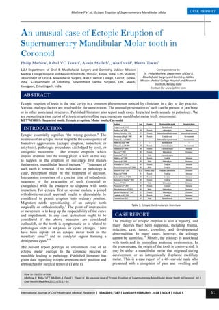
- 1. International Journal of Oral Health and Medical Research | ISSN 2395-7387 | JANUARY-FEBRUARY 2018 | VOL 4 | ISSUE 5 51 CASE REPORTMathew P et al.: Ectopic Eruption of Supernumerary Mandibular Molar Correspondence to: Dr. Philip Mathew, Department of Oral & Maxillofacial Surgery and Dentistry, Jubilee Mission Medical College Hospital and Research Institute, Kerala, India Contact Us: www.ijohmr.com An unusual case of Ectopic Eruption of Supernumerary Mandibular Molar tooth in Coronoid Philip Mathew1 , Rahul VC Tiwari2 , Aswin Mullath3 , Jisha David4 , Heena Tiwari5 Ectopic eruption of teeth in the oral cavity is a common phenomenon noticed by clinicians in a day to day practice. Various etiologic factors are involved for the same reason. The unusual presentation of teeth can be present in jaw bone or in other associated structures. Published literature also report such cases. Impacted tooth sequela to pathology. We are presenting a case report of ectopic eruption of the supernumerary mandibular molar tooth in coronoid. KEYWORDS: Impacted tooth, Ectopic eruption, Molar tooth, Coronoid AASSSAAsasasss Ectopic essentially signifies "the wrong position." The nearness of an ectopic molar might be the consequence of formative aggravations (ectopic eruption, impaction, or ankylosis), pathologic procedures (dislodged by cyst), or iatrogenic movement. The ectopic eruption, which implies eruption into the wrong place, is well on the way to happen in the eruption of maxillary first molars furthermore, mandibular lateral incisors.1,2 Treatment of such tooth is removal. If no indications or pathology is clear, perception might be the treatment of decision. Intercession comprises of a concise time of orthodontic treatment or the evacuation of teeth (essential or changeless) with the endeavor to dispense with tooth impaction. For ectopic first or second molars, a joined orthodontic-surgical approach ought to dependably be considered to permit eruption into ordinary position. Migration needs repositioning of an ectopic tooth surgically or orthodontically.3 The point of intercession or movement is to keep up the respectability of the curve and impediment. In any case, extraction ought to be considered if the above measures are considered outlandish, or the tooth is symptomatic or is related to pathologies such as ankylosis or cystic changes. There have been reports of an ectopic molar tooth in the maxillary sinus4,5 and in condylar region forming a dentigerous cysts.6–9 The present report portrays an uncommon case of an ectopic molar average to the coronoid process of mandible leading to pathology. Published literature has given data regarding ectopic eruptions their position and approaches for surgical removal (Table 1). The etiology of ectopic eruption is still a mystery, and many theories have been suggested, including trauma, infection, cyst, tumor, crowding, and developmental abnormalities. In many cases, however, the etiology cannot be identified.10 Mostly, the etiology is associated with tooth and its immediate anatomic environment. In the present case, the origin of the tooth is controversial. It may be either a mandibular molar that migrated during development or an iatrogenically displaced maxillary molar. This is a case report of a 46-year-old male who presented with a complaint of pain and swelling and How to cite this article: Mathew P, Rahul VCT, Mullath A, David J, Tiwari H. An unusual case of Ectopic Eruption of Supernumerary Mandibular Molar tooth in Coronoid. Int J Oral Health Med Res 2017;4(5):51-54. INTRODUCTION 1,2,4-Department of Oral & Maxillofacial Surgery and Dentistry, Jubilee Mission Medical College Hospital and Research Institute, Thrissur, Kerala, India. 3-PG Student, Department of Oral & Maxillofacial Surgery, KMCT Dental College, Calicut, Kerala, India. 5-Department of Dentistry, Government Dental Surgeon, CHC Makdi, Kondgaon, Chhattisgarh, India. ABSTRACT CASE REPORT Table 1: Ectopic third molars in literature
- 2. International Journal of Oral Health and Medical Research | ISSN 2395-7387 | JANUARY-FEBRUARY 2018 | VOL 4 | ISSUE 5 52 CASE REPORTMathew P et al.: Ectopic Eruption of Supernumerary Mandibular Molar difficulty in mouth opening. On clinical examination, there was intraoral swelling on the left side. It was also persistent in extra oral region. The periodontal health was compromised. Patient also gives a history of extraction and uneventful healing with maxillary left third molar. Orthopantomogram was advised which showed bilateral impacted supernumerary tooth was present (Figure 1). In the right side, it was present on ramus of mandible involving the inferior alveolar nerve. Tooth was incompletely formed according to its normal anatomical structure. On left side the supernumerary tooth was present on coronoid process of mandible forming radiolucent mass extending into ramus of mandible involving crown of supernumerary tooth. Clinicoradiographically it was diagnosed as dentigerous cyst. All the required pre-operative hematological investigations, chest X ray, electrocardiogram was obtained and surgical removal of impacted tooth was planned under general anesthesia. Intravenous antibiotics, analgesics and anti-inflammatory drugs were given before the procedure as prophylaxis. Intra oral approach was preferred using vertical incision extending distally to third molar towards external oblique ridge reaching the anterior border of ramus and coronoid was given. Tissue was reflected and lesion was exposed (Figure 2). Removal of infected mass was performed and impacted tooth was exposed in oral cavity (Figure 3). The supernumerary tooth was surgically removed (Figure 4). Surgical closure was done using 4-0 vicryl absorbable sutures. Postoperative instructions and medication was prescribed. Ectopic teeth are located in the jawbones or regions other than the alveolar arch. Ectopic eruption of a tooth is rare; however, there have been few reports of tooth in the nose, mandibular condylary and coronoid processes and maxillary sinus.11-14 Most of the cases in the mandibular coronoid and condylary regions had symptomatic signs; common symptoms of the clinical examination were pain, trismus, swelling, and temporomandibular joint problems. On the other hand, ectopic teeths are often discovered in routine clinical or radiographic examinations; as some of the cases were asymptomatic, like the case study above. The aetiology of ectopic eruption is still unclear, and reaches have suggested many theories, including trauma, infection, pathologic conditions, crowding and develop mental anomalies. However, it is likely to be an ectopic mandibular third molar for the following reasons: (1) the sinus discharge is in the mandible; (2) a pericoronal radiolucency can be seen around the crown; (3) if the root was located within the bony part of the coronoid process; absence of severe trismus after the maxillary molar extraction, and (5) an iatrogenically displaced maxillary third molar is usually positioned distal to the posterior maxillary wall and higher up in the infratemporal fossa. Figure 1: Orthopantomogram showing a mandibular third molar tooth present in Chondroid region of mandible. Figure 2:Incision and Exposure of Lesion Figure 3:Impacted mandibular third molar in the choronoid region of mandible. Figure 4:Extracted tooth DISCUSSION
- 3. International Journal of Oral Health and Medical Research | ISSN 2395-7387 | JANUARY-FEBRUARY 2018 | VOL 4 | ISSUE 5 53 CASE REPORTMathew P et al.: Ectopic Eruption of Supernumerary Mandibular Molar Odontogenesis is a complex process, and abnormal tissue interactions between the oral epithelium and the underlying mesenchymal tissue during development may potentially result in ectopic tooth development and eruption.15 A mandibular third molar may be displaced by a lesion such as a cyst or a tumor.16 The displacement of tooth buds by the expansion of progressively growing dentigerous cysts may result in the displacement of the tooth to other areas. In some of the reports in the literature review, the cysts associated with the ectopic third molars were very small like our case. Such cysts may have once occupied the entire ramus, but their walls may have been perforated, which resulted in drainage and decompression.16,17 This pathological process may support the idea that a dentigerous cyst was the etiologic factor of ectopic eruption in the subject of our case. For ectopic mandibular third molars associated with the condylar process of the mandible, various approaches have been used to gain access for retrieval of the tooth. Szerlip reported an ectopic third molar in the condylar process removed by an intraoral approach.18 Bux and Lisco reported a third molar associated with a dentigerous cyst in the subcondylar region and approached it through a cutaneous incision below the mandibular angle followed by a submasseteric dissection to create a subperiosteal tunnel leading to the surgical site.19 Tumer et al reported a third molar and a cyst in a similar position and excised them through a preauricular approach.20 Recently, Suarez-Cunqueiro et al used an endoscopic approach to remove an ectopic mandibular third molar in the condylar process and claimed advantages such as good illumination, clear and magnified visualization of the operating field, and more conservative surgery.21 Possible risk of damage to the facial nerve and scars on the skin could also be avoided with an endoscopic approach.22 They therefore advocated that endoscopy be used to remove ectopic third molars, not only in the condylar process but also in other ectopic locations, such as the maxillary sinus and nasal fossa. However, adequate training beforehand is essential. In this case, a pure intraoral approach was sufficient since the coronoid process is accessible via a wide incision along the ascending ramus. Adequate isolation was achieved by placing a sigmoid notch retractor buccally in the sigmoid notch, a Kocher artery forceps clamping the tip of the coronoid process, a channel retractor, and a periosteal elevator medially distal to the tooth. In this way, further displacement distally during manipulation was prevented. A coronoidectomy could be considered in case of inadequate access but was found to be unnecessary. Unusual presentation of impacted or ectopic tooth is a common phenomenon, but the presence of an ectopic supernumerary molar on coronoid process of mandible tooth is rare. In such condition the tooth is also associated with pathology and infection, surgical removal of tooth is indicated under proper antibiotic coverage and pre- operative hematological and radiological investigations. We acknowledge Doctors of Department of Oral and Maxillofacial Surgery, Department of Emergency Medicine and Department of Radiodiagnosis of Jubilee Mission Medical College Hospital and Research Centre for their help and support. 1. Bjerklin K, Kurol J. Ectopic eruption of the maxillary first permanent molar: Etiologic factors. Am J Orthod 1983;84:147–155. 2. Shapira Y, Kuftinec MM. The ectopically erupted mandibular lateral incisor. Am J Orthod 1982;82: 426–429. 3. Frank CA. Treatment options for impacted teeth. J Am Dent Assoc 2000;131:623–632. 4. Goh YH. Ectopic eruption of maxillary molar tooth—An unusual cause of recurrent sinusitis. Singapore Med J 2001;42:80–81. 5. Buyukkurt MC, Tozoglu S, Aras MH, Yolcu U. Ectopic eruption of a maxillary third molar tooth in the maxillary sinus: A case report. J Contemp Dent Pract 2005;6:104–110. 6. Szerlip L. Displaced third molar with dentigerous cyst—An unusual case.J Oral Surg 1978;36:551– 552. 7. Bux P, Lisco V. Ectopic third molar associated with a dentigerous cyst in the subcondylar region: Report of case. J Oral Maxillofac Surg 1994;52:630–632. 8. Tumer C, Eset AE, Atabek A. Ectopic impacted mandibular third molar in the subcondylar region associated with a dentigerous cyst: A case report. Quintessence Int 2002;33:231–233. 9. Suarez-Cunqueiro MM, Schoen R, Schramm A, Gellrich NC, Schmelzeisen R. Endoscopic approach to removal of an ectopic mandibular third molar. Br J Oral Maxillofac Surg 2003;41:340–342. 10. Debes RR, Miller SB. Molar in coronoid process. Oral Surg Oral Med Oral Pathol 1969;28:511. 11. Iglesias-Martin F, Infante-Cossio P, Torres-Carranza E, Prats-Golczer VE, Garcia-Perla-Garcia A. Ectopic third molar in the mandibular condyle: a review of the literature. Med Oral Patol Oral Cir Bucal. 2012;17:e1013-7. 12. Lambade P, Lambade D, Dolas RS, Virani N. Ectopic mandibular third molar leading to osteomyelitis of condyle: a case report with literature review. Oral Maxillofac Surg. 2013;17:127-30. 13. Baykul T, Doğru H, Yasan H, Cina Aksoy M. Clinical impact of ectopic teeth in the maxillary sinus. Auris Nasus Larynx. 2006;33:277- 81. 14. Verma RK, Bakshi J, Panda NK. Ectopic intranasal tooth: an unusual cause of epistaxis in a child. Ear Nose Throat J. 2012;91:242-4. 15. Srinivasa Prasad T, Sujatha G, Niazi T, Rajesh P. Dentigerous cyst associated with an ectopic third molar in the maxillary sinus: A rare entity. Indian J Dent Res. 2007;18:141-3 CONCLUSION ACKNOWLEDGEMENT REFERENCES
- 4. International Journal of Oral Health and Medical Research | ISSN 2395-7387 | JANUARY-FEBRUARY 2018 | VOL 4 | ISSUE 5 54 CASE REPORTMathew P et al.: Ectopic Eruption of Supernumerary Mandibular Molar 16. Medici A, Raho MT, Anghinoni M. Ectopic third molar in the condylar process: case report. Acta Biomed Ateneo Parmense. 2001;72:115-8. 17. Bux P, Lisco V. Ectopic third molar associated with a dentigerous cyst in the subcondylar region: report of case. J Oral Maxillofac Surg. 1994;52:630-2. 18. Szerlip L. Displaced third molar with dentigerous cyst—An unusual case.J Oral Surg 1978;36:551– 552. 19. Bux P, Lisco V. Ectopic third molar associated with a dentigerous cyst in the subcondylar region: Report of case. J Oral Maxillofac Surg 1994;52:630–632. 20. Tumer C, Eset AE, Atabek A. Ectopic impacted mandibular third molar in the subcondylar region associated with a dentigerous cyst: A case report. Quintessence Int 2002;33:231–233. 21. Suarez-Cunqueiro MM, Schoen R, Schramm A, Gellrich NC, Schmelzeisen R. Endoscopic approach to removal of an ectopic mandibular third molar. Br J Oral Maxillofac Surg 2003;41:340–342. 22. Debes RR, Miller SB. Molar in coronoid process. Oral Surg Oral Med Oral Pathol 1969;28:511. Source of Support: Nil Conflict of Interest: Nil