1. Ultrasound Obstet Gynecol 2008; 32: 860–864
Published online 6 October 2008 in Wiley InterScience (www.interscience.wiley.com). DOI: 10.1002/uog.6142
Fetal abdominal cysts in the first trimester: prenatal
detection and clinical significance
W. SEPULVEDA*†, K. DICKENS‡, A. CASASBUENAS*, J. GUTIERREZ† and V. DEZEREGA*
*Fetal Medicine Center, Department of Obstetrics and Gynecology, Clinica Las Condes, †Maternal-Fetal Medicine Unit, Department of
Obstetrics and Gynecology, San Jose Hospital, University of Santiago de Chile, Santiago, Chile and ‡Department of Ultrasound, Centro de
Salud No. 3, MSP, Guayaquil, Ecuador
K E Y W O R D S: abdominal cyst; fetal ultrasound; first trimester; prenatal diagnosis
ABSTRACT INTRODUCTION
The ultrasound detection of a fetal abdominal cyst in
Objective In order to determine the clinical significance
the second and third trimesters, although rare, is a
of fetal abdominal cysts detected in the first trimester, we
well characterized finding representing a wide variety of
reviewed our experience with such cases collected over a
clinical and surgical conditions1 – 4 . Among them, the main
5-year period.
differential diagnoses include cystic structures originating
Methods Five cases in which a fetal abdominal cyst from either the gastrointestinal tract (mesenteric or
was detected by ultrasound in the first trimester were omental cysts, intestinal duplication cysts, hepatic or
identified. Information on the ultrasound findings, choledochal cysts, and dilated bowel loop secondary to
antenatal course and perinatal outcome was obtained atresia or obstruction) or the genitourinary tract (ovarian,
in all cases. renal, urachal, and adrenal cysts)1 – 4 .
In recent years the progressive incorporation of
Results The abdominal cyst was confirmed by an early
first-trimester ultrasound screening into routine clinical
second-trimester scan at 14–16 weeks in all cases, at
practice has allowed the early detection of a significant
which time no associated anomalies were detected. The
number of fetal structural abnormalities5 – 7 . However,
standard detailed second-trimester scan at 18–22 weeks
there is a paucity of reports dealing with the diagnosis
demonstrated complete resolution in three cases. These
and significance of an abdominal cyst detected at this early
women had an uneventful antenatal course, and normal
gestational age. In this report we present our experience
newborn infants were delivered at term. However, one
with the prenatal diagnosis, subsequent management, and
of these infants had intestinal malrotation, chronic perinatal outcome in cases in which an abdominal cystic
abdominal distension and midgut volvulus requiring mass was detected by ultrasound in the first trimester.
surgery at the age of 7 months. Among the remaining two
cases in which the abdominal cyst persisted, one required
prenatal aspiration at 19 weeks owing to significant PATIENTS AND METHODS
enlargement and resolved. The other remained stable
in size and was managed conservatively, but the infant Cases in which an abdominal cystic mass was detected
required surgery at the age of 7 weeks owing to a by ultrasound in the first trimester were prospectively
choledochal cyst causing intermittent episodes of acholia. collected for this study. In order to be included, the
cystic structure had to be clearly differentiated from
Conclusion Abdominal cysts in early pregnancy often the fetal bladder by ruling out the presence of the
resolve spontaneously or remain small and are usually umbilical arteries alongside the mass with color-flow
associated with a good outcome. Nevertheless, as they can mapping8 . Color Doppler ultrasound was also used to
also be associated with serious underlying gastrointestinal exclude the possibility of a vascular structure by ruling
pathological conditions, close surveillance in the perinatal out the presence of blood flow within the cystic mass.
period is advocated. Copyright 2008 ISUOG. Published Once the diagnosis had been established, the parents
by John Wiley & Sons, Ltd. were informed on the prenatal findings, differential
Correspondence to: Prof. W. Sepulveda, Fetal Medicine Center, Clinica Las Condes, Casilla 208, Santiago 20, Chile
(e-mail: fetalmed@yahoo.com)
Accepted: 21 March 2008
Copyright 2008 ISUOG. Published by John Wiley & Sons, Ltd. ORIGINAL PAPER
2. First-trimester abdominal cyst 861
diagnoses, and probable natural course, and offered
ultrasound follow-up, including an early second-trimester
scan at 14–16 weeks and the standard detailed second-
trimester scan at 18–22 weeks. Information on maternal
demographics, ultrasound findings, subsequent antenatal
course and perinatal outcome was obtained by reviewing
the medical records, ultrasound reports and neonatal
charts. If the woman delivered in another institution,
the referring obstetrician was contacted to obtain the
pertinent perinatal information for analysis.
RESULTS
During the 5-year period from June 2002 to July
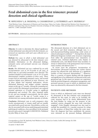
2007, five cases of first-trimester fetal abdominal cyst Figure 1 Ultrasound image of a first-trimester fetal abdominal cyst.
were diagnosed by the authors (estimated prevalence). The bladder is seen as a separate anechoic structure in the lower
Table 1 displays the most relevant clinical and ultrasound abdomen, caudal to the abdominal cyst. The infant had intestinal
findings in these cases. All the women were younger malrotation (Case 1).
than 35 years and two (40%) were primigravidas.
Gestational age at the time of diagnosis ranged from 10 +
4 weeks to 13 + 2 weeks, with the fetal crown–rump course was complicated with mesenteric thrombosis
length measuring between 36 and 77 mm. The nuchal requiring small bowel resection, which led to short
translucency thickness was measured in four cases and bowel syndrome. In the remaining two cases, the
reported to be within the normal range for gestational age abdominal cyst was again identified at the time of
in all of them. The cystic mass was single in all cases, with the detailed second-trimester scan. One was managed
the largest diameter at the time of detection measuring with a single percutaneous ultrasound-guided aspiration
between 5 and 11 mm (Figures 1–4). An early second- at 19 weeks because of significant enlargement of
trimester follow-up scan confirmed the presence of the the cyst, with subsequent resolution documented from
abdominal cystic mass, but no associated anomalies, in 20 weeks onwards. The further antenatal course was
all cases. uncomplicated, and the infant was delivered at 33 weeks
Spontaneous resolution of the cyst was documented by Cesarean section owing to rupture of membranes and
at the time of the detailed second-trimester scan in suspicion of chorioamnionitis unrelated to the prenatal
three cases. Among them, two newborn infants had invasive procedure. Subsequently the infant did well, and
an uneventful neonatal course, and after a normal at the time of writing was 12 months old and thriving. The
abdominal scan both were discharged with their mother, other case was managed expectantly; the cyst doubled in
and remained asymptomatic at the pediatric follow- size from 5 × 4 mm to 11 × 9 mm and remained stable in
up examinations. The other infant developed chronic size until term. The infant had an uncomplicated neonatal
abdominal distension, which was managed medically. course and was discharged with her mother. However,
However, at 7 months he had an intestinal pseudo- this infant underwent laparoscopic surgery at the age
obstruction and underwent surgery confirming intestinal of 7 weeks for excision of a choledochal cyst causing
malrotation and a midgut volvulus. The postoperative intermittent episodes of acholia.
Table 1 Fetal abdominal cyst in the first trimester: clinical cases
MA GA CRL NT Abdominal
Case (years) (weeks) (mm) (mm) cyst (mm) Remarks
1 27 13 + 2 77 2.3 8×7 Resolution. Term delivery of male infant with
intestinal malrotation, midgut volvulus, necrotizing
enterocolitis, mesenteric thrombosis, intestinal
resection, short bowel syndrome
2 20 13 + 1 72 1.1 5×4 Term delivery of female neonate. Surgery for
choledochal cyst at 7 weeks of postnatal life
3 17 12 + 1 60 0.7 10 × 8 Cyst aspiration at 19 weeks with subsequent
resolution. Preterm rupture of membranes and
delivery at 33 weeks. Normal female neonate
discharged on day 17
4 31 11 + 5 53 1.0 10 × 9 Resolution. Term delivery of normal female neonate
5 28 10 + 4 36 ND 11 × 11 Resolution. Term delivery of normal male neonate
CRL, crown–rump length; GA, gestational age; MA, maternal age; ND, no data available; NT, nuchal translucency thickness.
Copyright 2008 ISUOG. Published by John Wiley & Sons, Ltd. Ultrasound Obstet Gynecol 2008; 32: 860–864.
3. 862 Sepulveda et al.
Figure 2 Transverse views of the fetal upper abdomen of Case 2,
showing a cystic structure adjacent to the stomach at 13 weeks’
gestation (a) and at 18 weeks, when it had doubled in size (b). The Figure 3 A 12-week fetus (Case 3) with a large cyst located in the
infant was diagnosed neonatally with choledochal cyst. upper abdomen.Two-dimensional sagittal view (a) and
three-dimensional image (b), showing the location and relative size
of the abdominal cyst. The cyst increased in size, was aspirated at
19 weeks and resolved. The presumptive diagnosis was hepatic cyst.
DISCUSSION
This report describes the prenatal ultrasound detection
improvements in image resolution and a better system-
of an abdominal cyst in five first-trimester fetuses,
atic anatomic assessment protocol, evaluation of the early
together with its subsequent management and perinatal
fetal anatomy with greater detail is now possible9,10 , mak-
outcome. The abdominal cyst was confidently identified
independent of the fetal bladder and kidneys, and ing the detection of several structural anomalies at this
confirmed as such during the follow-up scan in the stage of pregnancy possible11 – 15 . Among them, mega-
early second trimester, thus reducing the possibility of cystis is the most common condition presenting as an
an ultrasound artifact. The subsequent detailed second- abdominal cystic mass in the first trimester, and the dif-
trimester scan demonstrated that the abdominal cyst ferential diagnosis can easily be established with color
resolved spontaneously in three cases, one of which Doppler ultrasound, as the bladder is normally sur-
was associated with serious gastrointestinal complications rounded by the intra-abdominal umbilical arteries8 . In
after birth. Two others persisted throughout the second contrast, there are only a few reports describing the
trimester, one of which required percutaneous cyst finding of an abdominal cyst in the first trimester, the
aspiration owing to significant enlargement, and the other majority of them being isolated case reports invariably
underwent surgery because of a symptomatic choledochal describing different associated gastrointestinal malforma-
cyst. The etiology of the abdominal cyst in three of our tions including distended sigmoid colon and intestinal
cases, in the absence of surgical or pathological proof, malrotation16,17 , hepatic cyst18 , ileal duplication cyst19 ,
remains therefore speculative. and anal atresia or imperforate anus17,20 . We are also
First-trimester ultrasound examination at 11–14 weeks aware of reports describing complete resolution of an
has been shown to be an important tool for the screen- abdominal cyst detected early in pregnancy in the pres-
ing of chromosomal abnormalities5 – 7 . With significant ence of a serious underlying gastrointestinal pathology
Copyright 2008 ISUOG. Published by John Wiley & Sons, Ltd. Ultrasound Obstet Gynecol 2008; 32: 860–864.
4. First-trimester abdominal cyst 863
spontaneously in utero, so close perinatal surveillance is
advocated.
ACKNOWLEDGMENTS
We are grateful to Drs R. Cassis, M. Ivankovic,
V. Rodriguez and C. Schnapp for their significant
contribution to the antenatal care of the patients reported
in this study. This work was supported by Sociedad
Profesional de Medecina Fetal ‘Fetalmed’ Limitada,
Chile.
REFERENCES
1. Nyberg DA, Neilson IR. Abdomen and gastrointestinal tract. In
Diagnostic Imaging of Fetal Anomalies, Nyberg DA, McGa-
han JP, Pretorius DH, Pilu G (eds). Lippincott Williams &
Wilkins: Philadelphia, PA, 2003; 547–602.
2. Hill LM. Ultrasound of fetal gastrointestinal tract. In
Ultrasonography in Obstetrics and Gynecology (4th edn),
Callen PW (ed.). W. B. Saunders: Philadelphia, PA, 2000;
457–487.
3. Gabrielli S, Rizzo N, Reece EA. Gastrointestinal and genitouri-
nary anomalies. In Clinical Obstetrics. The Fetus & Mother
(3rd edn). Reece EA, Hobbins JC (eds). Blackwell Publishing:
Malden, MA, 2007; 377–400.
4. McEwing R, Hayward C, Furness M. Foetal cystic abdominal
masses. Australas Radiol 2003; 47: 101–110.
5. Snijders RM, Noble P, Sebire N, Souka A, Nicolaides KH.
UK multicentre project on assessment of risk of trisomy
21 by maternal age and fetal nuchal-translucency thick-
ness at 10–14 weeks of gestation. Fetal Medicine Founda-
tion First Trimester Screening Group. Lancet 1998; 352:
343–346.
6. Nicolaides KH. Nuchal translucency and other first-trimester
sonographic markers of chromosomal abnormalities. Am J
Figure 4 Transverse (a) and parasagittal (b) ultrasound images of
Obstet Gynecol 2004; 191: 45–67.
Case 4 at 12 weeks’ gestation, showing a large cyst in the lower
7. Malone FD, Canick JA, Ball RH, Nyberg DA, Comstock CH,
fetal abdomen. There was complete resolution by the time of the
Bukowski R, Berkowitz RL, Gross SJ, Dugoff L, Craigo SD,
detailed second-trimester scan.
Timor-Tritsch IE, Carr SR, Wolfe HM, Dukes K, Bianchi DW,
Rudnicka AR, Hackshaw AK, Lambert-Messerlian G, Wald NJ,
D’Alton ME; First- and Second-Trimester Evaluation of Risk
manifesting only in the neonatal period. Teele et al.21 (FASTER) Research Consortium. First-trimester or second-
trimester screening, or both, for Down’s syndrome. N Engl
described two cases of intestinal malrotation present- J Med 2005; 353: 2001–2011.
ing as an abdominal cyst at 15 weeks that subsequently 8. Sepulveda W. Megacystis in the first trimester. Prenat Diagn
resolved in utero. Gilbert et al.22 also described the pres- 2004; 24: 144–149.
ence of a cystic mass in the lower fetal abdomen at 9. von Kaisenberg CS, Kuhling-von Kaisenberg H, Fritzer E,
Schemm S, Meinhold-Heerlein I, Jonat W. Fetal transabdom-
12 weeks, which had resolved by 17 weeks. At birth, the inal anatomy scanning using standard views at 11 to
male infant had multiple anomalies including anorectal 14 weeks’ gestation. Am J Obstet Gynecol 2005; 192:
agenesis, rectourethral fistula, horseshoe kidney and an 535–542.
anomalous sacrum. These authors hypothesized that an 10. Souka AP, Pilalis A, Kavalakis Y, Kosmas Y, Antsaklis P,
Antsaklis A. Assessment of fetal anatomy at the 11–14-week
intra-abdominal cyst in early pregnancy may have effects
ultrasound examination. Ultrasound Obstet Gynecol 2004; 24:
on the rotation and fixation of the bowel, and recom- 730–734.
mended close neonatal follow-up even if the cystic mass 11. Souka AP, Nicolaides KH. Diagnosis of fetal abnormalities at
disappears before birth21,22 . the 10–14-week scan. Ultrasound Obstet Gynecol 1997; 10:
In conclusion, the detection of a fetal abdomi- 429–442.
12. Whitlow BJ, Chatzipapas IK, Lazanakis ML, Kadir RA,
nal cyst in the first trimester is usually an isolated Economides DL. The value of sonography in early pregnancy for
finding and is associated with a good perinatal out- the detection of fetal abnormalities in an unselected population.
come in the majority of cases. In these cases, the Br J Obstet Gynaecol 1999; 106: 929–936.
abdominal cyst probably represents a transient find- 13. Carvalho MH, Brizot ML, Lopes LM, Chiba CH, Miyadahira S,
Zugaib M. Detection of fetal structural abnormalities at the
ing with no clinical significance. However, some other 11–14 week ultrasound scan. Prenat Diagn 2002; 22: 1–4.
cases can be associated with serious gastrointestinal 14. Fong KW, Toi A, Salem S, Hornberger LK, Chitayat D, Keating
malformations, even in cases in which the mass resolves SJ, McAuliffe F, Johnson JA. Detection of fetal structural
Copyright 2008 ISUOG. Published by John Wiley & Sons, Ltd. Ultrasound Obstet Gynecol 2008; 32: 860–864.
5. 864 Sepulveda et al.
abnormalities with US during early pregnancy. Radiographics cyst. Ultrasound Obstet Gynecol 2002; 19: 287–289.
2004; 24: 157–174. 19. Chen M, Lam YH, Lin CL, Chan KW, Hui PW, Tang MH,
15. Souka AP, Pilalis A, Kavalakis I, Antsaklis P, Papantoniou N, Lee CP, Khong PL. Sonographic features of ileal duplication
Mesogitis S, Antsaklis A. Screening for major structural abnor- cyst at 12 weeks. Prenat Diagn 2002; 22: 1067–1070.
malities at the 11- to 14-week ultrasound scan. Am J Obstet 20. Taipale P, Rovamo L, Hiilesmaa V. First-trimester diagnosis
Gynecol 2006; 194: 393–396. of imperforate anus. Ultrasound Obstet Gynecol 2005; 25:
16. Suzuki S. Megacolon in a fetus during the first trimester. Prenat 187–188.
Diagn 2001; 21: 422–423. 21. Teele RL, Pease PWB, Rowley RSH. Malrotation in newborns
17. Lam YH, Shek T, Tang MHY. Sonographic features of anal following antenatal diagnosis of intra-abdominal cyst. Pediatr
atresia at 12 weeks. Ultrasound Obstet Gynecol 2002; 19: Radiol 1998; 28: 717–721.
523–524. 22. Gilbert CE, Hamill J, Metcalfe RF, Smith P, Teele RL. Chang-
18. Berg C, Baschat AA, Geipel A, Krapp M, Germer U, Smrcek JM, ing antenatal sonographic appearance of anorectal atresia from
Sigge W, Gembruch U. First-trimester diagnosis of fetal hepatic first to third trimesters. J Ultrasound Med 2006; 25: 781–784.
Copyright 2008 ISUOG. Published by John Wiley & Sons, Ltd. Ultrasound Obstet Gynecol 2008; 32: 860–864.