Centralization of ulna by dr chiranjeevi
•Download as PPTX, PDF•
0 likes•78 views

centralization of ulna, rare case presentation

Recommended
More Related Content
What's hot
What's hot (20)
Similar to Centralization of ulna by dr chiranjeevi
Similar to Centralization of ulna by dr chiranjeevi (20)
More from rangaraya medical college
Recently uploaded
Recently uploaded (20)
Centralization of ulna by dr chiranjeevi
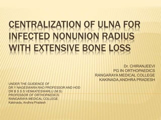
- 1. CENTRALIZATION OF ULNA FOR INFECTED NONUNION RADIUS WITH EXTENSIVE BONE LOSS Dr. CHIRANJEEVI PG IN ORTHOPAEDICS RANGARAYA MEDICAL COLLEGE KAKINADA,ANDHRA PRADESH UNDER THE GUIDENCE OF DR Y NAGESWARA RAO PROFESSOR AND HOD DR B S S S VENKATESWARLU (M.S) PROFESSOR OF ORTHOPAEDICS RANGARAYA MEDICAL COLLEGE Kakinada, Andhra Pradesh
- 2. INTRODUCTION Infected nonunion with extensive bone loss remains a challenging problem in orthopaedic traumatology. Acquired Radial club hand deformity, post osteomyelitis of radius bone is a very rare disease. It results in functional and cosmetic deficit of upper limb which is similar to congenital cases.
- 3. WHAT ARE THE VARIOUS METHODS TO RECONSTRUCT THE DEFORMITY 1. Bone grafting, plating, 2. Ilizarov, 3. Monorail external fixator, 4. Callus distraction etc
- 4. WHAT WE DID………? Keeping this in mind, to create a single bone forearm we planned for centralization of ulna on wrist to correct the deformity. This was originally devised by Hey-Groves in 1921, modified by Greenwood in 1932
- 5. CASE REPORT A 15years old boy came to OPD with deformity of left forearm since childhood. He had h/o trauma during childhood at the age of 8yrs managed conservatively with pop cast . Later he had purulent discharge from the same limb.
- 6. ON EXAMINATION I. Patient had marked atrophy of the both hand and forearm. II. No active infection and sinus tracts but old healed sinus tracts present. III. He had complete pronation movements but supination upto 90 degrees. IV. Fixed radial deviation 50 degrees. V. Fixed volar deviation 30 degrees. VI. No neurovascular deficit.
- 8. RADIOLOGICAL EXAMINATION Forearm AP and lateral views Showing distal radial metaphysis and distal 3rd diaphysis. No signs of active osteomyelitis Positive ulnar variance
- 9. PROCEDURE ‘C’-shaped dorsal midline incision given. Branches of superficial sensory radial nerve are preserved. The extensor tendons were identified, and a thorough dissection was done to release any tight radial structure. Distal end of ulna was osteotomised 1cm proximal to the distal physis.
- 10. INTRA OPERATIVE
- 11. The distal stump of the radius was exposed, freshened and a trough created in its proximal surface to accommodate the proximal ulnar fragment. The latter was translated radially and impacted into the distal radial remnant. This construct was held in place by k wires passed through the 3rd metacarpal. Above elbow pop cast was applied for 3 months. Later k-wires were removed and wrist movements were allowed.
- 12. RESULTS At 8months follow up 15-year-old boy had an infection free, cosmetically acceptable forearm with a stable wrist joint and a good hand grip. He lacked the last 10 degrees of palmar and dorsiflexion . The left forearm was 3 inches shorter than the right. It was also smaller in girth by 1 inch at mid-level and by 0.5 inch at wrist level.
- 13. Radiographs showed consolidation of the union between the hypertrophied centralized ulna and distal radius, A good wrist joint space with a mild ulnar negative variance and normal palmar tilt of the distal radial articular surface.
- 14. POST OPERATIVE
- 18. CONCLUSION We conclude that centralization of ulna showed good result in correcting the deformity and producing wrist stability reasonably. There was no detrimental effect on the growth of the distal ulnar epiphysis. The finding of the study need further follow-up, as a short follow-up period for evaluating such rare cases is not appropriate.
- 19. THANK YOU