Git j club be management advices22
•Download as PPTX, PDF•
0 likes•32 views

GIT J Club BE management advices

Recommended
More Related Content
What's hot
What's hot (20)
Similar to Git j club be management advices22
Similar to Git j club be management advices22 (20)
More from Case records of Sulaymaniah General Teaching Hospital.
More from Case records of Sulaymaniah General Teaching Hospital. (20)
Recently uploaded
Recently uploaded (20)
Git j club be management advices22
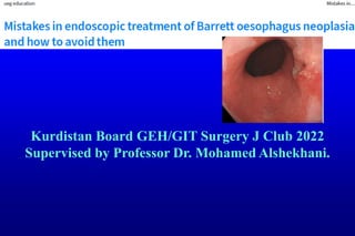
- 1. Kurdistan Board GEH/GIT Surgery J Club 2022 Supervised by Professor Dr. Mohamed Alshekhani.
- 2. Introduction : BE A premalignant condition of the distal oesophagus predisposing to EAC. Given the potential for malignant progression & the poor prognosis of EAC when diagnosed at a symptomatic stage, patients with known BE undergo regular endoscopic surveillance to detect neoplastic progression at an early& preferably endoscopically, treatable stage. Endoscopic management of early BE neoplasia consists of endoscopic imaging, endoscopic resection& endoscopic ablation. Below we discuss a number of mistakes that are frequently made when managing BE neoplasia and how to avoid them. Much of this discussion draws on existing guidelines&practically driven recommendations based on common sense &experience.
- 3. 1:Clean&inspect well With inadequate cleaning& immediately ‘jumping’ to obtain the inevitable random biopsy samples you will not detect the more subtle Barrett lesions. Use the waterjet of to clean the Barrett segment;takes 1–2 minutes. Switch to optical chromoscopy: if the oesophagus looks impeccably clean on narrow-band imaging (NBI), then cleaning is optimal. Spend 3–5 minutes inspecting the segment using white light endoscopy. Switching back-forth with optical chromoscopy, help to see more. Retroflex the endoscope to inspect the ‘danger zone’—the area where the Barrett segment transits into the hiatal hernia,area has the highest risk of neoplasia &highest risk of neoplasia being missed endoscopically. Look longer, biopsy less! After taking first biopsy sample most of imaging opportunities are lost. Detecting early neoplasia is all about recognizing how early neoplasia actually looks. Excellent training modules are available at [www.best-academia.eu].
- 7. 2:Think twice of biopsy results. When BE segment biopsy is diagnosed as non-dysplastic or inflammatory, this situation requires either endoscopic resection of the abnormality for optimal diagnosis or repeat endoscopy to document its regression, because a lesion that clearly looks neoplastic on endoscopy generally is neoplastic. The biopsy samples not to be misplaced&the histological assessment might not close to the squamocolumnar junction in the presence of a grade A or B reflux oesophagitis&provide this information as well.
- 8. 3:perform intervention after optimal evaluation. We prefer to use a diagnostic endoscope for most therapeutic work in patients with Barrett oesophagus. Optimal imaging makes the right decision regarding resection versus ablation& allows optimal delineation of lesions. A waterjet is essential to ensure adequate cleaning. During endoscopic resection, deal with bleeding before proceeding with ESD or piecemeal resection. For piecemeal resections, any bleeding from prior resections must be adequately treated& the surface cleaned of blood/ mucous&emptied the stomach of fluids / blood before you embark on your next resection.
- 9. 4:resect visible resectable lesions before ablation. The above is the most common reason for neoplastic progression under ablation. The endoscopist performing ablation in BE should be able to switch gears to endoscopic resection&ablation shouldn’t be used as an excuse for not having to do an endoscopic resection. Endoscopist should have skills in managing both; ablation& resection of visible resettable BE.
- 10. 5:Don’t ablate inflamed or swollen BE. Ablation sessions are generally scheduled at 3-month intervals & the 2nd session should not be done if there inflammation&swelling because: Ablation will not be effective, given the thickness of the epithelium, The likely inadequate acid suppression. Not be able to adequately inspect the segment for neoplastic progression. Not allow to detect neoplastic progression, especially when you have overlooked a visible lesion at the initial ablation.
- 12. 6:Start resection with marking. The outer margins of your neoplastic target area may not be sufficiently visible once the multiband mucosectomy (MBM) kit has been assembled& resections/ bleeding may hamper visualisation during piecemeal procedures. Given the importance of marking, generally use a diagnostic endoscope with a small distant attachment cap for this purpose to allows stabilize the endoscope tip onto the mucosa during the marking. The use of optical chromoscopy& a near-focus mode (with Olympus endoscopes) or zoom function (with Fujifilm or Pentax endoscopes) enables both the detection of the demarcation line& the controlled positioning of the electrocoagulation markers with the tip of the snare (for MBM procedures) or the ESD knife .
- 13. 7:carefully choose the ideal resection technique. Most of the early neoplastic lesions can be effectively removed by MBM,but a subgroup of lesions should not be resected by MBM because of the likelihood that there is deep submucosal invasion&/or a large intraluminal extent of the lesion. This will compromises any subsequent endoscopic resection being performed by more experienced endoscopists.
- 16. 8:Should be trained well with good case load. All guidelines state that adequate training& a yearly case volume of at least 10 new patients with HGD or early cancer are needed,soneeds to be centralized.
- 17. 9:Be prepared to manage significant bleeding. Most ESD bleeds can be managed by directing therapy to the vessel that has been accidentally cut7coagulation forceps will do the job here. After MBM procedures, where the resection is ‘blind’& less controlled than in ESD, the bleeding source may be more difficult to determine. Do not remove the MBM cap unless it is absolutely necessary, because most bleeds can be treated by touching the bleeding site with the tip of the snare& apply very gentle pressure with the tip of the snare. A careful snare tip coagulation generally suffices, but if the bleeding continues despite two or three applications you need to switch gears to coagulation forceps,which will require to release the remaining rubber bands in the stomach to allow passage of the forceps.
- 18. 10:don’t make thing worse after resection perforation.