2. clinical practice
tive enzymes causes pancreatic injury and results
in an inflammatory response that is out of propor-tion
to the response of other organs to a similar
insult. The acute inflammatory response itself
causes substantial tissue damage and may pro-gress
beyond the pancreas to a systemic inflam-matory
response syndrome, multiorgan failure,
n engl j med 354;20 www.nejm.org may 18, 2006 2143
or death.
Strategies and Evidence
Diagnosis
The clinical diagnosis of acute pancreatitis is based
on characteristic abdominal pain and nausea, com-bined
with elevated serum levels of pancreatic en-zymes.
In gallstone pancreatitis, the pain is typi-cally
sudden, epigastric, and knife-like and may
radiate to the back. In hereditary or metabolic cas-es
or in those associated with alcohol abuse, the
onset may be less abrupt and the pain poorly local-ized.
Serum amylase levels that are more than
three times the upper limit of normal, in the set-ting
of typical abdominal pain, are almost always
caused by acute pancreatitis. Lipase levels are also
elevated and parallel the elevations in amylase lev-els.
The levels of both enzymes remain elevated
with ongoing pancreatic inflammation, with amy-lase
levels typically returning to normal shortly
before lipase levels in the resolution phase.
Tests that are more specific for acute pancreati-tis
but less widely available evaluate levels of tryp-sinogen
activation peptide10 and trypsinogen-2.11
Abdominal imaging by computed tomography
(CT), magnetic resonance imaging (MRI), or trans-abdominal
ultrasonography is useful in confirm-ing
the diagnosis of pancreatitis or ruling out
other intraabdominal conditions as the cause of
pain or laboratory abnormalities. Such imaging
may also identify the cause of pancreatitis or its
associated complications.
Management
Determination of the cause is important for guid-ing
immediate management and preventing recur-rence.
An elevated alanine aminotransferase level
in a patient without alcoholism who has pancre-atitis
is the single best laboratory predictor of bili-ary
pancreatitis; a level of more than three times
the upper limit of normal has a positive predictive
value of 95 percent for gallstone pancreatitis.12
However, the presence of normal alanine amino-transferase
levels does not reliably rule out the di-agnosis.
4 Laboratory testing may reveal hypertri-glyceridemia
or hypercalcemia as possible causes
of pancreatitis, although pancreatitis may also
cause mildly elevated triglyceride levels.
Imaging Studies
CT or MRI can identify gallstones or a tumor (an
infrequent cause of pancreatitis), as well as local
complications. MRI may also identify early duct
disruption that is not seen on CT.13 Transabdomi-nal
ultrasonography is more sensitive than either
CT or MRI for identifying gallstones and sludge
and for detecting bile-duct dilatation, but it is in-sensitive
for detecting stones in the distal bile
duct.4,5 Endoscopic ultrasonography may be the
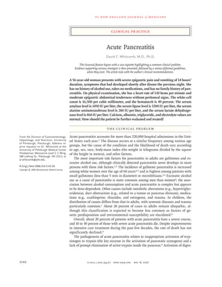
most accurate test for diagnosing or ruling out
biliary causes of acute pancreatitis (Fig. 1) and may
guide the emergency use of endoscopic retrograde
cholangiopancreatography (ERCP).14
A
B
Figure 1. Endoscopic Ultrasonography of the Gall-bladder
and Common Bile Duct from the Duodenum.
Microlithiasis (sludge) is shown within the gallbladder
(Panel A, arrow) and within the common bile duct
(Panel B, arrow). Also visible in Panel B are the head of
the pancreas (curved arrow) and the pancreatic duct
(arrowhead). (Images courtesy of Neeraj Kaushik, M.D.)
3. The new england journal o f medicine
Table 1. Scoring Methods for the Prediction of Severe Acute Pancreatitis.
Criterion and Marker Threshold Value Severe Pancreatitis
Atlanta criteria* Indicated by any positive factor listed
Ranson’s score† ≥3
APACHE II score‡ ≥8
Organ failure
Shock Blood pressure of <90 mm Hg
Pulmonary insufficiency Partial pressure of arterial oxygen of ≤60 mm Hg5
Renal failure Creatinine level of >177 μmol/liter (2 mg/dl)
after hydration
Systemic complications
Disseminated intravascular coagulation Platelet count of ≤100,000/mm3
Fibrinogen level of <1 g/liter
Fibrin-split products level of >80 μg/ml
Metabolic disturbance Calcium level of ≤7.5 mg/dl
Local complications
Pancreatic necrosis Present
Pancreatic abscess Present
Pancreatic pseudocyst Present
Ranson’s score† Indicated by a total score ≥3, with
Age >55 yr
Blood glucose level >200 mg/dl (10 mmol/liter)
White-cell count >16,000/mm3
Lactate dehydrogenase level >350 IU/liter
Alanine aminotransferase level >250 IU/liter
Within 48 hr after presentation
Hematocrit >10% decrease
Serum calcium <8 mg/dl (2 mmol/liter)
Base deficit >4 mEq/liter
Blood urea nitrogen >5 mg/dl (1.8 mmol/liter) increase
Fluid sequestration >6 liters
Partial pressure of arterial oxygen§ <60 mm Hg
ERCP
Persistent biliary obstruction worsens the outcome
and increases the severity of acute pancreatitis and
predisposes the patient to bacterial cholangitis.
ERCP is used with endoscopic sphincterotomy to
extract impacted gallstones and to drain infected
bile in severe acute pancreatitis.15-18 Although ERCP
has risks, including bleeding after sphincteroto-my
and causing acute pancreatitis, complications
are uncommon when the procedure is performed
by experienced endoscopists. Three randomized
trials involving a total of 511 patients with gall-stone
pancreatitis compared conservative manage-ment
1 point for each positive factor
with ERCP and endoscopic sphincterotomy
within 24 to 72 hours after admission. The stud-ies
showed a significantly lower risk of pancreati-tis-
associated complications in the ERCP group
(odds ratio, 0.27; 95 percent confidence interval,
0.14 to 0.53).16
Hospitalization
Patients who present with persistent or severe pain,
vomiting, dehydration, or signs of impending se-vere
acute pancreatitis (to be discussed later) should
be hospitalized. Clinical trials have failed to show
the efficacy of medications proposed to alter the
At presentation
2144 n engl j med 354;20 www.nejm.org may 18, 2006
4. clinical practice
Table 1. (Continued.)
Criterion and Marker Threshold Value Severe Pancreatitis
CT severity index¶ Indicated by a total score of >6
course of acute pancreatitis, including an inhibi-tor
of platelet-activating factor (lexipafant19), so-matostatin
and its analogues, and protease inhib-itors20;
treatment is primarily supportive. Patients
should receive nothing by mouth and receive in-travenous
pain medication and aggressive hydra-tion
to treat or prevent hemoconcentration (e.g.,
a bolus of fluids to achieve hemodynamic stabil-ity,
followed by 250 to 500 ml of crystalloid solu-tions
per hour in an average-sized patient without
substantial kidney or heart disease). Fluid balance
should be maintained and pulse oximetry should
be considered, especially when narcotic analgesics
are used.
Predicting Severe Acute Pancreatitis
The severity of acute pancreatitis is defined by the
presence or absence of organ failure, local com-plications,
or both21-25 (Table 1). It is critical to
identify patients who are at high risk for severe
(CT grade plus necrosis score)
disease, since they require close monitoring and
possible intervention. Recognized markers of the
risk of severe acute pancreatitis include specific
laboratory values that measure the systemic in-flammatory
response (such as C-reactive protein),
scoring systems that assess inflammation or or-gan
failure (such as Ranson’s score), and findings
on imaging studies13,23 (Table 2). The Acute Phys-iology
and Chronic Health Evaluation score (based
on initial values of 12 routine physiological mea-surements,
age, and previous health status) is
among the best predictors of severity on admission,
whereas elevated C-reactive protein levels are equal-ly
useful when measured 24 to 48 hours after the
onset of symptoms.27 Severity scores are useful in
predicting both complications and death (Table 3).
Other markers that are not included in standard
scoring systems should also be considered. Obe-sity
(a body-mass index of more than 30) is associ-ated
with an increase in the risk of a severe clinical
n engl j med 354;20 www.nejm.org may 18, 2006 2145
CT grade
Normal pancreas (grade A) 0 points
Focal or diffuse enlargement (grade B) 1 point
Intrinsic change; fat stranding (grade C) 2 points
Single, ill-defined collection of fluid (grade D) 3 points
Multiple collections of fluid or gas in or adja-cent
to pancreas (grade E)
4 points
Necrosis score
No pancreatic necrosis 0 points
Necrosis of one third of pancreas 2 points
Necrosis of one half of pancreas 4 points
Necrosis of >one half of pancreas 6 points
APACHE II score‡ Indicated by a score of ≥8
Initial values of 12 routine physiological measure-ments,
age, and previous health status
* Data are from Bradley.21 The Atlanta criteria were adopted in 1992 by the International Symposium on Acute Pancreatitis. The presence of
any condition in the five main categories indicates severe acute pancreatitis.
† Data are from Ranson et al.22 The original Ranson’s score is based on 11 clinical signs (5 measured on admission and 6 in the 48 hours af-ter
admission), with a higher score indicating greater correlation with the incidence of systemic complications and the presence of pancreat-ic
necrosis. The relationship between Ranson’s score and the CT severity index23 is given in Table 3.
‡ Data are from Knaus et al.24 and Larvin and McMahon.25 The Acute Physiology and Chronic Health Evaluation (APACHE II) score is based
on initial values of 12 routine physiological measurements, age, and previous health status, with a score of 8 or more commonly used as
the threshold for classification as severe pancreatitis.
§ The test was performed without the use of supplemental oxygen.
¶ The CT severity index23 is a combination of the sum of the necrosis score and points assigned to five grades of findings on CT. The index
ranges from 0 to 10, with higher scores indicating a greater severity of illness.
5. The new england journal o f medicine
Table 2. Value of Various Scoring Systems and Inflammatory Markers in the Prediction of Severe Acute Pancreatitis.*
Scoring System Sensitivity Specificity
course by a factor of 2 to 3.29 A hematocrit above
44 percent is a clear risk factor for pancreatic ne-crosis,
30 although it is a poor predictor of the sever-ity
of disease. Preliminary evidence suggests that
genetic factors, such as polymorphisms in the che-mokine
monocyte chemotactic protein 1 (MCP-1)
gene,31 may also predict severity, although such
genetic testing is not currently used in practice.
Several clinical findings — including thirst,
poor urine output, progressive tachycardia, tachyp-nea,
hypoxemia, agitation, confusion, a rising he-matocrit
level, and a lack of improvement in symp-toms
within the first 48 hours — are warning
signs of impending severe disease. If such symp-toms
develop, admission to an intensive care unit
should be considered. Intensive care may also be
warranted in patients at risk for rapid deteriora-tion
in their condition, including those over the
age of 55 years,22 those who need ongoing volume
resuscitation or invasive monitoring of fluid sta-tus
(e.g., central venous pressure monitoring), or
Positive
Predictive
Value
those with renal failure or respiratory compro-mise.
15
Pancreatic-Fluid Collections, Pseudocysts,
and Necrosis
Up to 57 percent of patients who are hospitalized
with acute pancreatitis will have fluid collections,
with 39 percent having two areas involved and 33
percent having three or more.32 Fluid collections
are initially ill defined,21 evolve over time, and are
usually managed conservatively. If the fluid col-lections
continue to enlarge, cause pain, become
infected (as suggested by the presence of unex-plained
fever, leukocytosis, or gas in the fluid col-lection),
or compress adjacent organs, then med-ical,
endoscopic, or surgical intervention may be
needed.33,34 Fluid collections with very high lev-els
of pancreatic enzymes are usually associated
with pancreatic-duct disruptions and may even-tually
form pseudocysts (usually over a period of
several weeks), ascites, or pleural effusions.34
2146 n engl j med 354;20 www.nejm.org may 18, 2006
Negative
Predictive
Value Accuracy
percent
On admission
APACHE II score†
≥6 83–99 33–54 28–40 80–97 45–65
≥8 68–71 48–67 30–40 84–87 53–68
≥10 52–63 66–81 32–64 81–89 63–77
Interleukin-6 level >400 pg/ml 89 87 80 93 88
At 24 hours
APACHE II score ≥8 63 73 38 88 71
C-reactive protein level >150 mg/dl 65 73 37 90 72
PMN elastase >300 μg/liter 93 99 97 98 98
Urinary TAP >35 nmol/liter 68 74 44 89 73
At 48 hours
APACHE II score ≥8 56–78 52–64 30–33 85–88 58–63
Ranson’s score ≥3 75–89 54–71 37–49 91–96 62–75
Modified Glasgow score ≥3‡ 45–75 63–89 28–66 79–93 59–84
C-reactive protein level >150 mg/dl 65 73 37 90 72
* PMN denotes polymorphonuclear leukocyte, and TAP trypsinogen activation peptide.
† Data are from Knaus et al.24 and Larvin and McMahon.25 The Acute Physiology and Chronic Health Evaluation
(APACHE II) score is based on initial values of 12 routine physiological measurements, age, and previous health status,
with a score of 8 or more commonly used as the threshold for the classification of severe pancreatitis.
‡ The Modified Glasgow score is similar to Ranson’s score but can be completed on admission. The score ranges from
0 to 8, with scores of 3 or more indicating a greater severity of illness.26 Data are from Papachristou and Whitcomb.27
6. clinical practice
Asymptomatic pseudocysts can be managed con-servatively,
whereas symptomatic pseudocysts can
often be drained endoscopically.35 ERCP may help
to define the anatomy of the pancreatic duct and
identify any duct disruptions to guide further in-tervention.
Table 3. Relationship between Severity Scores and Outcomes in Acute Pan-creatitis.*
Index Score
Ranson’s score 0–2 3–4 5–6 7–8
percent of patients
Intensive care for >7 days and
survival
1 24 53 NA
Death 3 16 40 100
Either intensive care for >7 days
or death
4 40 93 100
CT severity index 0–3 4–6 7–10 NA
Complications 8 35 92 NA
Death 3 6 17 NA
* Data are from Balthazar et al.23 and Ranson.28 NA denotes not applicable.
n engl j med 354;20 www.nejm.org may 18, 2006 2147
33,34
Pancreatic necrosis, occurring as diffuse or fo-cal
areas of nonviable pancreatic parenchyma,21
is an important complication that can develop dur-ing
the first few days of pancreatitis; the condition
is associated with late complications and death if
the necrotic tissue becomes infected. The devel-opment
of necrosis is associated with pancreatic
inflammation, hypovolemia, and hypotension from
the shunting of blood from other organs, vascu-lar
spasm, and hemoconcentration.30 Pancreatic
necrosis can be demonstrated by a loss of tissue
perfusion on contrast-enhanced CT.23
Infection of necrotic tissue is suspected when
there is fever, leukocytosis, and a failure to improve
or unexpected deterioration — usually after the
first week of illness. Visualization of gas bubbles
within the necrotic tissue on CT is evidence of in-fection.
The diagnosis of infected necrosis is usu-ally
made by fine-needle aspiration of the necrotic
area guided by either CT or ultrasonography, with
Gram’s staining and culture of the aspirate.36
Lack of Improvement
If the condition of a patient whose pancreatitis is
predicted to be mild fails to improve within two or
three days, then contrast-enhanced CT (“pancreas
protocol”) should be considered to identify fluid
collections, pancreatic necrosis, or other compli-cations
that may require intervention. Antibiotic
therapy and nutritional support also warrant con-sideration
in patients whose condition fails to im-prove
promptly or in whom complications develop.
Use of Antibiotics
The proper role of antibiotics in acute pancreati-tis
remains controversial. No antibiotics are indi-cated
in mild cases. However, infectious compli-cations
are an important concern in severe cases,
especially cases of pancreatic necrosis. A potential
role for prophylactic antibiotics in severe pancre-atitis
was initially given support by a randomized
trial demonstrating that the administration of
imipenem reduced infectious complications, in-cluding
central-line sepsis, pulmonary infection,
urinary tract infection, and infected pancreatic ne-crosis.
37 Subsequent trials yielded mixed, but gen-erally
confirmatory, results.38 However, a recent
randomized trial failed to demonstrate differenc-es
in outcome among patients treated with ci p-rofloxacin
plus metronidazole, as compared with
placebo, leading some experts to recommend
against the routine use of prophylactic antibiot-ics.
39 Some centers use antifungal therapy as well
as antibacterial therapy, but this practice has not
been validated by randomized trials.
Nutritional Support
Ensuring adequate nutrition is important in pa-tients
with severe or complicated pancreatitis, but
the optimal means of doing so remains contro-versial.
40 Two small trials involving a total of 70
patients showed a nonsignificant reduction in ad-verse
outcomes with enteral feeding through na-soenteric
feeding tubes, as compared with total
parenteral nutrition.41 More recent meta-analy-ses
of six randomized trials involving a total of
263 patients demonstrated improved outcomes
with enteral nutrition,42,43 including decreased
rates of infection42,44 and surgical intervention,42
a reduced length of hospital stay,42 and reduced
costs (20 percent of the costs associated with to-tal
parenteral nutrition).43 Enteral feeding is usu-ally
well tolerated in patients with ileus.40 How-ever,
total parenteral nutrition may be necessary
for patients who cannot obtain sufficient calories
through enteral nutrition or in whom enteral ac-cess
cannot be maintained.45
Surgery
Surgical intervention is indicated in patients with
infected pancreatic necrosis. In most cases, the di-
7. The new england journal o f medicine
agnosis is confirmed by fine-needle aspiration be-fore
surgical intervention, but because false neg-ative
results can occur (reported sensitivity, 88
percent),46 surgery also warrants consideration
when there is a high index of suspicion of infected
necrosis even if infection is not documented.
Surgery within the first few days after the onset
of severe acute pancreatitis is associated with rates
of death up to 65 percent.47 Furthermore, there is
no clear demarcation between viable and nonviable
tissue early in the course of acute pancreatitis.47
Observational data support delaying surgical dé-bridement
of necrotic tissue for at least two weeks
if possible while the patient’s medical condition is
optimized and viable pancreatic tissue becomes
evident.47 This approach appears to improve sur-vival
and maximize organ preservation.47
Discharge Planning
Whenever possible, the cause of pancreatitis should
be determined and plans to prevent recurrence
should be devised before the patient is discharged
from the hospital. In patients with acute pancre-atitis
caused by gallstones, cholecystectomy should
be considered before discharge in those with mild
cases or within a few months in those with more
severe or complicated cases to allow inflamma-tory
processes or fluid collections to organize or
resolve.47 ERCP with sphincterotomy is an alter-native
in patients who are not surgical candidates
or in whom surgery must be delayed.47 If the cause
is hypertriglyceridemia, then dietary measures,
cessation of alcohol intake, weight reduction, and
possibly, treatment with the administration of gem-fibrozil
or fenofibrate should be initiated.48 The
identification of hypercalcemia requires attention
to the underlying cause, such as hyperparathyroid-ism
or cancer. Medications associated with acute
pancreatitis should be discontinued.49 Recurrent
pancreatitis — in the absence of biliary disease,
alcoholism, and toxic or metabolic causes — sug-gests
other causes, such as strictures, pancreas di-visum,
duct-obstructing masses, autoimmune pan-creatitis,
and genetic susceptibility.50 Systematic
approaches to idiopathic and recurrent acute pan-creatitis
have been reviewed elsewhere.50-52
Patients can be discharged when their pain is
controlled with oral analgesics and they are able
to eat and drink. Oral feeding can be started when
abdominal tenderness diminishes and the patient
becomes hungry. Clinical experience provides sup-port
for a recommendation that patients eat small,
low-fat meals of carbohydrates and proteins, with
a gradual increase in quantity over a period of
three to six days as tolerated.40 Patients who are
unable to eat because of persistent pain or gastric
compression from a pseudocyst have been suc-cessfully
treated as outpatients with nasoenteric
feeding tubes, surgical jejunal tubes, or total par-enteral
nutrition.
Areas of Uncertainty
Data from randomized trials are needed to identify
ways to improve the management of acute pan-creatitis,
including the optimization of nutrition-al
support and the prevention and treatment of in-fections
and other complications.
Guidelines
The prophylactic use of antibiotics in patients with
pancreatic necrosis is supported by the guidelines
of the International Association of Pancreatology
for the surgical management of acute pancreati-tis47
and the Japanese Society of Abdominal Emer-gency
Medicine53 but is discouraged by an expert
panel of the American Thoracic Society and other
organizations.15 No consensus was reached by the
United Kingdom Working Party on Acute Pancre-atitis.
17 The last three organizations15,17,53 favor
the use of enteral nutrition over total parenteral
nutrition in patients with severe acute pancreati-tis
whenever possible. Early intervention for gall-stone
pancreatitis with bile-duct obstruction with
the use of ERCP with endoscopic sphincterotomy
is consistently recommended.
Summary and Recommendations
In a patient presenting with acute pancreatitis,
such as the woman in the vignette, immediate con-siderations
include assessment of the severity and
cause of the condition. The patient in the vignette
has a Ranson’s score that indicates a high risk of
severe disease on the basis of her age, white-cell
count, and levels of lactate dehydrogenase and ala-nine
aminotransferase. She should be admitted to
the hospital, receive aggressive hydration, and be
closely monitored. Given her sex, age, absence of
alcohol intake, and alanine aminotransferase lev-els,
gallstones are the likely cause, and transab-dominal
or endoscopic ultrasonography should be
performed to look for stones or sludge in the gall-
2148 n engl j med 354;20 www.nejm.org may 18, 2006
8. clinical practice
bladder. If the findings on imaging or the clinical
presentation provide support for a biliary cause,
consultation or transfer to a facility with an expe-rienced
therapeutic endoscopist is warranted, since
emergency treatment with ERCP is useful in such
patients. Nasoenteric feedings are recommended
for most patients with severe pancreatitis; among
patients whose condition is stable, such feedings
should be started within two to three days after
presentation. Data and clinical guidelines con-flict
with respect to whether antibiotics are indi-cated
in severe acute pancreatitis. Pending more
data to inform this decision, the use of antibiotics
should be reserved for patients with necrosis of
more than 30 percent of the pancreas, since small
areas of necrosis seldom become infected; the
use of imipenem was associated with the preven-tion
of infectious complications in two random-ized
trials.37,54
Dr. Whitcomb reports having received consulting and lecture
fees from Solvay Pharmaceuticals. He reports being listed on a
patent for genetic testing for hereditary pancreatitis, which is
licensed to Ambry Genetics. No other potential conflict of inter-est
relevant to this article was reported.
I am indebted to Drs. Veronique Morinville, Kevin McGrath,
Georgios Papachristou, Scott Cooper, and Adam Slivka for their
critical review of the manuscript.
21.
22.
23.
24.
25.
26.
27.
28.
29.
30.
31.
n engl j med 354;20 www.nejm.org may 18, 2006 2149
References
DeFrances CJ, Hall MJ, Podgornik MN.
2003 National Hospital Discharge Survey.
Advance data from vital and health statis-tics.
No. 359. Hyattsville, Md.: National
Center for Health Statistics, 2005.
Lankisch PG, Lowenfels AB, Maison-neuve
P. What is the risk of alcoholic pan-creatitis
in heavy drinkers? Pancreas 2002;
25:411-2.
Venneman NG, Buskens E, Besselink
MG, et al. Small gallstones are associated
with increased risk of acute pancreatitis:
potential benefits of prophylactic chole-cystectomy?
Am J Gastroenterol 2005;100:
2540-50.
Chwistek M, Roberts I, Amoateng-
Adjepong Y. Gallstone pancreatitis: a com-munity
teaching hospital experience. J Clin
Gastroenterol 2001;33:41-4.
Levy P, Boruchowicz A, Hastier P, et
al. Diagnostic criteria in predicting a bili-ary
origin of acute pancreatitis in the era
of endoscopic ultrasound: multicentre pro-spective
evaluation of 213 patients. Pan-creatology
2005;5:450-6.
Drinking in the United States: main
findings from the 1992 National Longi-tudinal
Alcohol Epidemiologic Survey
(NLAES). Vol. 6. Bethesda, Md.: National
Institutes of Health, 1998. (NIH publica-tion
no. 99-3519.)
Whitcomb DC, Lowe ME. Pancreatitis:
acute and chronic. In: Walker WA, Goulet
O, Kleinman RE, Sherman PM, Shneider
BL, Sanderson IR, eds. Pediatric gastroin-testinal
disease: pathophysiology, diag-nosis,
management. 4th ed. Hamilton,
Ont., Canada: B.C. Decker, 2004:1584-97.
Whitcomb DC. Value of genetic test-ing
in management of pancreatitis. Gut
2004;53:1710-7.
McKay CJ, Imrie CW. The continuing
challenge of early mortality in acute pan-creatitis.
Br J Surg 2004;91:1243-4.
Neoptolemos JP, Kemppainen EA,
Mayer JM, et al. Early prediction of sever-ity
in acute pancreatitis by urinary tryp-sinogen
activation peptide: a multicentre
study. Lancet 2000;355:1955-60.
Kemppainen E, Hedstrom J, Puolak-
1.
2.
3.
4.
5.
6.
7.
8.
9.
10.
11.
kainen P, et al. Increased serum trypsino-gen
2 and trypsin 2-alpha 1 antitrypsin
complex values identify endoscopic retro-grade
cholangiopancreatography induced
pancreatitis with high accuracy. Gut 1997;
41:690-5.
Tenner S, Dubner H, Steinberg W. Pre-dicting
gallstone pancreatitis with labora-tory
parameters: a meta-analysis. Am J
Gastroenterol 1994;89:1863-6.
Arvanitakis M, Delhaye M, De Maer-telaere
V, et al. Computed tomography and
magnetic resonance imaging in the as-sessment
of acute pancreatitis. Gastroen-terology
2004;126:715-23.
Romagnuolo J, Currie G. Noninvasive
vs. selective invasive biliary imaging for
acute biliary pancreatitis: an economic
evaluation by using decision tree analysis.
Gastrointest Endosc 2005;61:86-97.
Nathens AB, Curtis JR, Beale RJ, et al.
Management of the critically ill patient with
severe acute pancreatitis. Crit Care Med
2004;32:2524-36.
Ayub K, Imada R, Slavin J. Endoscop-ic
retrograde cholangiopancreatography
in gallstone-associated acute pancreati-tis.
Cochrane Database Syst Rev 2004;4:
CD003630.
Working Party of the British Society
of Gastroenterology, Association of Sur-geons
of Great Britain and Ireland, Pan-creatic
Society of Great Britain and Ire-land,
Association of Upper GI Surgeons of
Great Britain and Ireland. UK guidelines
for the management of acute pancreatitis.
Gut 2005;54:Suppl 3:iii1-iii9.
NIH state-of-the-science statement on
endoscopic retrograde cholangiopancrea-tography
(ERCP) for diagnosis and therapy.
NIH Consens State Sci Statements 2002;
19(1):1-26.
Johnson CD, Kingsnorth AN, Imrie
CW, et al. Double blind, randomised, pla-cebo
controlled study of a platelet activat-ing
factor antagonist, lexipafant, in the
treatment and prevention of organ failure
in predicted severe acute pancreatitis. Gut
2001;48:62-9.
Steinberg W, Schlesselman S. Treat-
12.
13.
14.
15.
16.
17.
18.
19.
20.
ment of acute pancreatitis: comparison of
animal and human studies. Gastroenter-ology
1987;93:1420-7.
Bradley EL III. A clinically based clas-sification
system for acute pancreatitis:
summary of the International Symposium
on Acute Pancreatitis, Atlanta, Ga., Sep-tember
11 through 13, 1992. Arch Surg
1993;128:586-90.
Ranson JH, Rifkind KM, Turner JW.
Prognostic signs and nonoperative perito-neal
lavage in acute pancreatitis. Surg
Gynecol Obstet 1976;143:209-19.
Balthazar EJ, Robinson DL, Megibow
AJ, Ranson JH. Acute pancreatitis: value
of CT in establishing prognosis. Radiolo-gy
1990;174:331-6.
Knaus WA, Draper EA, Wagner DP,
Zimmerman JE. APACHE II: a severity of
disease classification system. Crit Care
Med 1985;13:818-29.
Larvin M, McMahon MJ. APACHE-II
score for assessment and monitoring of
acute pancreatitis. Lancet 1989;2:201-5.
Blamey SL, Imrie CW, O’Neill J, Gil-more
WH, Carter DC. Prognostic factors in
acute pancreatitis. Gut 1984;25:1340-6.
Papachristou GI, Whitcomb DC. Pre-dictors
of severity and necrosis in acute
pancreatitis. Gastroenterol Clin North
Am 2004;33:871-90.
Ranson JH. Etiological and prognos-tic
factors in human acute pancreatitis:
a review. Am J Gastroenterol 1982;77:633-
8.
Martinez J, Sanchez-Paya J, Palazon
JM, Suazo-Barahona J, Robles-Diaz G,
Perez-Mateo M. Is obesity a risk factor in
acute pancreatitis? A meta-analysis. Pan-creatology
2004;4:42-8.
Baillargeon JD, Orav J, Ramagopal V,
Tenner SM, Banks PA. Hemoconcentra-tion
as an early risk factor for necrotizing
pancreatitis. Am J Gastroenterol 1998;93:
2130-4.
Papachristou GI, Sass DA, Avula H, et
al. Is the monocyte chemotactic protein-1
–2518 G allele a risk factor for severe
acute pancreatitis? Clin Gastroenterol
Hepatol 2005;3:475-81.