💚😋Mumbai Escort Service Call Girls, ₹5000 To 25K With AC💚😋
KimFracture2016.pdf
1. Cracked Teeth: Distribution, Characteristics, and Survival
after Root Canal Treatment
Sung Hyun Kang, DDS, MSD,* Bom Sahn Kim, MD, MS, PhD,†
and Yemi Kim, DDS, MSD, PhD*
Abstract
Introduction: The aims of this study were to analyze
the distribution and characteristic features of cracked
teeth and to evaluate the outcome of root canal treat-
ments (RCTs) for cracked teeth. The prognostic factors
for tooth survival were investigated. Methods: Over
the 5-year study period, 175 teeth were identified as
having cracks. Data were collected regarding the pa-
tients’ age, sex, tooth type, location and direction of
cracks, probing depth, pulp vitality, type of restoration,
cavity classification, opposing teeth, and previous end-
odontic treatment history. Cracked teeth were managed
via various treatment methods, and the 2-year survival
rate after RCT was analyzed using the Kaplan-Meier
method in which significance was identified using the
log-rank test. Possible prognostic factors were investi-
gated using Cox multivariate proportional hazards
modeling. Results: One hundred seventy-five teeth
were diagnosed with cracks. Most of the patients
were aged 50–60 years (32.0%) or over 60 (32.6%).
The lower second molar was the most frequently
(25.1%) affected tooth. Intact teeth (34.3%) or teeth
with class I cavity restorations (32.0%) exhibited a
higher incidence of cracks. The 2-year survival rate of
88 cracked teeth after RCT was 90.0%. A probing depth
of more than 6 mm was a significant prognostic factor
for the survival of cracked teeth restored via RCT. The
survival rate of root-filled cracked teeth with a probing
depth of more than 6 mm was 74.1%, which is signifi-
cantly lower than that of teeth with probing depths of
less than 6 mm (96.8%) (P = .003). Conclusions:
Cracks were commonly found in lower second molars
and intact teeth. RCT was a reliable treatment for
cracked teeth with a 2-year survival rate of 90.0%.
Deep probing depths were found to be a significant clin-
ical factor for the survival of cracked teeth treated with
RCT. (J Endod 2016;-:1–6)
Key Words
Cracked teeth, Korean population, probing depth, root
canal treatment, tooth survival
Cracked teeth may be described as teeth with crack lines present in the vertical plane
(1, 2). Many terminologies and classifications have been proposed to describe the
characteristics and conditions of cracked teeth (3–5). The American Association of
Endodontists (AAE) categorizes cracks into 5 types: craze lines, fractured cusp,
cracked tooth, split tooth, and vertical root fracture (VRF) (6). Cracked teeth may result
in sharp pain upon biting, unexplained cold sensitivity, pain on release of pressure, or
deep probing depths associated with the crack (7–9). The diagnosis of cracked teeth is
not straightforward because the symptoms are diverse, and crack lines may be difficult
to locate; dye staining, transillumination, or microscopy may be necessary to identify
cracks (2, 10). The determination of the severity of a crack is often a prediction
rather than an accurate diagnosis, and there are no accurate methods to predict the
prognosis of a cracked tooth based on clinical examinations (11).
Cracked teeth represent a restorative dilemma and a source of frustration for both
clinicians and patients because of their complicated and vague symptoms and unpre-
dictable prognosis.Treatmentplans forcrackedteeth dependon the extentandlocation
of the cracks and the severity of the symptoms (12). If the size of the involved portion of
the tooth is relatively small and the crack avoids the pulp, the tooth could be restored
conventionally using resins, inlays, or crowns (13). If the crack is extensive with pro-
longed symptoms, thermal hypersensitivity, and pulpal and periapical pathology, root
canal treatment (RCT) is required before crown placement. There are some cases in
which the crack extends into the pulpal floor, deep down to the bone, or symptoms
persist even after RCT; in such situations, extraction is usually the only viable option
(13, 14). RCT is among the most important treatment options to salvage
symptomatic cracked teeth diagnosed with irreversible pulpitis or pulp necrosis.
However, there is a lack of information regarding the endodontic prognosis of
cracked teeth; only in 1 study did the authors apply survival analysis to evaluate the
outcome of RCT in cracked teeth at a tertiary institute, and the sample size was small
(15). The aims of this study were to analyze the distribution and characteristic features
of cracked teeth, to evaluate the survival rate of cracked teeth after RCT, and to inves-
tigate prognostic factors for tooth survival.
Materials and Methods
This study was approved by the ethics committee of the Ewha Womans University
Hospital, Seoul, Korea. Patients who visited the Department of Conservative Dentistry at
Ewha Womans University Dental Hospital between 2009 and 2014 and were suspected
of having cracked teeth were examined thoroughly by 2 examiners. Examinations by the
naked eye, with staining using methylene blue dye, and through the use of microscopy
were performed to detect cracks. There were 1977 teeth examined during a 5-year
period. Cracks were observed in 175 teeth, and the patients’ age, sex, tooth number,
location and direction of cracks, crack type, probing depth, pulp vitality, results of
From the Departments of *Conservative Dentistry and †
Radiology, Ewha Womans University School of Medicine, Seoul, Korea.
Address requests for reprints to Dr Yemi Kim, Department of Conservative Dentistry, Ewha Womans University, 1071, Anyangcheon-ro, Yangcheon-gu, Seoul 07985,
Korea. E-mail address: yemis@ewha.ac.kr
0099-2399/$ - see front matter
Copyright ª 2016 American Association of Endodontists.
http://dx.doi.org/10.1016/j.joen.2016.01.014
Clinical Research
JOE — Volume -, Number -, - 2016 Survival Rate of Cracked Teeth after RCT 1
2. bitetestand percussiontest, presence ofrestoration, typeof restoration,
cavity classification, opposing teeth, and previous endodontic treatment
history were recorded. Four types of cracks described by the AAE were
used for classification. Craze lines can easily be mistaken for cracks;
however, if a tooth has a craze line, the entire tooth will appear bright
under transillumination, whereas only a segment of the tooth will do so
when cracks are present. Teeth with craze lines were not included in
this study.
The following clinical signs and symptoms of cracked teeth were
recorded: probing depths, bite pain, bite tests, percussion tests, and
pulp vitality tests. Probing depths were classified along 3-mm intervals
(ie, <3 mm, 3–6 mm, 6–9 mm, and >9 mm). Patients were asked if
they experienced pain during mastication or the release of food, and
the results were recorded as bite pain. The bite test was performed
with cotton rolls to reproduce the bite experience. Percussion tests
were conducted by tapping teeth with mirror shanks. For pulp vitality,
ice stickswereused, andthe results wererecordedusing the following 4
categories: normal pulp, mild cold sensitivity, severe cold sensitivity,
and nonvital.
Thecracked teeth were treatedbasedon symptomsand pulpal and
periapical diagnoses (16). For teeth diagnosed with normal pulp or
reversible pulpitis (17), resin fillings or inlays were performed. Provi-
sional crowns were placed on teeth with mild cold sensitivity or bite
pain. If the symptoms resolved, permanent crowns were placed. If
the symptoms did not improve after the placement of a provisional
crown, RCT was performed. For cracked teeth diagnosed with irrevers-
ible pulpitis or pulp necrosis or that have been previously treated, RCT
was performed. The teeth diagnosed with VRF or split teeth were ex-
tracted. Of 175 cracked teeth, 88 were treated with RCT. RCT was per-
formed by an endodontist at the Department of Conservative Dentistry at
Ewha Womans University Hospital. The teeth that underwent RCT
received provisional coronal restoration such as a temporary crown
or stainless steel orthodontic band.
Patients were recalled at 3, 6, and 12 months for checkups. Prob-
ing depths, bite tests, percussion tests, and periapical radiographic ex-
amination were performed in the routine examination. If the tooth was
present at the time of checkup, survival was recorded (15).
Statistical Analysis
Statistical analysis was performed using the SPSS software pack-
age (SPSS statistics 21.0; SPSS, Inc, Chicago, IL). The 2-year survival
rate of cracked teeth after RCT was analyzed using the Kaplan-Meier
method, and significance was identified using the log-rank test.
Possible prognostic factors were investigated using the Cox multivar-
iate proportional hazards regression model. Backward selection was
used to identify the most relevant prognostic factor. The prognostic
power of variables was expressed by calculating hazard ratios
(HRs) with 95% confidence intervals (CIs). Statistical significance
was considered as P < .05.
Results
Distribution and Characteristics of Cracked Teeth
Of the 1977 teeth examined, 175 teeth were diagnosed as cracked
teeth (8.9%). The majority of patients with cracked teeth were in the age
ranges of 50–59 years (32.0%) and >60 years (32.6%) (Table 1).
Cracks were more prevalent among men (61.1%) than women. The
lower second molar was most frequently cracked (25.1%).
Sixty cracked teeth were intact with no restoration (Table 2).
Among restored teeth, gold inlay restorations (25.1%) and class I res-
torations (32.0%) were common. According to the AAE crack types, of
the 175 cracks, 25 were fractured cusps (14.3%), 111 were cracked
teeth (63.4%), 21 werediagnosedwith VRF (12.0%), and 18 werediag-
nosed with split tooth (10.3%). Half of the cracks were in the mesiodis-
tal orientation (50.9%), and 150 teeth exhibited a single crack line
(85.7%). Cracks frequently extended subgingivally (66.9%). The
cracked teeth were mostly occluded natural teeth without restorations
(50.9%) followed by teeth with gold crowns (23.4%) and teeth with in-
lays (18.3%). RCT had been performed previously in 34 of the cracked
teeth (19.4%).
Regarding the clinical signs and symptoms of the cracked teeth, 22
had probing depths of >9 mm (12.6%) (Table 3). Most patients expe-
rienced bite pain (72.6%) and gave a positive response to the bite test
(56.0%). Negative responses to the percussion test were recorded in 99
teeth (56.6%), 15 exhibited severe (8.6%) cold sensitivity, and 74 were
nonvital (42.3%).
With respect to treatment for cracked teeth, 10 were managed us-
ing resins (5.7%), and another 10 were treated with inlays (5.7%)
(Table 4). Of the 38 teeth in which a provisional crown was placed,
27 (71.1%) remained vital and asymptomatic and could be restored us-
ing a permanent restoration. Eleven (28.9%) required RCT after provi-
sional crown placement. Of the 175 cracked teeth, 88 (50.3%) were
treated with RCT and 40 (22.9%) were extracted. The most common
reasons for extraction were VRF (21 teeth) and split tooth (18 teeth).
One tooth was diagnosed with a fractured cusp; however, the coronal
tooth structure was so scarce that the tooth was extracted for the pros-
thetic reason. When the tooth was diagnosed as hopeless with a definite
split or VRF, the tooth extraction was performed. When there were mul-
tiple sinus tracts and J-shaped radiolucency, VRF was strongly
TABLE 1. The Distribution of Cracked Teeth
Location of tooth
Sex Age (y)
Total, n (%)
Male Female 20–29 30–39 40–49 50–59 #60
Maxillary
1st premolar 9 0 0 1 1 5 2 9 (5.1)
2nd premolar 17 7 0 2 4 10 8 24 (13.7)
1st molar 19 15 2 6 4 10 12 34 (19.4)
2nd molar 6 19 0 1 10 2 12 25 (14.3)
Total 51 39 2 10 19 27 34 92 (52.6)
Mandibular
1st premolar 0 1 0 0 0 0 1 1 (0.6)
2nd premolar 3 0 0 0 1 0 2 3 (1.7)
1st molar 21 14 1 3 8 12 11 35 (20.0)
2nd molar 32 12 1 8 9 17 9 44 (25.1)
Total 56 27 2 11 18 29 23 83 (47.4)
Total n (%) 107 (61.1) 68 (38.9) 4 (2.3) 21 (12.0) 37 (21.1) 56 (32.0) 57 (32.6) 175 (100)
Clinical Research
2 Kang et al. JOE — Volume -, Number -, - 2016
3. suspected. In those cases, intentional replantation was tried first; how-
ever, VRF was finally diagnosed during the operation, and tooth extrac-
tion was the only reasonable treatment.
Survival Analysis of Cracked Teeth after RCT
Of the 88 teeth treated with RCT, 13 were previously root canal–
treated teeth (13.7%), and 10 were extracted. The most common rea-
sons for extraction were VRF or split tooth. The average follow-up time
was 19.86 months (median = 12.2 months). The Kaplan-Meier survival
curve showed that the cumulative survival rate for cracked teeth that had
received RCT was 90.0% over 2 years (Fig. 1).
Univariate Survival Analysis of Root-filled Cracked Teeth
Survival analyses based on clinical variables were assessed us-
ing the Kaplan-Meier method, and significance was identified using
the log-rank test (Table 5). Prognostic variables for univariate anal-
ysis included crack direction, subgingival extension, probing depth,
preoperative pain, sex, dental arch, type of tooth, terminal tooth in
the arch, and the presence of class II cavities. Kaplan-Meier analysis
and log-rank tests revealed that a probing depth of >6 mm
(P = .003) and the presence of class II cavities (P = .027)
were correlated with significantly worse survival rates for cracked
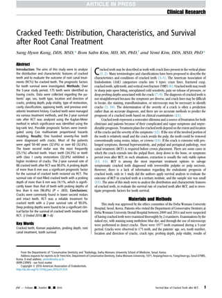
teeth after RCT. Figure 2 shows the Kaplan-Meier survival curve
as a function of the probing depth. For cracked teeth with initial
probing depths <6 mm, the 2-year survival rate after RCT was
96.8%. For cracked teeth with initial probing depths >6 mm, the
2-year survival rate after RCT was 74.1%. This difference was sta-
tistically significant (P = .003).
Prognostic Factors for Survival of Root-filled Cracked
Teeth Using the Multivariate Model
The following clinically relevant factors for the survival of root-
filled cracked teeth were selected as candidate variables: multiple crack
directions, subgingival extension of cracks, deep probing depth, preop-
erative pain, sex (female), dental arch (mandible), tooth type (molars),
terminal tooth in the arch, pulp necrosis at the initial examination, and
the presence of class II cavities. Table 6 lists the HRs and CIs for the
selected variables calculated using the Cox proportional hazards model.
Backward stepwise selection identified deep probing depth as the var-
iable with the strongest association with reduced survival of cracked
teeth after RCT (HR = 19.67; 95% CI, 2.02–175.82; P = .01).
Discussion
We have analyzed the distribution and characteristics of cracked
teeth, evaluated the survival rates of cracked teeth after RCT, and
TABLE 2. Characteristics of the Cracked Teeth
Variables n (%)
Type of restoration
No restoration 60 (34.3)
Direct filling 21 (12.0)
Amalgam 11 (6.3)
Resin 9 (5.1)
Glass ionomer 1 (0.6)
Inlay 46 (26.3)
Resin 2 (1.1)
Gold 44 (25.1)
Temporary filling 24 (13.7)
Crown 24 (13.7)
Porcelain 6 (3.4)
Gold 16 (9.1)
Zirconia 1 (0.6)
Innovium 1 (0.6)
Cavity classification
No treatment 60 (34.3)
Class I 56 (32.0)
Class II 32 (18.3)
Class V 3 (1.7)
Crown 24 (13.7)
AAE crack category
Fractured cusp 25 (14.3)
Cracked tooth 111 (63.4)
Split tooth 18 (10.3)
Vertical root fracture 21 (12.0)
Direction of crack
Mesiodistal 89 (50.9)
Buccolingual 34 (19.4)
Both 52 (29.7)
Number of cracks
1 150 (85.7)
2 20 (11.4)
$3 5 (2.9)
Subgingival extension
Yes 117 (66.9)
No 58 (33.1)
Opposing tooth
No restoration 89 (50.9)
Amalgam 2 (1.1)
Resin 4 (2.3)
Porcelain 3 (1.7)
Zirconia 1 (0.6)
Gold inlay 32 (18.3)
Gold crown 41 (23.4)
Implant 3 (1.7)
Previous endodontic treatment
Yes 34 (19.4)
No 141 (80.6)
AAE, American Association of Endodontists.
TABLE 3. Clinical Signs and Symptoms of the Cracked Teeth
n (%)
Probing depth
<3 mm 94 (53.7)
36 mm 37 (21.1)
69 mm 22 (12.6)
9 mm 22 (12.6)
Bite pain
Positive 127 (72.6)
Negative 48 (27.4)
Bite test
Positive 98 (56.0)
Negative 77 (44.0)
Percussion test
Positive 76 (43.4)
Negative 99 (56.6)
Pulp vitality test
Normal pulp 54 (30.9)
Moderate cold
sensitivity
32 (18.3)
Severe cold sensitivity 15 (8.6)
Nonvital tooth 74 (42.3)
TABLE 4. Treatments for the Cracked Teeth
Treatment n (%)
Resin filling 10 (5.7)
Inlay 10 (5.7)
Provisional crown / permanent crown 27 (15.4)
Provisional crown / RCT / permanent crown 11 (6.3)
RCT / permanent crown 77 (44.0)
Extraction 40 (22.9)
RCT, root canal treatment.
Clinical Research
JOE — Volume -, Number -, - 2016 Survival Rate of Cracked Teeth after RCT 3
4. investigated prognostic factors for tooth survival. The incidence of
cracked teeth in this study was in agreement with the previous study
(7). The results show that cracked teeth occurred primarily in patients
over 50, whereas previous studies have reported the occurrence of
cracked teeth mainly in patients aged 30–50 years (4, 13, 18–20). It
has been shown that the fatigue resistance of human dentin
decreases with age (14, 21). The suggested reasons for a higher
incidence of cracks in older patients are a loss of dentin elasticity
and increased stress fatigue over time (2, 20).
We found that the lower second molars were the most commonly
affected teeth followed by lower first molars. The high incidence of
cracks in the lower second molars may be related to their proximity
to the temporomandibular joint (13). Based on the lever effect, we
may expect the masticatory force on the tooth to be larger closer to
the temporomandibular joint, and most studies show that cracks are
most prevalent among mandibular molars (22–25). It has been
suggested that the lingual cusps of maxillary molars may function as
plungers, leading to structural fatigue in the lower antagonists.
Furthermore, mandibular molars have a deeper central fossa than
maxillary molars, and the oblique ridge of the maxillary molar
increases resistance to crack formation (26). However, upper premo-
lars were more affected than lower premolars, which may be associated
with the deep cusp-fossa relationships of upper premolars.
Figure 1. The Kaplan-Meier survival curve of root-filled cracked teeth. The
2-year survival rate was 90.0%.
TABLE 5. Two-year Survival Analysis of Cracked Teeth after Root Canal
Treatment Depending on the Clinical Variables
Variables
No. of
teeth
Extracted
teeth
2-year
survival (%)
P value
(log-rank)
Crack direction
Single 75 7 91.2 .951
Multiple 13 1 83.3
Subgingival extension
Yes 52 6 88.3 .985
No 36 2 92.9
Probing depth
6 mm 67 4 96.8 .003
$6 mm 21 4 74.1
Preoperative pain
Absent 20 2 85.7 .339
Present 68 6 90.9
Sex
Male 55 7 86.2 .680
Female 33 1 80.0
Dental arch
Maxilla 45 3 92.3 .467
Mandible 43 5 81.6
Type of tooth
Premolar 22 2 85.7 .331
Molar 66 6 85.2
Terminal tooth in the arch
Yes 31 4 73.8 .165
No 57 4 96.8
Class II cavity
Yes 21 3 70.0 .027
No 67 5 94.1
Figure 2. Kaplan-Meier survival of root-filled cracked teeth as a function of
probing depth. For cracked teeth with initial probing depths of 6 mm (blue),
the 2-year survival rate after RCT was 96.8%. For cracked teeth with initial
probing depths of 6 mm (green), the 2-year survival rate after RCT was
74.1%. The difference in survival rates was significant (log-rank P = .003).
TABLE 6. Cox Proportional Hazards Regression Model Showing the
Association of Variables with the Survival of Cracked Teeth after Root Canal
Treatment
Variables HR 95% CI P value
Multiple crack direction 3.78 0.34–42.29 .28
Subgingival extension
of cracks
1.29 0.14–12.01 .83
Probing depth $6 mm 19.67 2.02–175.82 .01
Preoperative pain 1.44 0.124–16.58 .77
Female 8.08 0.086–756.32 .37
Mandible 5.00 0.048–516.05 .50
Molars 9.08 0.029–2809.65 .45
Terminal tooth in the arch 1.29 0.067–24.93 .87
Pulp necrosis at initial
examination
5.69 0.519–62.39 .16
Class II cavity 1.63 0.070–38.07 .76
CI, confidence interval; HR, hazard ratio.
Clinical Research
4 Kang et al. JOE — Volume -, Number -, - 2016
5. In this study, 64.6% of cracks were found in intact teeth or in those
with class I restorations. This result contrasts with a previous study,
which found that cracks were more common in heavily restored teeth
(2). However, there have also been reports of high incidences of cracks
in unrestored teeth (4, 16, 18). Internal structural weakness may exist
at the coalescence of calcification sites (4, 26). Masticatory forces on
teeth with untreated caries lesions can also lead to the formation of
cracks (27). Thermal cycling and parafunctional habits have also
been reported to be responsible for the progression of cracks in intact
teeth (3, 13).
Cracks were often found in teeth restored with gold inlays. The
sharp internal angle required for the retention of nonbonded restora-
tions may explain this phenomenon. Furthermore, the relatively large
thermal expansion coefficients of gold (14.20 ppm/
C) and amalgam
(22–28 ppm/
C) are associated with the development of cracks (2).
If there is a large difference between the thermal expansion coefficients
of the dental restoration and the tooth, an increase in temperature may
result in expansion of the restorative material causing tooth fracture or
it may lead to chronic pulpitis.
When probing a VRF, the clinician typically finds a deep, narrow,
isolated periodontal pocket over the bony dehiscence that was created
secondary to the VRF (28). Among the 21 teeth diagnosed with VRF, 12
had a history of RCT. It has been reported that root canal procedures
may result in stresses, potentially causing cracks in the root (29).
Excessive removal of intraradicular dentin and overinstrumentation
of canals may increase the risk of VRF (2, 30). Clinicians should
ensure that the internal wedging forces are small and should
carefully control the condensation of the root canal filling materials
(6). Furthermore, the use of posts should be carefully considered.
The proportion of teeth that were sensitive to the bite test was
smaller than has been reported previously (2, 18). Difficulties in
positioning the cotton roll over a specific cusp of the tooth may have
affected these results. The use of a special plastic bite block such as
the Tooth Slooth (Professional Results Inc, Laguna Niguel, CA) may
result in improved accuracy (12). The Tooth Slooth is designed to apply
force to a specific cusp to identify damaged cusps and is a useful tool for
the differential diagnosis of partial crown fractures.
Several teeth were restored using resin fillings or inlays. Despite
the preference for resin fillings over other treatment options for the
cracked teeth, resin fillings were only applied in certain cases. In
many cases, the depth of the crack was so severe that it could not be
completely removed, and further removal would have exposed the
pulp. A previous investigation of 40 cracked teeth restored using direct
composite resin showed that 90% of the teeth maintained pulp vitality
after 7 years (31). Furthermore, bonded indirect resin composite on-
lays for painful cracked teeth have been reported to have a 6-year sur-
vival rate of 93.02% (32). These studies suggest that bonded composite
resin restorations can be an effective treatment for cracked teeth; how-
ever, an important difference between these studies and our work is that
the subjects in our study were patients in the tertiary institute who pre-
sented more severe cases.
The proportion of cracked teeth treated via RCT was larger than in
other studies because patients referred to the university dental hospital
had typically experienced prolonged symptoms (Table 3). The large
proportion of nonvitalteeth (42.3%) or teeth with severe cold sensitivity
(8.6%) is associated with the larger proportion of RCT. A previous
investigation of histopathology and histobacteriology reported that
cracks were colonized by bacterial biofilms (33). In this study, there
were 2 cases involving teeth that were diagnosed with pulp necrosis
in the absence of caries, restoration, or trauma. It is thought that
pulp necrosis is caused by cracks, which is termed fracture necrosis
(11). The 2 teeth in this study suspected of fracture necrosis survived
until follow-up days 1209 and 1460.
RCT was required in 28.9% of cases in which cracks were identi-
fied early and a provisional crown was placed. A previous study of 127
patients reported that 21% of teeth diagnosed with reversible pulpitis
and cracks eventually required RCT in a 6-year evaluation (7). Another
study reported that, among 21 teeth with provisional crowns, 9 (42.9%)
required RCT (16). The early detection of cracks may enable conserva-
tive treatment before the symptoms and extent of cracks progress to the
point of required RCT. Early diagnosis is important to maintain pulp vi-
tality and prevent further propagation of cracks.
The survival rate of RCTs in 88 cracked teeth after 2 years was
90.0%, which is remarkably high, considering that cracked teeth typi-
cally have an unfavorable prognosis with possible postoperative compli-
cations (15). However, clinicians should properlyinform patientsof the
potential for failure because the cracks may continue to progress,
causing the tooth to separate at some point in the future. It is desirable
for the clinician to inform patients of the prognosis of cracks and pro-
vide treatment alternatives. A previous study reported a survival rate of
85.5% over 2 years in 50 root-filled cracked teeth in which significant
prognostic factors were multiple cracks, terminal teeth, and pretreat-
ment probing depth (15).
Among the potential prognostic factors that were evaluated, the re-
sults of the log-rank test and Cox proportional hazard analysis indicate
that only probing depth was significantly correlated with tooth survival
rate. A deep probingdepth impliesthat the crackcan progress deep into
the root, adversely affecting the support of the periodontium (15, 34). It
has been reported that cracked teeth with probing depths of 4 mm
have poor prognoses and that the proportion of teeth requiring RCT
increases with increasing probing depths (16). Thus, careful assess-
ment of the impact of the crack on the periodontal status is important,
not only for the proper management of the supporting tissue but also for
the determination of the prognosis of the anticipated treatment proce-
dures. Multivariate analysis revealed a weak correlation between mul-
tiple crack directions and pulp necrosis at initial examination
although the relationship was not statistically significant. Our data sug-
gest that these variables may have potential as additional significant
prognostic factors. Further study with a larger data set may show signif-
icant relationships between these variables and the survival rate of
cracked teeth.
Prognosis assessment of teeth with cracks is a challenging task
for clinicians. RCT is considered to be the last nonsurgical treatment
option for salvaging cracked teeth. Information regarding endodontic
prognosis assessment is scarce, and only 1 previous study has con-
ducted an evaluation of survival rates of root-filled cracked teeth
(15); however, we evaluated a larger number of teeth and included
a greater variety of prognostic factors. Further study with a larger
sample size and longer follow-up duration is desirable to investigate
the prognostic factors that contribute to the survival of root-filled
cracked teeth. Additional work is required to determine the crack
characteristics that are predictive of prognosis as well as clinical clas-
sifications of the severity of cracks and evaluation of the outcome of
different treatment modalities.
Conclusions
The incidence of cracks was higher in lower second molars and
intact teeth. RCT was a reliable treatment for cracked teeth, with a 2-
year survival rate of 90.0%. Deep probing depths of 6 mm were
significantly associated with reduced survival of cracked teeth after
RCT.
Clinical Research
JOE — Volume -, Number -, - 2016 Survival Rate of Cracked Teeth after RCT 5
6. Acknowledgments
Supported by grants from the National Research Foundation
(NRF-2015R1C1A1A01054030, NRF-2015R1C1A1A02037051, and
NRF-2012M3A9B6055379).
The authors deny any conflicts of interest related to this study.
References
1. Rivera EM, Williamson A. Diagnosis and treatment planning: cracked tooth. Tex
Dent J 2003;83:38–41.
2. Seo DG, Yi YA, Shin SJ, et al. Analysis of factors associated with cracked teeth.
J Endod 2012;38:288–92.
3. Ellis SG. Incomplete tooth fracture–proposal for a new definition. Br Dent J 2001;
190:424–8.
4. Hiatt WH. Incomplete crown-root fracture in pulpal-periodontal disease.
J Periodontol 1973;44:369–79.
5. Ritchey B, Mendenhall R, Orban B. Pulpitis resulting from incomplete tooth frac-
ture. Oral Surg Oral Med Oral Pathol 1957;10:665–70.
6. American Association of Endodontists. Cracking the cracked tooth code. End-
odontics: Colleagues for Excellence. Chicago: American Association of
Endodontists; 2008.
7. Krell KV, Rivera EM. A six year evaluation of cracked teeth diagnosed with reversible
pulpitis: treatment and prognosis. J Endod 2007;33:1405–7.
8. Homewood CI. Cracked tooth syndrome—incidence, clinical findings and treat-
ment. Aust Dent J 1998;43:217–22.
9. Banerji S, Mehta SB, Millar BJ. Cracked tooth syndrome. Part 1: aetiology and diag-
nosis. Part 2: restorative options for the management of cracked tooth syndrome. Br
Dent J 2010;208:459–63. 503–14.
10. Ozer SY. Detection of vertical root fractures by using cone beam computed tomog-
raphy with variable voxel sizes in an in vitro model. J Endod 2011;37:75–9.
11. Berman LH, Kuttler S. Fracture necrosis: diagnosis, prognosis assessment, and treat-
ment recommendations. J Endod 2010;36:442–6.
12. T€
urp JC, Gobetti JP. The cracked tooth syndrome: an elusive diagnosis. J Am Dent
Assoc 1996;127:1502–7.
13. Lynch CD, McConnell RJ. The cracked tooth syndrome. J Can Dent Assoc 2002;68:
470–5.
14. Lubisich EB, Hilton TJ, Ferracane J, et al. Cracked teeth: a review of the literature.
J Esthet Restor Dent 2010;22:158–67.
15. Tan L, Chen NN, Poon CY, et al. Survival of root filled cracked teeth in a tertiary insti-
tution. Int Endod J 2006;39:886–9.
16. Kim SY, Kim SH, Cho SB, et al. Different treatment protocols for different pulpal and
periapical diagnoses of 72 cracked teeth. J Endod 2013;39:449–52.
17. Recommended terms. AAE Consensus Conference Recommended Diagnostic Termi-
nology. J Endod 2009;35:1634.
18. Roh BD, Lee YE. Analysis of 154 cases of teeth with cracks. Dent Traumatol 2006;22:
118–23.
19. Snyder DE. The cracked-tooth syndrome and fractured posterior cusp. Oral Surg
Oral Med Oral Pathol 1976;41:698–704.
20. Udoye CI, Jafarzadeh H. Cracked tooth syndrome: characteristics and distribution
among adults in a Nigerian teaching hospital. J Endod 2009;35:334–6.
21. Bajaj D, Sundaram N, Nazari A, et al. Age, dehydration and fatigue crack growth in
dentin. Biomaterials 2006;27:2507–17.
22. Abou-Rass M. Crack lines: the precursors of tooth fractures - their diagnosis and
treatment. Quintessence Int Dent Dig 1983;14:437–47.
23. Bader JD, Martin JA, Shugars DA. Incidence rates for complete cusp fracture. Com-
munity Dent Oral Epidemiol 2001;29:346–53.
24. Eakle WS, Maxwell EH, Braly BV. Fractures of posterior teeth in adults. J Am Dent
Assoc 1986;112:215–8.
25. Gher ME, Dunlap RM, Anderson MH, et al. Clinical survey of fractured teeth. J Am
Dent Assoc 1987;114:174–7.
26. Ehrmann EH, Tyas MJ. Cracked tooth syndrome. Aust Dent J 1990;35:390–1.
27. Rosen H. Cracked tooth syndrome. J Prosthet Dent 1982;47:36–43.
28. Cohen S, Blanco L, Berman L. Vertical root fractures: clinical and radiographic diag-
nosis. J Am Dent Assoc 2003;134:434–41.
29. Adorno CG, Yoshioka T, Jindan P, et al. The effect of endodontic procedures on api-
cal crack initiation and propagation ex vivo. Int Endod J 2013;46:763–8.
30. Tang W, Wu Y, Smales RJ. Identifying and reducing risks for potential fractures in
endodontically treated teeth. J Endod 2010;36:609–17.
31. Opdam NJ, Roeters JJ, Loomans BA, et al. Seven-year clinical evaluation of painful
cracked teeth restored with a direct composite restoration. J Endod 2008;34:
808–11.
32. Signore A, Benedicenti S, Covani U, et al. A 4- to 6-year retrospective clinical study of
cracked teeth restored with bonded indirect resin composite onlays. Int J Prostho-
dont 2007;20:609–16.
33. Ricucci D, Siqueira JF, Loghin S, et al. The cracked tooth: histopathologic and his-
tobacteriologic aspects. J Endod 2015;41:343–52.
34. Gutmann JL, Rakusin H. Endodontic and restorative management of incompletely
fractured molar teeth. Int Endod J 1994;27:343–8.
Clinical Research
6 Kang et al. JOE — Volume -, Number -, - 2016