
Recommended
More Related Content
What's hot
What's hot (20)
Similar to Venous and lymphatic drainage of head
Similar to Venous and lymphatic drainage of head (20)
More from Abhishek Roy
More from Abhishek Roy (12)
Recently uploaded
Recently uploaded (20)
Venous and lymphatic drainage of head
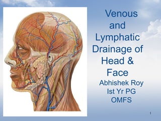
- 1. 1 Venous and Lymphatic Drainage of Head & Face Abhishek Roy Ist Yr PG OMFS
- 2. 22 ContentsContents • IntroductionIntroduction • Venous systemVenous system • Veins vs ArteriesVeins vs Arteries • Veins of Head and FaceVeins of Head and Face • Anatomical VariationsAnatomical Variations • Introduction to LymphIntroduction to Lymph • Lymphatic SystemLymphatic System • Flow of LymphFlow of Lymph • Classification of Lymph nodesClassification of Lymph nodes • Lymph nodes of Head and NeckLymph nodes of Head and Neck • Causes of Lymph node enlargementCauses of Lymph node enlargement • ConclusionConclusion • ReferencesReferences
- 3. 33 IntroductionIntroduction •The venous system is a network of conduits i.e.The venous system is a network of conduits i.e. veins which transport deoxygenated blood fromveins which transport deoxygenated blood from tissues to the heart.tissues to the heart. •Exceptions are the Pulmonary and UmbelicalExceptions are the Pulmonary and Umbelical veins, both of which carry oxygenated blood toveins, both of which carry oxygenated blood to the heart. Equally important ,they serve as majorthe heart. Equally important ,they serve as major reservoirs of blood.reservoirs of blood.
- 4. 44
- 5. 55 Introduction(contd.)Introduction(contd.) • About 84% of entire blood volume of body is inAbout 84% of entire blood volume of body is in systemic circulation and 16% in heart and lungs.systemic circulation and 16% in heart and lungs. • Of the 84 % in systemic circulation,64%is inOf the 84 % in systemic circulation,64%is in veins,13%in arteries and 7% in systemicveins,13%in arteries and 7% in systemic arterioles and capillaries.arterioles and capillaries.
- 6. 66 Functioning of VenousFunctioning of Venous systemsystem • The venous system in the body returns deoxygenated blood from all parts of the body, including the organs, to the right side of the heart, and then on to the lungs, to be oxygenated. From the lungs, the oxygenated blood passes to the left part of the heart, to be pumped to all the tissues and organs of the body.
- 7. 77 Types of VeinsTypes of Veins Superficial Veins are thoseSuperficial Veins are those whosewhose coursecourse isis closeclose toto thethe surface of the body and have no corresponding arteries.surface of the body and have no corresponding arteries. Deep Veins are deeper in the body and have correspondingDeep Veins are deeper in the body and have corresponding arteriesarteries Communicating veins (or perforator veins) are veins thatCommunicating veins (or perforator veins) are veins that directly connect superficial veins to deep veins.directly connect superficial veins to deep veins. The pulmonary veins are a set of veins that deliver oxygenatedThe pulmonary veins are a set of veins that deliver oxygenated blood from the lungs to the heartblood from the lungs to the heart Systemic Veins drain the tissues of the body and deliverSystemic Veins drain the tissues of the body and deliver deoxygenated blood to the heart.deoxygenated blood to the heart.
- 8. 88 Superficial Veins of HandSuperficial Veins of Hand
- 10. 1010 Valves of VeinsValves of Veins
- 12. 1212 Embryology of VeinsEmbryology of Veins
- 13. 1313
- 14. 1414 Veins of Head and FaceVeins of Head and Face
- 15. 1515 Veins of head and faceVeins of head and face • Supratrochlear veinSupratrochlear vein • Supraorbital veinsSupraorbital veins • Facial veinFacial vein • Superficial temporal veinSuperficial temporal vein • Pterygoid venous plexusPterygoid venous plexus • Maxillary veinMaxillary vein • Retromandibular veinRetromandibular vein • Posterior auricular veinPosterior auricular vein • Occipital veinOccipital vein
- 16. 1616 Supratrochlear VeinSupratrochlear Vein •This starts on the forehead . Veins from this form a single trunk, descending near the midline parallel to radix nasi . •The veins then diverge, each joining supraorbital vein near medial canthus to form facial vein.
- 17. 1717 Supraorbital VeinSupraorbital Vein ORIGINORIGIN:: Zygomatic process ofZygomatic process of frontal bonefrontal bone EMPTIESEMPTIES:: Angular VeinAngular Vein DRAINS: Eyebrows and forehead
- 18. 1818 Anterior Facial VeinAnterior Facial Vein Is formed by the union of theIs formed by the union of the supraorbital andsupraorbital and supratrochlear veins to formsupratrochlear veins to form the angular veinthe angular vein Communicate with theCommunicate with the cavernous sinus through thecavernous sinus through the ophthalmic vein via theophthalmic vein via the supraorbital veinsupraorbital vein
- 19. 1919 Anterior Facial VeinAnterior Facial Vein • Runs downwards and backwardsRuns downwards and backwards behind the facial artery to thebehind the facial artery to the lower border of the mandiblelower border of the mandible • To be joined by the anteriorTo be joined by the anterior division of the retromandibulardivision of the retromandibular veinvein Joins the: Pterygoid plexus through deepPterygoid plexus through deep facial veinfacial vein Cavernous sinus throughCavernous sinus through superior ophthalmic veinsuperior ophthalmic vein
- 20. 2020 TributariesTributaries-- Anterior facial vein receives a branch of deepAnterior facial vein receives a branch of deep facialfacial vein from pterygoid venous plexus.vein from pterygoid venous plexus. Joined by superior and inferior palpebral, superiorJoined by superior and inferior palpebral, superior and inferior labial, buccinator and masseteric veins.and inferior labial, buccinator and masseteric veins. Below mandible receives submental, palatine,Below mandible receives submental, palatine, submaxillary, and vena comitans of hypoglossal nerve.submaxillary, and vena comitans of hypoglossal nerve.
- 21. 2121 Dangerous Area of FaceDangerous Area of Face The facial vein is devoid of valves and rests directly on the facial muscles. Absence of deep fascia The movement of facial muscles might facilitate the spread of septic emboli from the infected area of upper lip and lower part of the nose in retrograde direction. Cause thrombosis of cavernous sinus with serious complication.
- 22. 2222 Applied ImportanceApplied Importance Facial veins have no valves and it connects to cavernousFacial veins have no valves and it connects to cavernous sinus by two routes –sinus by two routes – 1. via ophthalmic vein or supraorbital vein1. via ophthalmic vein or supraorbital vein 2. via deep facial vein to pterygoid plexus2. via deep facial vein to pterygoid plexus and hence to cavernous sinusand hence to cavernous sinus Thus infective thrombosis of facial vein may extend toThus infective thrombosis of facial vein may extend to intracranial venous sinusintracranial venous sinus
- 23. 2323 Cavernous Sinus ThrombosisCavernous Sinus Thrombosis • severe headachessevere headaches • neck stiffnessneck stiffness • altered consciousness levelsaltered consciousness levels • epileptic fitsepileptic fits • high grade feverhigh grade fever • rigorsrigors • headachesheadaches • a reduced conscious levela reduced conscious level
- 24. 2424 Superficial temporal VeinSuperficial temporal Vein •This begins in a widespread network joined across the scalp. •Frontal and Parietal branches emerge which combine to form trunk of vein above Zygomatic arch •It crosses posterior root of zygoma and enters the parotid gland to join the maxillary vein, to form retromandibular vein. •Drains : side of scalp, temporalis muscle and ear.
- 25. 2525 PTERYGOID VENOUS PLEXUSPTERYGOID VENOUS PLEXUS LOCATIONLOCATION:: Partly b/w temporalis & lateralPartly b/w temporalis & lateral pterygoid & partly b/w pterygoidspterygoid & partly b/w pterygoids CONNECTSCONNECTS:: Deep facial vein withDeep facial vein with cavernous sinus via.cavernous sinus via. foramen ovale andforamen ovale and foramen lacerumforamen lacerum DRAINAGEDRAINAGE:: Chiefly: Maxillary VeinChiefly: Maxillary Vein Retromandibular VeinRetromandibular Vein
- 26. 2626 Applied ImportanceApplied Importance •Applied importance :Applied importance : - Needle track communications can also result in infection to- Needle track communications can also result in infection to pterygoid plexus.pterygoid plexus. - PSA block- PSA block hematoma , black eyehematoma , black eye - serve as a media for spread of external infection to eye.- serve as a media for spread of external infection to eye.
- 27. 2727 Maxillary veinMaxillary vein Unites with superficialUnites with superficial temporal to formtemporal to form retromandibular veinretromandibular vein
- 28. 2828 Retromandibular VeinRetromandibular Vein Formed by the union of maxillaryFormed by the union of maxillary vein and superficial temporal vein.vein and superficial temporal vein. Leaving parotid gland divides intoLeaving parotid gland divides into anterior branch and posterioranterior branch and posterior branch.branch. Anterior branch joins facial veinAnterior branch joins facial vein which drains into common facialwhich drains into common facial vein to internal jugular veinvein to internal jugular vein Posterior branch joins posteriorPosterior branch joins posterior auricular vein to form externalauricular vein to form external jugular vein.jugular vein.
- 29. 2929 Posterior auricular veinPosterior auricular vein ORIGINORIGIN:: Parieto occipital plexus of veinsParieto occipital plexus of veins DESCENDSDESCENDS:: Behind auricle,Behind auricle, Joins posterior facial vein to formJoins posterior facial vein to form external jugular veinexternal jugular vein Applied importanceApplied importance : receives mastoid: receives mastoid emissary veins from sigmoid sinus.emissary veins from sigmoid sinus. Infection here can be dangerous orInfection here can be dangerous or fatal from retrograde thrombosis offatal from retrograde thrombosis of cerebellar and medullary veinscerebellar and medullary veins
- 30. 3030 Occipital veinOccipital vein ORIGIN:ORIGIN: •Posterior network inPosterior network in scalpscalp •Joins deep cervical &Joins deep cervical & vertebral veinsvertebral veins COURSE:COURSE: •Follows occipital arteryFollows occipital artery ENDS:ENDS: •Internal Jugular veinInternal Jugular vein •Sometimes joins posterior auricularSometimes joins posterior auricular vein which joins EJVvein which joins EJV
- 31. 3131 Anatomical VariationsAnatomical Variations The common facial vein does not exist. TheThe common facial vein does not exist. The retromandibualr vein continues into the externalretromandibualr vein continues into the external jugular vein, the facial vein opens into the internaljugular vein, the facial vein opens into the internal jugular vein.jugular vein. The retromandibular vein opens into the internalThe retromandibular vein opens into the internal jugular vein, the facial vein continues into thejugular vein, the facial vein continues into the anterior jugular vein, and a common facial veinanterior jugular vein, and a common facial vein again is absent.again is absent.
- 32. 3232 Anatomical Variations(contd.)Anatomical Variations(contd.) The common facial vein loses its connection with theThe common facial vein loses its connection with the internal jugular vein and empties instead into theinternal jugular vein and empties instead into the external jugular veinexternal jugular vein • Retromandibular vein continues into the externalRetromandibular vein continues into the external jugular vein, facial vein continues into the anteriorjugular vein, facial vein continues into the anterior jugular vein.jugular vein.
- 33. 3333 Anatomical Variations(contd.)Anatomical Variations(contd.) •In someIn some cadaverscadavers the followingthe following variationvariation waswas observed, theobserved, the facial vein was found running downwardfacial vein was found running downward & backwards at the anterior border of masseter& backwards at the anterior border of masseter,where,where thethe submental vein united with the facial vein to form asubmental vein united with the facial vein to form a common venous channelcommon venous channel.. •The common venous channel united with theThe common venous channel united with the retromandibular vein at the angle of the mandible toretromandibular vein at the angle of the mandible to form the common facial vein.form the common facial vein. • Before this common facial vein joins the internalBefore this common facial vein joins the internal jugular vein , a venous channel originating from thejugular vein , a venous channel originating from the anterior jugular vein comes & joins the common facialanterior jugular vein comes & joins the common facial vein &which then joins the internal jugular vein.vein &which then joins the internal jugular vein. ( j anat.soc.india,52(1),64-65 (2003))( j anat.soc.india,52(1),64-65 (2003))
- 34. 34 Lymphatic Drainage of Head and Face
- 35. 3535 LymphLymph DefinitionDefinition:: Transparent, slightly yellowish liquid of alkalineTransparent, slightly yellowish liquid of alkaline reaction found in lymphatic vessels and derived from tissuereaction found in lymphatic vessels and derived from tissue fluidfluid Rate of lymph flowRate of lymph flow:: • About 100ml/hr lymph flows via. thoracic duct of a restingAbout 100ml/hr lymph flows via. thoracic duct of a resting human.human. • Approximately 20ml/hr flows into circulation.Approximately 20ml/hr flows into circulation. • Total estimated lymph flow is about 120ml/hrTotal estimated lymph flow is about 120ml/hr
- 36. 3636 Lymphatic SystemLymphatic System •The lymphatic system is a endotheliun – linedThe lymphatic system is a endotheliun – lined network of blind ended capillaries found in nearlynetwork of blind ended capillaries found in nearly all tissues, draining via collecting vessels into largeall tissues, draining via collecting vessels into large vascular trunks that eventually empty into bloodvascular trunks that eventually empty into blood circulatory system.circulatory system. •The lymphatic system is a part of immune system .The lymphatic system is a part of immune system .
- 37. 3737 Lymphatic SystemLymphatic System Consist of: 1)Lymphatic channels- a) lymphatic capillaries b) lymphatic vessels 2) Lymph 3 )Lymph nodes 4) Lymphoid organs and tissues
- 38. 3838 Embryonic developmentEmbryonic development Begins to develop by end of 5Begins to develop by end of 5thth wk. IU lifewk. IU life Develop from lymph sacs thatDevelop from lymph sacs that arise from developing veins,arise from developing veins, derived from mesoderm.derived from mesoderm. 6 primary lymph sacs formed6 primary lymph sacs formed First lymph sacs to appear areFirst lymph sacs to appear are paired jugular lymph sacs .paired jugular lymph sacs .
- 39. 3939 Capillary plexuses enlargeCapillary plexuses enlarge Form lymphatic vesselsForm lymphatic vessels Each jugular lymphEach jugular lymph sac retains at leastsac retains at least one connection withone connection with its jugular veinits jugular vein Left one developingLeft one developing into superior portioninto superior portion of thoracic duct.of thoracic duct.
- 40. 4040 88thth week of IUL - Retroperitoneal lymph sacs forms.week of IUL - Retroperitoneal lymph sacs forms. 99thth week of IUL cisterna chyli develops-lower part ofweek of IUL cisterna chyli develops-lower part of thoracic duct develops from left jugular sac.thoracic duct develops from left jugular sac. Later stages-lymph sacs are invaded byLater stages-lymph sacs are invaded by lymphocytes.lymphocytes. Transformed into group of lymph nodesTransformed into group of lymph nodes
- 41. 4141 FunctionsFunctions •The lymphatic system has three functions : 1. Fluid recovery 2. Immunity 3. Lipid absorption
- 42. 4242 Route of LymphRoute of Lymph
- 43. 4343 WALDEYER’S LYMPHATIC RINGWALDEYER’S LYMPHATIC RING • Ringed arrangement of lymphoid tissue in theRinged arrangement of lymphoid tissue in the pharynxpharynx • Waldeyer's ring surrounds the naso- andWaldeyer's ring surrounds the naso- and oropharynx, with some of its tonsillar tissueoropharynx, with some of its tonsillar tissue located above and some below the soft palatelocated above and some below the soft palate • Presence ofPresence of MALTMALT • Aggregations are :Aggregations are : Pharyngeal tonsil - 1Pharyngeal tonsil - 1 Tubal tonsils - 2Tubal tonsils - 2 Palatine tonsils - 2Palatine tonsils - 2 Lingual tonsil - 1Lingual tonsil - 1
- 44. 44 Classification by American Joint Committee on Cancer and the American Academy of Otolaryngology- Head and Neck Surgery
- 45. 4545 Classification byClassification by American JointAmerican Joint Committee on Cancer and the AmericanCommittee on Cancer and the American Academy of OtolaryngologyAcademy of Otolaryngology- Head and- Head and Neck SurgeryNeck Surgery
- 46. 4646 Level I - all nodes above hyoid bone, belowLevel I - all nodes above hyoid bone, below mylohyoid muscle, and anterior to posterior edge ofmylohyoid muscle, and anterior to posterior edge of submandibular glandsubmandibular gland Level IA - all nodes between medial margins ofLevel IA - all nodes between medial margins of anterior digastric muscles, above hyoid bone, belowanterior digastric muscles, above hyoid bone, below mylohyoid musclemylohyoid muscle
- 47. 4747 Level IB - all nodes below mylohyoid muscle,Level IB - all nodes below mylohyoid muscle, above hyoid bone, posterior and lateral toabove hyoid bone, posterior and lateral to medial anterior digastric musclemedial anterior digastric muscle and anteriorand anterior to submandibular glandto submandibular gland
- 48. 4848 Level II - all nodes below skull base at jugularLevel II - all nodes below skull base at jugular fossa to hyoid bone, anterior to posterior edgefossa to hyoid bone, anterior to posterior edge of sternocleidomastoid muscle and posterior toof sternocleidomastoid muscle and posterior to submandibular glandsubmandibular gland
- 49. 4949 Level IIA - all nodes that lie posterior to internal jugular veinLevel IIA - all nodes that lie posterior to internal jugular vein and are inseparableand are inseparable from vein or lie anterior, lateral or medialfrom vein or lie anterior, lateral or medial to veinto vein Level IIB - all nodes that lie posterior to internal jugular veinLevel IIB - all nodes that lie posterior to internal jugular vein and have a fat plane separating nodes and veinand have a fat plane separating nodes and vein
- 50. 5050 Level III - all nodes between hyoid bone and cricoid cartilageLevel III - all nodes between hyoid bone and cricoid cartilage arch and anterior to posterior sternocleidomastoid muscle, andarch and anterior to posterior sternocleidomastoid muscle, and lateral to ICAlateral to ICA Level IV - all nodes between cricoid cartilage arch and clavicle,Level IV - all nodes between cricoid cartilage arch and clavicle, anterior to posterior sternocleidomastoid muscle and posteroanterior to posterior sternocleidomastoid muscle and postero lateral to anterior scalene muscle and lateral to CCAlateral to anterior scalene muscle and lateral to CCA
- 51. 5151 Level V - all nodes from skullLevel V - all nodes from skull base posterior down to posteriorbase posterior down to posterior border of sternocleidomastoidborder of sternocleidomastoid muscle to level of clavicle,muscle to level of clavicle, anterior to trapezius muscleanterior to trapezius muscle
- 52. 5252 Level VA - all nodes between skull base and cricoid cartilageLevel VA - all nodes between skull base and cricoid cartilage arch, behind posterior edge of sternocleidomastoid musclearch, behind posterior edge of sternocleidomastoid muscle Level VB - all nodes between cricoid cartilage arch and clavicle,Level VB - all nodes between cricoid cartilage arch and clavicle, behind sternocleidomastoid musclebehind sternocleidomastoid muscle
- 53. 5353 Level VI - all nodes inferior toLevel VI - all nodes inferior to hyoid bone and above top ofhyoid bone and above top of manubrium, between medialmanubrium, between medial margins of bilateral CCA and ICAmargins of bilateral CCA and ICA Level VII - all nodes behindLevel VII - all nodes behind manubrium between medialmanubrium between medial margins of CCA bilaterally,margins of CCA bilaterally, extending inferiorly to level ofextending inferiorly to level of innominate veininnominate vein
- 54. 5454
- 55. 5555 SUPERFICIAL NODESSUPERFICIAL NODES •Sub-mental nodesSub-mental nodes •Sub-mandibular nodesSub-mandibular nodes •Buccal nodesBuccal nodes •PreauricularPreauricular •PostauricularPostauricular •OccipitalOccipital •Anterior cervicalAnterior cervical •Superficial cervicalSuperficial cervical
- 56. 5656 b) The deep cervical nodes :b) The deep cervical nodes : - The upper deep cervical ( jugulo - digastric group ): lie- The upper deep cervical ( jugulo - digastric group ): lie along the upper part of internal jugular vein deep toalong the upper part of internal jugular vein deep to sternocleidomastoid muscle.sternocleidomastoid muscle. - The lower deep cervical ( jugulo- omohyoid group):- The lower deep cervical ( jugulo- omohyoid group): arranged along the lower part of internal jugular vein.arranged along the lower part of internal jugular vein. DEEP LYMPH NODESDEEP LYMPH NODES •Prelaryngeal and PretrachealPrelaryngeal and Pretracheal •ParatrachealParatracheal •RetropharyngealRetropharyngeal
- 57. 5757 SUBMENTAL LYMPH NODESSUBMENTAL LYMPH NODES •Lies b/w chin & hyoid bone & b/wLies b/w chin & hyoid bone & b/w anterior bellies of digastric muscles inanterior bellies of digastric muscles in submental triangles.submental triangles. •Receives lymph from tip of tongue,Receives lymph from tip of tongue, floor beneath tongue, lower incisors,floor beneath tongue, lower incisors, central part of lower lip, and skin overcentral part of lower lip, and skin over chinchin •Drains into submandibular & deepDrains into submandibular & deep cervical nodescervical nodes
- 58. 5858 SUBMANDIBULAR LYPMH NODESSUBMANDIBULAR LYPMH NODES a) Superficial surface of submandibulara) Superficial surface of submandibular salivary gland.salivary gland. b) Beneath investing layer of deep cervicalb) Beneath investing layer of deep cervical fascia.fascia. • They are divided into anterior, middle, deepThey are divided into anterior, middle, deep group.group. •Anterior group found along submental veinAnterior group found along submental vein close to chin.close to chin. •Middle group situated around facial vein&Middle group situated around facial vein& facial artery above submandibular salivaryfacial artery above submandibular salivary gland.gland. •Posterior group located behind facial vein.Posterior group located behind facial vein.
- 59. 5959 Receives lymph from:Receives lymph from: •Front of scalp.Front of scalp. •Anterior part of nasal cavity, palate & adjacentAnterior part of nasal cavity, palate & adjacent cheek.cheek. •Upper & lower lip except central part.Upper & lower lip except central part. •Frontal, maxillary, ethmoidal air sinuses.Frontal, maxillary, ethmoidal air sinuses. •Upper& lower teeth except lower incisors.Upper& lower teeth except lower incisors. •Anterior 2/3Anterior 2/3rdrd of tongue.of tongue. •Floor of mouth, vestibule.Floor of mouth, vestibule. Drains into deep cervical nodes.Drains into deep cervical nodes.
- 60. 6060 BUCCAL LYPMH NODESBUCCAL LYPMH NODES •Situated over buccinator muscleSituated over buccinator muscle close to facial vein.close to facial vein. •Receives lymph from Eyelids,Receives lymph from Eyelids, cheek, mid portion of face rarelycheek, mid portion of face rarely gums & palategums & palate •Drains into submandibular lymphDrains into submandibular lymph nodesnodes
- 61. 6161 PAROTID LYMPH NODESPAROTID LYMPH NODES •Situated on/ within parotid gland.Situated on/ within parotid gland. •Receives lymph fromReceives lymph from a) Strip of scalp above parotid salivary gland.a) Strip of scalp above parotid salivary gland. b) Lateral surface of auricle.b) Lateral surface of auricle. c) Anterior wall of external auditory meatusc) Anterior wall of external auditory meatus d) Lateral wall of external auditory meatus.d) Lateral wall of external auditory meatus. e) Lateral wall of eyelide) Lateral wall of eyelid •Drains into deep cervical nodesDrains into deep cervical nodes
- 62. 6262 MASTOID / RETROAURICULAR LYMPH NODESMASTOID / RETROAURICULAR LYMPH NODES •Situated over lateral surface of mastoid process of temporalSituated over lateral surface of mastoid process of temporal bonebone •Receives lymph fromReceives lymph from a)Strip of scalp above auricle.a)Strip of scalp above auricle. b)Posterior wall of external auditory meatusb)Posterior wall of external auditory meatus •Drains into deep cervical lymph nodesDrains into deep cervical lymph nodes
- 63. 6363 OCCIPITAL NODESOCCIPITAL NODES •Situated at the apex of posteriorSituated at the apex of posterior triangle of necktriangle of neck •Receives lymph from back of scalpReceives lymph from back of scalp •Drains into deep cervical lymphDrains into deep cervical lymph nodesnodes
- 64. 6464 Anterior cervical nodesAnterior cervical nodes •Lie along anterior jugular vein.Lie along anterior jugular vein. •Drains skin of anterior part ofDrains skin of anterior part of neckneck •Efferents pass to deep cervicalEfferents pass to deep cervical nodesnodes
- 65. 6565 Superficial cervical nodes :Superficial cervical nodes : • Lie along external jugular vein superficial toLie along external jugular vein superficial to sternocleidomastoid.sternocleidomastoid. • Drains lobule of auricle, floor of external acousticDrains lobule of auricle, floor of external acoustic meatus, skin over lower parotid region and angle of jaw.meatus, skin over lower parotid region and angle of jaw. • It drains into deep cervical group of lymph nodesIt drains into deep cervical group of lymph nodes
- 66. 6666 B)B) LYMPHATIC DRAINAGE OF DEEP TISSUES OFLYMPHATIC DRAINAGE OF DEEP TISSUES OF HEAD AND NECK:HEAD AND NECK: RETROPHARYNGEAL NODES:RETROPHARYNGEAL NODES: • Lie in front of prevertebralLie in front of prevertebral fascia and behind buccopharyngeal fasciafascia and behind buccopharyngeal fascia • Extend laterally in front of lateralExtend laterally in front of lateral mass of atlas.mass of atlas. • Drains pharynx, auditory tube, soft palate.Drains pharynx, auditory tube, soft palate. • Efferents pass to upper deep cervical nodes.Efferents pass to upper deep cervical nodes.
- 67. 6767 b)b) PARATRACHEAL NODES:PARATRACHEAL NODES: • Lie on sides of trachea andLie on sides of trachea and esophagusesophagus • Receives lymph from esophagus,Receives lymph from esophagus, trachea and larynx.trachea and larynx. • Efferent drains into lower jugularEfferent drains into lower jugular group ,anterior mediastinal lymphgroup ,anterior mediastinal lymph nodesnodes
- 68. 6868 c)c) PRELARYNGEAL AND PRETRACHEAL NODES:PRELARYNGEAL AND PRETRACHEAL NODES: • Lie deep to investing fascia, on cricothyroid membrane .Lie deep to investing fascia, on cricothyroid membrane . -Drains:-Drains: upper node supra glottic larynxupper node supra glottic larynx lower nodes infra glottic larynxlower nodes infra glottic larynx
- 69. 6969 • Pretracheal nodes in front ofPretracheal nodes in front of trachea below isthmus of thyroidtrachea below isthmus of thyroid gland.gland. -Drains: region of thyroid gland and-Drains: region of thyroid gland and tracheatrachea
- 70. 70 d)d) LINGUAL NODESLINGUAL NODES:: • SmallSmall • Situated on external surface of hyoglossus and alsoSituated on external surface of hyoglossus and also between genioglossi.between genioglossi. • Drain into upper deep cervical nodes.Drain into upper deep cervical nodes.
- 71. 7171 LYMPHATIC DRAINAGE OF INDIVIDUALLYMPHATIC DRAINAGE OF INDIVIDUAL ORGANS OF HEAD AND NECKORGANS OF HEAD AND NECK Lymphatic drainage of scalp and earLymphatic drainage of scalp and ear • Anterior part of scalpAnterior part of scalp pre-auricular lymphpre-auricular lymph nodesnodes • Posterior part of scalp post-auricular (mastoid) ,Posterior part of scalp post-auricular (mastoid) , occipital lymph nodesoccipital lymph nodes • External acoustic meatus, middle ear and auditoryExternal acoustic meatus, middle ear and auditory tube deep parotid lymph nodestube deep parotid lymph nodes
- 72. 7272 Lymphatic drainage of faceLymphatic drainage of face • Upper part Parotid Lymph nodesUpper part Parotid Lymph nodes • Middle partMiddle part Submandibular lymph nodesSubmandibular lymph nodes • Lower partLower part Submental lymph nodesSubmental lymph nodes
- 73. 7373 Lymphatic drainage of deep tissue of head and neckLymphatic drainage of deep tissue of head and neck - Head and neck tissues to deep fascia- Head and neck tissues to deep fascia Deep cervical lymph nodesDeep cervical lymph nodes - Nasopharynx, pharyngotympanic membrane.- Nasopharynx, pharyngotympanic membrane. RetropharyngealRetropharyngeal - Trachea, esophagus, thyroid- Trachea, esophagus, thyroid ParatrachealParatracheal
- 74. 7474 Lymphatic drainage of mouth, teeth, tonsil, tongueLymphatic drainage of mouth, teeth, tonsil, tongue GingivaGingiva Submandibular lymph nodesSubmandibular lymph nodes Hard palateHard palate Superficial deep cervical andSuperficial deep cervical and retropharyngealretropharyngeal Soft palateSoft palate RetropharyngealRetropharyngeal Floor of the mouthFloor of the mouth Submental (deep cervical)Submental (deep cervical) TeethTeeth Submandibular and deep cervicalSubmandibular and deep cervical TonsilTonsil Jugulo digastric nodesJugulo digastric nodes
- 75. 7575 TongueTongue • Anterior 2/3rd submandibularAnterior 2/3rd submandibular and deep cervicaland deep cervical • Tip Submental (deep cervical)Tip Submental (deep cervical) • Posterior 1/3rdPosterior 1/3rd JuguloJugulo digastric lymph nodes.digastric lymph nodes.
- 76. 7676 Lymphatic drainage of nasal cavity and paranasalLymphatic drainage of nasal cavity and paranasal sinus:sinus: • Mucous membrane of nasal cavity and anterior partMucous membrane of nasal cavity and anterior part Submandibular and deep cervical lymph nodesSubmandibular and deep cervical lymph nodes • Posterior partPosterior part Deep cervicalDeep cervical
- 77. 7777 EXAMINATION OF LYMPH NODESEXAMINATION OF LYMPH NODES 1. Lymph nodes should be examined from patients behind.1. Lymph nodes should be examined from patients behind. 2. Examination is done by asking patient to flex his neck2. Examination is done by asking patient to flex his neck slightly to reduce tension of musclesslightly to reduce tension of muscles 3. To palpate, use the pads of all four fingertips.3. To palpate, use the pads of all four fingertips. 4. Examine both sides of head simultaneously while applying4. Examine both sides of head simultaneously while applying steady gentle pressure.steady gentle pressure.
- 78. 7878 ANTERIOR/POSTERIOR CERVICAL LYMPH NODESANTERIOR/POSTERIOR CERVICAL LYMPH NODES •They lie anterior & posterior to sternomastoid muscle.They lie anterior & posterior to sternomastoid muscle. •Tip of fingers are used to palpate anterior nodes, medial toTip of fingers are used to palpate anterior nodes, medial to sternomastoid muscle and posterior nodes behind the musclesternomastoid muscle and posterior nodes behind the muscle while patient’s head tipped slightly forwards.while patient’s head tipped slightly forwards.
- 79. 7979 SUBMANDIBULAR NODESSUBMANDIBULAR NODES Palpated from behind the patient, with patient’s chin tippedPalpated from behind the patient, with patient’s chin tipped slightly towards the chest.slightly towards the chest.
- 80. 8080 SUBMENTAL NODESSUBMENTAL NODES Roll the fingers below the chin(in the midline) with patient’sRoll the fingers below the chin(in the midline) with patient’s head tilted forwardshead tilted forwards
- 81. 8181 PAROTID NODES/PREAURICULAR NODESPAROTID NODES/PREAURICULAR NODES Roll the finger in front of ear , against the maxillaRoll the finger in front of ear , against the maxilla
- 82. 8282 POSTAURICULAR/ MASTOID NODESPOSTAURICULAR/ MASTOID NODES Roll the finger behind the earRoll the finger behind the ear
- 83. 8383 CAUSES OF ENLARGEMENT OF LYMPH NODESCAUSES OF ENLARGEMENT OF LYMPH NODES InflammatoryInflammatory (a) Acute Lymphadenitis(a) Acute Lymphadenitis (b) Chronic Lymphadenitis(b) Chronic Lymphadenitis (c) Lymphogranuloma inguinale(c) Lymphogranuloma inguinale NeoplasticNeoplastic (a) Benign – non-existent(a) Benign – non-existent (b) Malignant(b) Malignant 1. Primary1. Primary (i) Lymphosarcoma(i) Lymphosarcoma (ii) Hodgkin’s disease.(ii) Hodgkin’s disease. 2. Secondary2. Secondary (i) Sarcoma(i) Sarcoma (ii) Malignant melanoma(ii) Malignant melanoma (iii) Carcinoma(iii) Carcinoma
- 84. 8484 Autoimmune DisordersAutoimmune Disorders (i)(i)AIDSAIDS (ii) Juvenile rheumatoid arthritis(ii) Juvenile rheumatoid arthritis (iii) Other collagen diseases such as Polyarteritis(iii) Other collagen diseases such as Polyarteritis nodosa and scleroderma.nodosa and scleroderma.
- 85. 8585 CAUSES OF INDIVIDUAL LYMPH NODECAUSES OF INDIVIDUAL LYMPH NODE ENLARGEMENTENLARGEMENT Sub mandibular NodesSub mandibular Nodes • ConjunctivitisConjunctivitis • SinusitisSinusitis • TonsillitisTonsillitis • PharyngitisPharyngitis Sub mental NodesSub mental Nodes •PeriodontitisPeriodontitis •EBV infectionsEBV infections •CMV infectionsCMV infections •ToxoplasmosisToxoplasmosis
- 86. 8686 Deep cervical nodesDeep cervical nodes •PharyngitisPharyngitis •RubellaRubella •LymphomaLymphoma •TuberculosisTuberculosis •Head and neck cancerHead and neck cancer Occipital nodesOccipital nodes •Local infectionLocal infection •Secondary SyphilisSecondary Syphilis •NeoplasmNeoplasm
- 87. 8787 Postauricular nodesPostauricular nodes •Otitis ExternaOtitis Externa •Secondary SyphilisSecondary Syphilis •RubellaRubella Preauricular nodesPreauricular nodes •Local infectionLocal infection •Herpes ZosterHerpes Zoster •RubellaRubella • SyphilisSyphilis •TuberculosisTuberculosis
- 88. 8888 REFERENCESREFERENCES GRAY’S ANATOMY- 38GRAY’S ANATOMY- 38thth editionedition CUNNINGHAM’S TEXTBOOK OF ANATOMY-12CUNNINGHAM’S TEXTBOOK OF ANATOMY-12thth editionedition NETTER’S ATLAS OF EMBRYOLOGY- LARRYNETTER’S ATLAS OF EMBRYOLOGY- LARRY COCHARDCOCHARD TEXTBOOK OF HUMAN HISTOLOGY – INDERBIRTEXTBOOK OF HUMAN HISTOLOGY – INDERBIR SINGHSINGH B. D. CHAURASIA – 4B. D. CHAURASIA – 4thth editionedition ESSENTIALS OF MEDICAL PHYSIOLOGY-ESSENTIALS OF MEDICAL PHYSIOLOGY- K SEMBULIGAM-5K SEMBULIGAM-5thth editionedition
- 89. 8989