1. An Allelic Variant of mTOR Leads to Decreased DNA Damage
Response in Mouse Embryonic Fibroblasts
mTOR:
– Serine/threonine kinase involved in cell growth and survival
– Dysregulation of P13K/AKT/mTOR pathway observed in cancer
– Plasmacytoma susceptibility gene in BALB/c mice (Mock et al, Proc
Natl Acad Sci, 1993)
Zaw Phyo1,2, Joy Gary1,3, Nicholas Watson1, Shuling Zhan1, James Mitchell4, and Beverly Mock1
1- Laboratory of Cancer Biology and Genetics, CCR, NCI, NIH, 2- University of California, Los Angeles 3- Department of Pathobiology and Diagnostic Investigation, College of Veterinary Medicine,
Michigan State University, 4-Radiation Biology Branch, CCR, NCI, NIH
Figure 1: mTOR Pathway
Allelic Variant and R628C KI Mouse
Goals
• Compare proliferation pattern of WT and KI primary MEFs post irradiation.
• Examine expression of proteins associated with the G1-S checkpoint
regulation in response to DNA damage.
Microarray
- 647 differentially expressed genes between WT and KI in bone marrow
- Ingenuity Pathway Analysis: DNA Replication, Recombination and
Repair determined as a significantly enriched network.
Figure 3:
(A) 628C KI mice (homozygous for the variant allele), heterozygotes and wild type mice
were treated once with lethal total body irradiation (TBI) dose of 8 Gy gamma radiation
and mouse survival was observed.
(B) WT and KI mice were exposed to fractionated doses of 1.75 Gy, once weekly for 4 weeks and
survival and thymic lymphoma formation was observed.
628C KI Mice have decreased survival post irradiation
Figure 2:
(A) In BALB/c mouse, a single-nucleotide polymorphism (SNP) is observed in exon 12 of
Mtor on chromosome 4, which leads to a single amino acid substitution at 628 in the
HEAT domain of the mTOR protein (R628C). 15 human cancer mutations found
within 100 amino acids of BALB/c variant.
(B) B6;129 mice undergo gene knock-in via homologous recombination to produce 628C
Knock-in mouse expressing the SNP (R628C KI)
(C)Polyphen-2 analysis of 1977T SNP and predicted the impact of the single amino acid
substitution to be deleterious to protein function.
Future Direction
• Utilize flow cytometry to detect cell cycle differences post irradiation
between WT and KI MEFs.
• Elucidate the role of p27kip1 in KI MEFs proliferation – examine if
overexpression of p27kip1 inhibit cell proliferation.
• Examine p27kip1 localization in WT and KI MEFs.
• Further understanding of mTOR’s downstream targets and binding partners
contribute to development of mTOR inhibiting drugs.
Acknowledgements
• Joy Gary and Nicholas Watson (Research Mentors)
• Beverly Mock (Principal Investigator) and the Mock Lab
• Laboratory of Cancer Biology & Genetics, National Cancer Institute
• Vi Black and the Cancer Research Internship (CRI) Program
• Sharon Milgram and the Office of Intramural Training & Education (OITE)
Conclusions
• 628C variant allele is associated with decreased survival upon treatment
with ionizing radiation.
• MEFs carrying the 628C allele may be more susceptible to DNA damage
and experience less DNA repair following ionizing radiation.
• KI MEFs proliferation at a higher rate than WT MEFs, irrespective of
radiation treatment.
• KI MEFs have lower levels of the cyclin dependent kinase inhibitor p27.
Introduction
Dancey, J. (2010) mTOR signaling and drug
development in cancer
Nat. Rev. Clin. Oncol. doi:10.1038/nrclinonc.2010.21
A
C
Homologous
recombination
B6;129 mouseBALB/c mouse
B
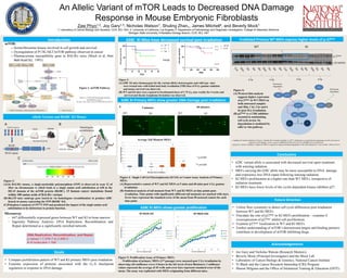
Irradiated Primary WT MEFs express higher levels of p-27kip1
Figure 6:
(A) Western blot analysis
suggests higher expression
of p-27kip1 in WT MEFs in
both untreated samples
and 30m, 1 hr, 3 hr and 6
hr post 2Gy irradiation.
(B) p27kip1 is a CDK inhibitor
essential in maintaining
cell cycle arrest. Its
degradation is mediated by
cdk2 or Akt pathway.
WT
p-27kip1
KI
2 Gy 4 Gy 2 Gy 4 Gy
α β tubulin
*
0
10
20
30
40
50
60
70
80
Unt 5 60 Unt 5 60
WT WT WT KI KI KI
AverageTailMoment
Minutes Post Irradiation
Average Tail Moment MEFs
*
*
* p=0.001
WT KI
** p<0.001
5 minutes 60 minutes
WT KI
**
**
628C KI Primary MEFs show greater DNA Damage post irradiation
Figure 4: Single Cell Gel Electrophoresis (SCGE) or Comet Assay Analysis of Primary
MEFs
(A) Representative comets of WT and KI MEFs at 5 mins and 60 mins post 4 Gy gamma
irradiation.
(B) Statistical analysis of tail moment from WT and KI MEFs at time points post-
irradiation. Time points with significantly different tail moments are marked with stars.
Error bars represent the standard error of the mean from 50 assessed comets for each
time point.
A
B
628C KI MEFs shows greater proliferation
Figure 5: Proliferation Assay of Primary MEFs
Proliferation of primary MEFs (2nd passage) were assessed post 2 Gy irradiation by
observing cell confluence every 6 hours in the InCucyte (Essen Biosience). Confluence
values represent the average of 45 wells and error bars represent standard error of the
mean. The assay was replicated with MEFs originating from different mice.
Cuadrado M, Gutierrez-Martinez P, Swat A, Nebreda AR, Fernandez-Capetillo O. p27kip1 stabilization is essential for the maintenance of cell cycle arrest in
response to DNA damage. Cancer research. 2009;69(22):8726-8732. doi:10.1158/0008-5472.CAN-09-0729.
Carrano AC, Eytan E, Hershko A, Pagano M. SKP2 is required for ubiquitin-mediated degradation of the CDK inhibitor p27. Nat Cell Biol. 1999;1(4):193-9.
N=51
N=37
P=0.05