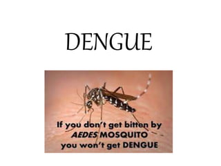
3. AROUND THE WORLD…
• About 2.5 billion people, or 40% of the world’s
population, live in areas where there is a risk of
dengue transmission.
• An estimated 50 million dengue infections occur
worldwide annually and about 500,000 people with
DHF require hospitalisation every year.
• 90 percent of those affected are children below 5
years, and about 2.5 percent of those affected die.
4. The incidence of dengue has grown dramatically around
the world in recent decades.
The actual numbers of dengue cases are underreported
and many cases are misclassified.
One recent estimate indicates :
390 million dengue infections per year
(95% credible interval 284–528 million)
96 million (67–136 million) manifest clinically
(with any severity of disease).
Another study, of the prevalence of dengue, estimates
that 3.9 billion people, in 128 countries, are at risk of
infection with dengue viruses.
The number of cases reported increased from 2.2 million
in 2010 to 3.2 million in 2015.
5.
6.
7. • Before 1970, only 9 countries had experienced severe
dengue epidemics.
• The disease is now endemic in more than 100 countries
in the WHO regions of Africa, the Americas, the Eastern
Mediterranean, South-East Asia and the Western
Pacific.
• The America, South-East Asia and Western Pacific
regions are the most seriously affected.
• Not only is the number of cases increasing as the
disease spreads to new areas, but explosive outbreaks
are occurring.
• Among travellers returning from low- and middle-
income countries, dengue is the second most
diagnosed cause of fever after malaria.
8.
9. YEAR 2014-2016….
• In 2014, there was an increase in trends in the number of
cases in China, Fiji, Malaysia.
• The year 2016 was characterized by large dengue outbreaks
worldwide.
• The Region of the Americas, reported more than 2.38
million cases in 2016, where Brazil alone contributed slightly
less than 1.5 million cases, approximately 3 times higher
than in 2014. 1032 dengue deaths were also reported in the
region.
• The Western Pacific Region reported more than 375 000
suspected cases of dengue in 2016, of which the Philippines
reported 176, 411 and Malaysia 100, 028 cases,
representing a similar burden to the previous year for both
countries.
10. IN MALAYSIA…
• Selangor, Kuala Lumpur and Johor are the areas that
have been largely affected by the disease and are
reporting high numbers of cases.
• Dengue is predominantly an urban disease due to the
abundance of the principle vector Aedes aegypti.
• The notification of dengue was made mandatory in
Malaysia since early 1970’s and dengue epidemic
activity in Malaysia has been increasing in frequency
and intensity over the past 40 years.
11.
12. • Dengue epidemics in Malaysia have been observed to occur every 5 to 8 years,
that is in 1974, 1982, 1987, 1991, 1998 and have been increasing since 2001
with the highest number of dengue cases was reported in 2008, totalling
49,335 cases with an incidence rate of 177/100,000 population.
• Subsequently a decrease of dengue cases was observed in 2011 and 2012,
which was followed by a sharp rise in 2013 with a total of 43,346 dengue cases
registered with an incidence rate of 150 /100,000 population.
• Dengue in Malaysia was predominantly confined to the densely populated and
urbanised areas of Peninsular Malaysia which cater for 20 million of the
country’s 29 million inhabitants. This contributed to 90% of all notifications
received nationwide..
• Selangor had the highest number of cases reported in 2013, with 23,852 cases
with an incidence rate 457/100,000 population, followed by Johor 4,828 cases,
Kuala Lumpur & Putrajaya 2,570 cases and Perak 2,519 cases.
• The total number of deaths reported due to dengue in 2013 was 92 with a Case
Fatality Rate of 0.21%. The states with the highest numbers of deaths were
Selangor and Johor with 24 deaths each.
• In 2014, a total of 108,698 cases were reported equivalent to IR 362 cases per
100,000 population and 215 dengue deaths reported equivalent to Case Fatality
Rate of 0.20%.
13.
14. 2. The causative organisms,
the types and their modes
of transmission.
Presenter : Harish
15. • Dengue is caused by Dengue virus (DENV), a mosquito-borne
flavivirus.
• DENV is an ssRNA positive-strand virus.
• The virus cannot be transmitted from human to human.
• In order to spread the disease needs a mosquito as alternate
host.
• The virus multiplies within the organism of the female
mosquitoes and is transmitted by bite.
i. Aedes aegyptii (yellow fever mosquito)
ii. Aedes albopticus (Asian tiger mosquito)
CAUSATIVE AGENT
17. • Mode of transmission for dengue fever is vector-borne
(mosquito).
• The virus is transmitted to humans through the bites of infected
female mosquitoes.
• After virus incubation for 4–10 days, an infected mosquito is
capable of transmitting the virus for the rest of its life.
• Infected symptomatic or asymptomatic humans are the main
carriers and multipliers of the virus, serving as a source of the
virus for uninfected mosquitoes.
• Patients who are already infected with the dengue virus can
transmit the infection (for 4–5 days; maximum 12)
via Aedes mosquitoes after their first symptoms appear.
MODES OF TRANSMISSION
18. a. The Aedes aegypti mosquito is the primary
vector of dengue.
• The Aedes aegypti mosquito lives in urban
habitats and breeds mostly in man-made
containers.
• Unlike other mosquitoes, Ae. aegypti is a day-time
feeder; its peak biting periods are early in the
morning and in the evening before dusk.
• Female Ae. aegypti bites multiple people during
each feeding period.
19. b. Aedes albopictus mosquito is a
secondary dengue vector in Asia, has
spread to North America and more than
25 countries in the European Region
• Largely due to the international trade in
used tyres (a breeding habitat) and
other goods (e.g. lucky bamboo).
• Ae. albopictus is highly adaptive and,
therefore, can survive in cooler
temperate regions of Europe.
• Its spread is due to its tolerance to
temperatures below freezing,
hibernation, and ability to shelter in
microhabitats.
21. • Goal & Objectives: To reduce the burden of dengue
i. To reduce dengue mortality by at least 50% by
2020
ii. To reduce dengue morbidity by at least 25% by
2020
iii. To estimate the true burden of the disease by
2015
PREVENTION & CONTROL
23. B. Immunization
• Dengvaxia (CYD-TDV), 1st dengue vaccine was registered in
several countries for use in individuals 9-45 years of age living
in endemic areas.
• WHO recommends that countries should consider
introduction of the dengue vaccine CYD-TDV only in
geographic settings (national or subnational) where
epidemiological data indicate a high burden of disease.
• Tetravalent live-attenuated vaccines are under development
in phase III clinical trials
• Other vaccine candidates (based on subunit, DNA and
purified inactivated virus platforms) are at earlier stages of
clinical development
24. • Park’s Textbook of Preventive and Social medicine(23rd edition)
• Graphs from WHO
https://www.who.int/denguecontrol/en/
REFERENCES
25. 4. How would you diagnose
each of the above? (What
laboratory tests and clinical
signs?)
Presenter : Huang Ruo Hui
27. • Dengue virus infection may be asymptomatic or may
cause undifferientiated febrile illness (viral syndrome),
dengue fever (DF), or dengue haemorrhagic fever (DHF)
including dengue shock syndrome (DSS)
1. Undifferentiated fever
- Infected with dengue virus, especially for the first time
(eg. primary dengue infection).
- May develop a simple fever indistinguishable from other
viral infection. Maculopapular rashes may accompany the
fever. Upper respiratory and gastrointestinal symptoms are
common.
28. 2. Dengue Fever
- All ages and both sexes are susceptible to dengue fever. Children usually
have a milder disease than adults.
- The illness is characterized by an incubation period of 3 to 10 days
(commonly 5-6 days).
- The onset is sudden, with chills and high fever (39°C-40°C), intense
headache, muscle and joint pains, which prevent all movement.
- Within 24 hours retroorbital pain, particularly on eye movements or eye
pressure and photophobia develops.
- Other common symptoms include extreme weakness, anorexia,
constipation, altered taste sensation, colicky pain and abdominal tenderness,
dragging pain in inguinal region, sore throat and general depression, rashes.
- Fever lasts for about 5 days, rarely more than 7 days after which recovery is
usually complete although convalescence may be protracted.
- The case fatality is exceedingly low.
29. 3. Dengue Hemorrhagic Fever
- Severe form of dengue fever.
- The course of dengue illness can be divided into three
phases: febrile phase, critical phase and recovery
phase.
30. Febrile Phase
• Following an incubation period of four to six days, the illness commonly begins
abruptly with high fever accompanied by facial flushing and headache.
Anorexia, vomiting, epigastric discomfort, tenderness at the right costal margin
and generalized abdominal pain are common. During the first few days the
illness usually resembles classical dengue fever, but maculopapular rash is less
common. It may appear early or late in the course of the illness. Occasionally,
the temperature may be 40°C to 41°C and febrile convulsions may occur
particularly in infants.
• The major pathophysiologic changes that determine the severity of disease in
DHF and differentiate it from DF are plasma leakage and abnormal
haemostasis, as manifested by a rising haematocrit value and moderate to
marked thrombocytopenia. These two clinical laboratory changes are
distinctive and constant findings.
• A positive tourniquet test is the most common haemorrhagic phenomenon.
31. Critical Phase
• Around the time of defervescence, when the temperature drops to 37.5°C -
38°C or less, and remains below this level, usually on days 3-7 of illness, an
increase in capillary permeability in parallel with increasing haematocrit
levels may occur. This marks the beginning of the critical phase. The period
of clinically significant plasma leakage usually lasts 24-48 hours.
• Progressive leukopenia followed by a rapid decrease in platelet count
usually precedes plasma leakage. At this point patients without an increase
in capillary permeability will improve, while those with increased capillary
permeability may become worse as a result of lost plasma volume. The
degree of plasma leakage varies. Pleural effusion mostly on right side. and
ascites may be clinically detectable depending on the degree of plasma
leakage and the volume of fluid therapy. Gall bladder oedema has been
found to precede plasma leakage. Hence chest X-ray and abdominal
ultrasound can be useful tools for diagnosis. The degree of increase above
the baseline haematocrit often reflects the severity of plasma leakage.
32. Recovery Phase
• If the patient survives the 24-48 hour critical phase, a gradual reabsorption of
extravascular compartment fluid takes place in the following 48-72 hours.
General well-being improves, appetite returns, gastrointestinal symptoms abate,
haemodynamic status stabilizes and diuresis ensues. Some patients may have a
rash of "isles of white in the sea of red” with generalized pruritus. Bradycardia
and electrocardiographic changes are common during this stage.
• The haematocrit stabilizes or may be lower due to the dilutional effect of
reabsorbed fluid. White blood cell count usually starts to rise soon after
defervescence but the recovery of platelet count is typically later than that of
white blood cell count.
• Respiratory distress from massive pleural effusion and
ascites will occur at any time if excessive intravenous fluids
have been administered. During the critical and/or recovery phases, excessive
fluid therapy is associated with pulmonary oedema or congestive heart failure.
35. LABORATORY TESTS
1. Virus isolation
Isolation of dengue virus from clinical specimens is possible provided the
specimen is taken during the first six days of illness and processed without
delay.
(Takes 2 weeks to complete and expensive)
2. Viral nucleic acid detection
- Reverse transcriptase polymerase chain reaction (RT-PCR) assay and real
time RT-PCR.
They offer better specificity and sensitivity compared to virus isolation with
a much more rapid turnaround time.
(Can determine dengue serotypes, but limited centres with facilities,
expensive, and need special storage)
36. 3. Immunological response and serological tests :
a. Haemagglutination inhibition assay (HIA);
b. Complement Fixation (CF);
c. Neutralization test (NT);
d. IgM capture enzyme-linked immunosorbent assay (MAC-ELISA);
e. Indirect lgG- ELISA,
f. IgM/IgG ratio
4. Viral antigen detection
ELISA and dot blot assays directed against the envelop/membrane (EM) antigens and
nonstructural protein 1 (NSl) can be detected in both patients with primary and secondary
dengue infection up to 6 days after the onset of the illness.
Commercial kits for the detection of NSl antigens are now available; however, these kits do
not differentiate between the serotypes.
Provides an early diagnostic marker for clinical management.
37. 5. Rapid diagnostic test (RDT)
A number of commercial rapid format serological test-kits
for anti-dengue IgM and IgG antibodies have become
available in the past few years, some of these producing
results within 15 minutes.
6. Analysis of haematological parameters
Standard haematological parameters such as platelet count
and haematocrit are important and are part of the
diagnosis of dengue infection. They should be closely
monitored.
38. Lab Test for Provisional Diagnosis / Screening Criteria
and Disease Monitoring Purpose
•Full Blood Count (FBC)
White cell count shows -
1 Leucopenia
2. Thrombocytopenia
3. Normal or raised hematocrit
For disease monitoring purpose, FBC have to be taken each and everyday once the
patient is admitted .
Platelet count should be closely monitored as it shows the severity of the disease .
39. Diagnostic Lab Test = (ELISA)
Dengue IgM test
- is significantly higher in primary infections, compared to secondary infections.
Once the IgM is detectable, it rises quickly and peaks at about 2 weeks after the
onset of symptoms, and it wanes to undetectable levels by 60 days.
Indirect IgG ELISA test
- primary and secondary dengue infection, dengue IgG was detected in
100% of patients after day 7 of onset of fever. Therefore dengue IgG is
recommended if dengue IgM is still negative after day 7 with the negative
IgG in the initial test sample.
40. Non Structural Protein (NS1 antigen) Test
• Latest diagnostic tool for diagnosing dengue
• Useful in the diagnosing in the early phase (Day 3 to 4 of illness)
Sometimes even from second day of illness
• But it is not useful after 5 days of illness.
Criteria for primary infection
• Positive NS1 antigen
Criteria for secondary infection
• Usually negative NS1 antigen, rarely can be positive
41. Rapid Test Combo Kit
• SD BIOLINE Dengue Duo
(To detect Dengue NS1 Ag and IgG/IgM in a
single test )
47. 7. What are the criteria for
hospital referral/ admission
of dengue?
Presenter : Chiew Ted Shon
48. CRITERIA FOR HOSPITAL REFERRAL / ADMISSION
• A) Referral from Primary Care Providers to Hospital
Reference: CPG Management of Dengue Infection In Adults (Third Edition) pg16
Decision for referral
and admission must
NOT be based on a
single clinical
parameter but
should depend on
the Total Assessment
of the patient.
52. B) Referral from Hospitals Without Specialist To Hospitals With Specialists
• Nearest physician should be consulted for all cases of
severe dengue, those who are pregnant and patients with
comorbidities.
Reference: CPG Management of Dengue Infection In Adults (Third Edition) pg 17
53. 8. What are the challenges
faced in dengue control?
Presenter : Chiew Ted Shon
54. 1) Rise in number and size of densely populated urban cities -conducive :
• A) for the spread of the disease
• B) the adaptation and proliferation of dengue vectors, esp. the primary
carrier of dengue virus, Aedes aegypti.
2) increased global travel has facilitated the spread of the virus:
• Cause increase in transmission of the viruses & genetic expansion of
virus-provide successful selections of viral variants of high epidemic
potential or virulence
3) Geographical expansion of the vector, Aedes aegypti:
• by invasion or reinvasion into temperate regions, such as Nepal and into
rural areas in Indonesia and Cambodia.
4) Dengue vaccine is not available
• its development is hindered by the lack of suitable animal models and the
requirement for a robust tetravalent vaccine that covers all four serotypes
of dengue
5) Cost and operational delivery shortfalls
• As such, Vector control remains the key strategy in dengue prevention
and control.
Reference: https://www.ncbi.nlm.nih.gov/pmc/articles/PMC3730958/
55. 6) Chemical treatment of breeding sites (larvicide)
• infectious adult mosquitoes are not affected and transmission
may continue
7) Insecticide spraying
• insecticidal effects of spraying are transient
• depend on persistence of the insecticide used and method of
application.
• long time lag between reporting of human cases and
commencement of spraying
• Poor functionality of sprayers
• Insufficient coverage of spraying
• Incorrect dosage of chemical insecticides
• Both: extensive and often indiscriminate use of insecticides has
resulted in a global pandemic of insecticide resistance.
Challenges and future perspective for dengue vector control in
the Western Pacific Region PDF PG 3-5
56. 8)Poor case reporting
• Clinicians at lower levels of the health systems may not
recognize dengue symptoms
• Poor surveillance system in rural areas .
9) Environmental management and vector control
• discarded containers, tyres and other vessels collect
rainwater during the rainy season
• Other examples: empty land, industry buildings,
construction sites, blocked cement drains and septic
tanks
57. 10) Community mobilization (challenge: community ignorance)
• By:
• simple dengue prevention messages through community
outreach teams
• distribution of larvicide for dengue outbreak intervention.
• school-based dengue control activities/campaign
• Dengue volunteers delivering dengue prevention messages in the
community
• communities may be reluctant /ignorant to take appropriate
dengue preventive measures except during outbreaks when the
effects of dengue are clearest.
58. 9. What is the definition for
“outbreak locality” and
“hotspot” in reference to
dengue and chikungunya?
Presenter : Chiew Ted Shon
59. • Outbreak: occurrence of cases of disease in excess of
what would normally be expected in a defined
community, geographical area or season.
(http://www.searo.who.int/topics/disease_outbreaks/e
n/)
• Hotspot: areas of elevated incidence or prevalence,
higher transmission efficiency or risk, or higher
probability of disease emergence
(https://www.ncbi.nlm.nih.gov/pmc/articles/PMC54625
59/)
• Cluster : an aggregation of cases grouped in place and
time that are suspected to be greater than the number
expected.
60. • Outbreak in Malaysia( Dengue) : Selangor, Kuala
Lumpur, and Johor
• (https://www.iamat.org/country/malaysia/risk/dengue
) /International association for medical assistance to
travelers
• In Malaysia:
• Dengue hotspot was defined by the Health Ministry’s
Crisis Preparedness and Response Centre as any locality
reported to have dengue outbreak continuously up to
30 days or more.
• Outbreak in Malayisa (Chikungunya) :
• Kedah (Alor Setar, near City Plaza), Selangor
• (https://www.nst.com.my/news/nation/2019/06/4983
87/14-suspected-chikungunya-alor-setar)
• (http://outbreaknewstoday.com/chikungunya-11-cases-
diagnosed-selangor-malaysia-73767/ )
61. REFERENCES
• https://www.ncbi.nlm.nih.gov/pmc/articles/PMC3730958/
• Challenges and future perspective for dengue vector control
in the Western Pacific Region PDF PG 3-5
• http://www.searo.who.int/topics/disease_outbreaks/en/
• https://www.ncbi.nlm.nih.gov/pmc/articles/PMC5462559/
• https://www.iamat.org/country/malaysia/risk/dengue
• https://www.nst.com.my/news/nation/2019/06/498387/14-
suspected-chikungunya-alor-setar
• http://outbreaknewstoday.com/chikungunya-11-cases-
diagnosed-selangor-malaysia-73767/
• CPG Management of Dengue Infection In Adults (Third
Edition) -pg6,15,16,17
64. WORLDWIDE
Malaria Cases :
• In 2017, an estimated 219 million cases of malaria
occurred worldwide compared with 239 million cases in
2010 and 217 million cases in 2016.
• Most malaria cases in 2017 were in the WHO African
Region (200 million or 92%), followed by the WHO
South-East Asia Region with 5% of the cases and the
WHO Eastern Mediterranean Region with 2%.
• Fifteen countries in sub-Saharan Africa and India
carried almost 80% of the global malaria burden. Five
countries accounted for nearly half of all malaria cases
worldwide: Nigeria (25%), Democratic Republic of the
Congo (11%), Mozambique (5%), India (4%) and Uganda
(4%). Reference : WHO , World Malaria
Report, 2018
65. WORLDWIDE
Malaria Cases :
• The incidence rate of malaria declined globally between
2010 and 2017, from 72 to 59 cases per 1000
population at risk. Although this represents an 18%
reduction over the period, the number of cases per
1000 population at risk has stood at 59 for the past 3
years.
• Plasmodium falciparum is the most prevalent malaria
parasite in the WHO African Region, accounting for
99.7% of estimated malaria cases in 2017, as well as in
the WHO regions of South-East Asia (62.8%), the
Eastern Mediterranean (69%) and the Western Pacific
(71.9%).
• P. vivax is the predominant parasite in the WHO Region
of the Americas, representing 74.1% of malaria cases.
Reference : WHO , World Malaria
Report, 2018
70. WORLDWIDE
Malaria Deaths :
• In 2017, there were an estimated 435 000 deaths from malaria
globally, compared with 451 000 estimated deaths in 2016, and
607 000 in 2010.
• Children aged under 5 years are the most vulnerable group
affected by malaria. In 2017, they accounted for 61% (266 000) of
all malaria deaths worldwide.
• Nearly 80% of global malaria deaths in 2017 were concentrated in
17 countries in the WHO African Region and India; 7 of these
countries accounted for 53% of all global malaria deaths: Nigeria
(19%), Democratic Republic of the Congo (11%), Burkina Faso
(6%), United Republic of Tanzania (5%), Sierra Leone (4%), Niger
(4%) and India (4%).
• All WHO regions except the WHO Region of the Americas
recorded reductions in mortality in 2017 compared with 2010.
The largest declines occurred in the WHO regions of South- East
Asia (54%), Africa (40%) and the Eastern Mediterranean (10%).
Despite these gains, the malaria mortality reduction rate has also
slowed since 2015, reflecting the estimated trends in malaria case
incidence.
Reference : WHO , World Malaria
Report, 2018
73. MALAYSIA
• In 2017, the country reported a total of 508 cases (local
and imported) of the human type of malaria, down
substantially from 6 141 cases in 2010.
• Overall, malaria transmission in Malaysia is largely
confined to Sabah and Sarawak, two states located on
the island of Borneo, where a significant proportion of
the population is at risk of the disease.
Reference : WHO , World Malaria
Report, 2018
89. Full Blood Count (FBC) :
• In falciparum malaria, the WBC count is generally normal, with relative
lymphopenia.
• Neutrophilia suggests secondary bacterial infection or severe disease.
• Thrombocytopenia is present in more than 90% of nonimmune patients.
Blood Urea & Serum Electrolytes (BUSE) :
• Sodium, calcium and albumin are low ( usually resolve spontaneously with
treatment)
CRP level : C-reactive protein (CRP) is raised.
Liver profile :
• Bilirubin is often raised[ due to haemolysis}.
• mild elevations of liver enzymes is normal
** jaundice + liver enzyme abnormalities may suggest that other diagnoses (e.g. viral
hepatitis) should be considered.
Blood glucose :
• low in severe cases (with high parasitaemia)
• during quinine treatment in adults.
LABORATORY FINDINGS
90.
91.
92. 3. Species-specific PCR diagnosis of malaria
4.Concentration technique.
5.Staining of nucleic acid.
93. 5. What is the national
strategic plan for
elimination of malaria
(2011 – 2020) and what
are the 7 strategies
outlined there?
Presenter : Vaschala Appalasamy
94. NATIONAL STRATEGIC PLAN FOR ELIMINATION
OF MALARIA (2011-2020)
• In 2011, the Malaria Control Programme was
reoriented from control to elimination, and MOH
formulated the National Strategic Plan for the
Elimination of Malaria (NSPEM) (2011-2020) with
the objective of eliminating locally acquired
human-only malaria by 2020.
• In 2016, WHO identified 21 countries with the
potential to eliminate malaria by the year 2020,
known as “E-2020 countries” – Malaysia is one of
them.
• In 2017, Malaysia has successfully achieved 0
human-only malaria deaths.
Reference : Management
Guidelines of Malaria in Malaysia,
First Edition (2014)
95. Seven strategies outlined in the NSPEM (2011 – 2020) :
• strengthen Malaria Surveillance System
• intensify control activities using Integrated Vector
Management approach
• ensure early detection of cases and prompt treatment
• heighten preparedness and early response to outbreak
• enhance awareness and knowledge on malaria towards
social mobilisation and empowerment
• strengthen human resource capacity and
• conduct relevant researches.
Reference : Management
Guidelines of Malaria in Malaysia,
First Edition (2014)
98. •Chikungunya is a local word meaning ‘double-
up’ due to excruciating joint pains.
•Chikungunya is rarely fatal.
•Symptoms are generally self-limiting
•Chikungunya shares some clinical signs with
dengue and can be misdiagnosed in areas
where dengue is common.
•Recovery from an infection will confer life-long
immunity.
99. • Chikungunya was first identified in Tanzania in the
early 1952 and has caused periodic outbreaks in
Asia and Africa since the 1960s.
• Outbreaks are often separated by periods of more
than 10 years. Between 2001 and 2011, a number
of countries reported on chikungunya outbreaks.
• Chikungunya has been identified in nearly 40
countries.
• Prior to 2013, chikungunya virus cases and
outbreaks had been identified in countries in Africa,
Asia, Europe, and the Indian and Pacific Oceans.
103. 2. The causative organisms,
the types and their modes
of transmission
Presenter : Lo Fang Ying
104. Causative agent:
Chikungunya virus
Genus : alphavirus
Family: Togavirus
Transmission:
infected mosquitoes – including Aedes aegypti and Aedes
albopictus.
Viruses spread by insects are collectively referred to as
arthropod-borne viruses, or arboviruses
References: Kumar & Clark
106. TREATMENT
• There is no specific antiviral drug treatment for
chikungunya.
• Treatment is directed primarily at relieving the symptoms,
including the joint pain using anti-pyretics, optimal
analgesics and fluids.
References: K.park pg 289,
https://www.who.int/emergencies/diseas
es/chikungunya/en/
107. (a) VECTOR CONTROL
The Aedes aegypti mosquito should be the main target of
control activities.
1. Keep water storage containers free of mosquitoes and to
eliminate the other breeding places of mosquitoes in and
around houses and dwellings
2. Organophosphorus insecticide(Abate) : used as a
larvicide prevent breeding for upto 3 months when
applied on sand granules; does not harm man and does
not affect the taste of water.
3. Aerosol spray of ultra low-volume (ULV) quantities of
malathion or sumithion (250 ml/hectare) : effective in
interrupting transmission and stopping epidemics of DHF.
The tiny droplets kill the mosquitoes in the air as well as
on water. References: K.park pg 289,
https://www.who.int/emergencies/diseases/chikungunya/en/
108. • Antilarval measures can prevent an epidemic, but do not
give immediate results when an epidemic has already
broken out.
• In such cases, anti-adult measures alone can bring about a
rapid interruption of transmission.
References: K.park pg 289,
https://www.who.int/emergencies/diseas
es/chikungunya/en/
109. (b) VACCINE
• No vaccine has yet been developed that is
considered suitable for use.
References: K.park pg 289,
https://www.who.int/emergencies/diseas
es/chikungunya/en/
110. (c) OTHERS
1. clothing which minimizes skin exposure to the day-
biting vectors is advised.
2. Repellents can be applied to exposed skin or to
clothing in strict accordance with product label
instructions.
3. insecticide-treated mosquito nets : Net treated with
permethrin (pyrethroid insecticide).
References: K.park pg 289,
https://www.who.int/emergencies/diseas
es/chikungunya/en/
111. 4. How would you diagnose
each of the above? (What
laboratory tests and clinical
signs?)
Presenter : Chok Mei Yan
112. CLINICAL FEATURES
• Incubation period: 4-7 days
• Sudden onset of high fever (40°C/ 104°F)
• Chill
• Cephalagia (Headache, pain in the region of the head or
neck)
• Anorexia
• Lumbago (pain in the lower /lumbar portion of the back.)
• Conjunctivitis
• Adenopathy
• Morbiliform rash, occasionally with purpura on trunk &
limb (60-80% of patient)
References: K. park pg289 , Kumar & Clark
113. • Cutaneous eruption may recur every 3-7 days.
• Prominent symptoms, especially in adult patient:
Arthropathy
• Appears between 3rd - 5th day after the onset of clinical
symptoms.
• Arthropathy is manifested by pain, swelling & stiffness
especially of metacarpophalangeal, wrist, elbow, shoulder,
knee, ankle and metatarsal joints.
• After 1 year, >20% of patients still suffer recurrent joint
pains.
• Other symptoms included:
Epixtaxis, Petechiae, Coffee-coloured vomiting
References: K. park pg289 , Kumar & Clark
114. DIAGNOSIS
1. Isolation of virus: from blood of febrile patient by
intracerebral inculation in suckling mice/ VERO cell
2. Serologic diagnosis: most commonly used. Sero-
conversion by comparing acute & conveslecent
phase sera in haemagglutination inhibition, serum
neutralization, complement fixation test.
3. ELIZA: detect IgM
4. Reverse transcription polymerase chain reaction
(RT-PCR)
References: K. park pg289 , Kumar & Clark
117. • The global incidence of JE is unknown because the
intensity and quality of JE surveillance and the
availability of diagnostic laboratory testing vary
throughout the world.
• Countries that have implemented high-quality
childhood JE vaccination programmes have seen a
dramatic decline in JE incidence.
• JE cases are seen presently in North Australia,
Bangladesh, Burma, Cambodia, China, Guam, India,
Indonesia, Japan, Laos, Malaysia, Nepal, North and
South Korea, Pakistan, New Guinea Papua,
Philippines, Russia, Saipan, Singapore, Sri Lanka,
Taiwan, Thailand, Timor-Leste and Vietnam
Reference: Bulletin of the World Health
Organization: Estimated global incidence of
Japanese encephalitis: a systematic review
WORLDWIDE
118. • Epidemic activity in Northern India, Central India,
and Nepal has increased since the early 1970s.
• In 1990s, the virus continued to spread in Pakistan,
Nepal and also in continental Australia
• In unvaccinated populations in endemic areas, JE is
largely a paediatric disease and most people have
acquired active immunity by adulthood.
• Conversely, in areas with long-standing, high-
quality childhood vaccination programmes, JE is
usually a rare disease of non-immune adults,
especially the elderly.
Reference: Bulletin of the World Health
Organization: Estimated global incidence of
Japanese encephalitis: a systematic review
WORLDWIDE
119. Hence for better understanding JE affected areas were
classified into various categories as follows:
Group A:
Historically high incidence areas with high quality vaccination programmes. It includes Japan,
Korea and Taiwan. Here overall incidence is reduced to 0.003 per 100 000 and the child (≤ 14
years) to adult (>14 years) case frequency ratio is 7:1
Group B:
Extremely low incidence areas with rare human cases and minimal or no vaccination
programmes. It includes Australia, Pakistan, Russia and Singapore. JE is rare and an overall
incidence is 0.003 per 100 000 with the child to adult case frequency ratio of 7:1
Group C:
Historically medium to high incidence areas with expanding vaccination programmes as seen
in China. Overall incidence is around 3.3 per 100 000 and the child to adult case frequency
ratio is 3:1
Group D:
High incidence areas with minimal or no vaccination programmes. It includes Cambodia,
Indonesia, Laos, Malaysia, Myanmar, Philippines, Timor Leste. Incidence in these areas is
10.6 per 100 000 and the child to adult case frequency ratio is 7:1
Reference: Bulletin of the World Health
Organization: Estimated global incidence of
Japanese encephalitis: a systematic review
WORLDWIDE
120. Group E:
Medium incidence areas with no vaccination programmes. It includes Malaysia
(Peninsular) and New Guinea Papua. Here incidence is assumed to be 5.3 per
100000 population.
Group F:
Historically high incidence areas with expanding vaccination programmes. It
includes India (high incidence stratum) and Nepal. Overall Incidence in these
areas is 2.8 per 100 000 and the child to adult case frequency ratio is 5:4
Group G:
Low incidence areas with minimal or no vaccination programmes. It includes
Bangladesh, Bhutan, Brunei and Nepal (lower incidence stratum). Incidence in
these areas is 1 per 100 000 and the child to adult case frequency ratio is 4:1
Group H:
Historically medium to high incidence areas with expanding vaccination
programmes. It includes India (medium incidence stratum), Malaysia (Sarawak),
Korea, SriLanka, Thailand and Vietnam. Overall Incidence in these areas is 1.5 per
100 000 and the child to adult case frequency ratio is 7:1
Reference: Bulletin of the World Health
Organization: Estimated global incidence of
Japanese encephalitis: a systematic review
WORLDWIDE
121. • WHO estimated that approximately 67 900 JE cases
occur annually in the 24 JE-endemic countries with
an overall incidence of 1.8 per 100 000 and 50% of
these cases occur in China (excluding Taiwan).
• Approximately 55 000 (81%) occur in areas with
well established or developing JE vaccination
programmes, while approximately 12 900 (19%)
occur in areas with minimal or no JE vaccination
programmes. 51 000 (75%) of these cases occur in
children aged 0–14 years, which gives an estimated
overall annual incidence of 5.4 per 100 000 in this
age group
Reference: Bulletin of the World Health
Organization: Estimated global incidence of
Japanese encephalitis: a systematic review
WORLDWIDE
122. IN MALAYSIA
• JE in Malaysia is considered an important disease among
children. However, JE is not considered a serious public
health problem in Malaysia, except Sarawak
• There have been four main outbreaks of JE reported in
Malaysia over the years: 1974 in Pulau Langkawi; 1988 in
Pulau Pinang; 1992 in Serian Sarawak; and 1998–1999 in
Perak and Negeri Sembilan
• In 7th July 2014, 17 JE cases were reported nationwide
consisting of 8 JE cases from Sarawak, 4 cases from Sabah, 3
cases from Penang, and one case each from Selangor and
Kelantan.
• In 2016, three JE cases were reported in Negeri Sembilan,
Jelebu and Kuala Pilah. In 2017, WHO reported that
Malaysia had 59 JE cases documented in 2016 (World Health
Organization, 2017) Reference :Japanese encephalitis in Malaysia:
An overview and timeline
https://www.sciencedirect.com/science/article/
pii/S0001706X18302407
123. 2. The causative organisms,
the types and their modes
of transmission
Presenter : Chong Wei Xun
124. • Japanese encephalitis (JE) is a mosquito-borne
encephalitis caused by a group B arbovirus
(Flavivirus).
• Transmitted by culicine mosquitoes.
• It is a zoonotic disease, i.e., infecting mainly animals
and incidentally man.
CAUSATIVE ORGANISM
Reference : K. Park-Park's Textbook of
Preventive and Social Medicine 23rd ed pg.284
125. • The envelope glycoprotein of the JE virus contains
specific as well as cross-reactive, neutralizing epitopes.
• The major genotypes of this virus have different
geographical distribution, but all belong to the same
serotype and are similar in terms of virulence and host
preference.
Reference : K. Park-Park's Textbook of
Preventive and Social Medicine 23rd ed pg.284
126. • Culicine mosquito species such as Culex
tritaeniorhyncus, Cx. gelidus, Cx.vishnui, Cx.
pseudovishnui and Cx fuscocephala are the prominent
vectors.
• Infects several extrahuman hosts, e.g., animals and birds.
• Basic cycles of transmission :
(a) Pig Mosquito Pig
(b) The Ardeid bird Mosquito Ardeid bird
• Humans become infected when they are bitten by
mosquitoes infected with JE virus.
• Man is an incidental "dead-end" host.
• Man to man transmission has not so far been recorded.
MODES OF TRANSMISSION
Reference : K. Park-Park's Textbook of
Preventive and Social Medicine 23rd ed pg.285
127. Animal hosts :
• Among the animal hosts, pigs have been incriminated as the major vertebrate hosts.
• Infected pigs do not manifest any overt symptoms of illness but circulate the virus thus
considered as "amplifiers" of the virus.
• Horses are the only domestic animals so far known which show signs of encephalitis
due to JE virus infection.
Incubation period: 4 – 16 days
129. 1.Vaccination
Currently, the three types of JE vaccines in large-scale use
are :
(i) Mouse brain-derived, purified and inactivated vaccine,
which is based on either the Nakayama or Beijing strains of
the JE virus and produced in several Asian countries;
(ii) the cell culture-derived, inactivated JE vaccine based on the
Beijing P-3 strain, and
(iii) the cell culture-derived, live attenuated vaccine based on
the SA 14-14-2 strain of the JE virus.
• The vaccine should be considered for all travelers to
rural endemic zones if they intend to stay there for at
least 2 weeks.
PREVENTION
Reference : K. Park-Park's Textbook of
Preventive and Social Medicine 23rd ed pg.286
130. Type of vaccine: Inactivated mouse-brain-derived
• Number of doses: Standard 3 doses schedule or reduced
2 dose schedule , subcutaneous
• Schedule: 3 doses at days 0, 7 and 28 OR 2 doses given 1
– 4 weeks apart (1.0ml for adults, 0.5ml for children)
• Booster: After 1 year and then 3 yearly
• Contraindications: Hypersensitivity to previous dose or
to the vaccine preservative thiomersal
• Adverse reactions: Occasional mild local or systemic
reaction, occasional severe reaction with generalized
urticaria, hypotension and collapse
• Before departure: At least two (2) doses before
departure
• Recommended for: Travellers over 1 year of age and
staying in endemic rural areas for more than 2 weeks
Reference :
http://www.myhealth.gov.my/en/prime-years-
japanese-encephalitis-je-2/
131. 2. Minimize exposure to bites by modifying activities to
avoid exposure to vector bites.
3. Avoid mosquito bite by applying mosquito repellent
to exposed skin. Active ingredient in a repellent DEET
(N, N-diethylmetatoluamide) repels but does not kill
insects.
• It is toxic when ingested and may cause skin irritation.
• Permethrin is highly effective both as an insecticide and as a
repellent.
• There is little potential for toxicity from Permethrin-treated
clothing.
Reference:http://www.myhealth.gov.my/en/pri
me-years-japanese-encephalitis-je-2/
132. 4. Use long sleeved clothes and long pant. Avoid
wearing dark colours (attract mosquitoes).
5. Close windows or shutters at night when indoors.
Use pyrethrum insecticide spray (aerosol insecticides),
pyrethroid coils or insecticide impregnated tablets in
evening before sleep.
6. Avoid strong perfumes, hair sprays or after-shaves
7. Use air-conditioning or good mosquito net
especially treated with Permethrin.
Reference:http://www.myhealth.gov.my/en/pri
me-years-japanese-encephalitis-je-2/
133. WHO responds to JE by:
• providing global recommendations for JE control,
including the use of vaccines. WHO recommends JE
immunization in all regions where the disease is a
recognized public health priority and supports
implementation.
• providing technical support for JE surveillance, JE
vaccine introduction and large-scale JE vaccination
campaigns, and evaluation of JE vaccine effectiveness
and programmatic impact.
PROMOTION
Reference : https://www.who.int/news-
room/fact-sheets/detail/japanese-encephalitis
134. 1. Preventive measures
2. Control of contacts
• The aim of identifying contacts is to:
-Alert them to the possibility that they could develop disease
-Recommend that a subset be offered preventive treatment, if
appropriate.
-Any unimmunised person who has travelled through a
Japanese encephalitis endemic country should be screened for
illness and potentially placed in isolation to avoid possible
contact with mosquitoes.
CONTROL MEASURES
Reference: https://www2.health.vic.gov.au
135. 3. Control of environment
• Control measures may include:
-searching for and eliminating breeding sites of mosquito
vectors
-avoiding having domestic pigs near residential areas
-using mosquito repellents, mosquito nets and other methods
of personal protection.
4. Health Education and communication for behavioural
change:
• Information for general public on how to avoid exposure
to mosquito bites and means and ways to encourage
and motivate communities to engage in vector control
activities
Reference: https://www2.health.vic.gov.au
136. 4. How would you diagnose
each of the above? (What
laboratory tests and clinical
signs?)
Presenter : Chong Wei Xun
137. LABORATORY TESTS
Presumptive: Detection of an acute phase anti-viral
antibody response through one of the following:
• Elevated and stable serum antibody titres to JE
virus through ELISA,
• Haemagglutination-inhibition or virus
neutralization assays or
• IgM antibody to the virus in the serum.
138. Confirmatory:
• JE virus-specific IgM in the CSF, or
• Fourfold or greater rise in the JE virus-specific
antibody in paired sera (acute and convalesent
phases) through IgM /IgG, ELISA, haemagglutination
inhibition test or virus neutralization test,
• Detection of the JE virus, antigen or genome in tissue,
blood or other body fluid by immunochemistry or
immunofluroscence or PCR.
139. • Febrile illness of variable severity
• Neurological symptoms ranging from headache to
meningitis or encephalitis.
The course of the disease is conveniently divided into
three stages namely:
1. Prodromal
2. Acute encephalitic
3. Late stage and sequelae
CLINICAL SIGNS
140. 1. Prodromal Phase
• The onset of illness is usually acute and is heralded by
fever, headache, gastrointestinal disturbances, lethargy
and malaise.
• The duration of this stage is usually 1-6 days.
2. Acute encephalitis phase
• Fever is usually high, 38 to 40.7 ̊C.
• The prominent features are fever, nuchal rigidity, focal
CNS signs, convulsions signs of raised intracranial
pressure, difficulty of speech, dystonia, ocular palsies,
hemiplegia, quadriplegia, extra-pyramidal signs like
coarse tremors and altered sensorium progressing in
many cases to coma.
141. 3. Late Stage and Sequelae
• Active inflammation is at an end, i.e., the temperature
and ESR touch normal.
• Neurological signs become stationary or tend to
improve.
• Convalescence may be prolonged and residual
neurological deficits may not be uncommon.
• The case fatality rate varies between 20-40%.
• The average period between the onset of illness and
death is about 9 days.
144. In a study conducted in 2015, in the worldwide there
are 1,030,000 cases and 58,900 deaths due to
leptospirosis annually .
• The majority of leptospirosis cases and deaths
occur in tropical regions; 73% of the world’s
leptospirosis cases and deaths occur in countries
situated between the Tropics.
• Highest morbidity occurred among males with 20–
29 years of age (35.27 cases per 100,000), while
highest estimated mortality occurred in older males
with 50–59 years of age (2.89 deaths per 100,000).
145. • There were 3,665 and 4,457 probable and laboratory
confirmed leptospirosis cases notified in 2012 and 2013,
respectively.
• In the 2-year period, the most common age group of
patients was 19 years old or less (23.3%) with male:female
ratio of 2.6:1. Students consisted about 16.9% of patients,
followed by agriculture-based or plantation workers
(14.7%). Overall age-standardized incidence rate of
leptospirosis in Malaysia for 2012 and 2013 was 29.02 per
100,000.
• Overall case fatality rate was 1.47% for 2-year period and
overall age-standardized mortality rate was 0.45 per
100,000.
• Leptospirosis is an emerging public health concern in
Malaysia and may pose a significant health impact and
burden to the nation in the coming years if not well
controlled.
146. 2. The causative organisms,
the types and their modes
of transmission
Presenter : Dhasney
147. • Spirochetes are a group of six genera of spiral-
shaped, slender bacteria of varying length.
• The most significant spirochetal infections for
human disease are Borrelia burgdorferi (Lyme
disease), also known as B. burgdoferi, the
treponemes (syphilis and endemic
treponematoses) and Leptospira interrogans
(leptospirosis).
• They are all resistant to rifampin, and this is often
used in microbiological isolation.
• Treponema are anaerobic, borrelia micro-aerophilic
and leptospires aerobic.
150. • Control of infection source (eg. Rodent control,
animal vaccinations)
• Interrupt mode of transmission (eg. Wear
protective clothing , provide clean drinking water,
avoid contacting with infected animal, or refrain
from dirty swimming areas)
• Prevent infection or disease in human host (eg.
Antibiotic prophylaxis , vaccination)
151. 4. How would you diagnose
each of the above? (What
laboratory tests and clinical
signs?)
Presenter : Kavisneha
152. Bacteraemic
leptospirosis
• Bacteraemia with any serogroup can
produce a nonspecific illness with high
fever, weakness, muscle pain and
tenderness (especially of the calf and
back), intense headache and
photophobia, and sometimes diarrhoea
and vomiting.
• Conjunctival congestion is the only
notable physical sign.
Aseptic meningitis
• Classically associated with L. canicola
infection, this illness is very difficult to
distinguish from viral meningitis.
• The conjunctivae may be congested
• Laboratory :neutrophil leucocytosis,
abnormal LFTs, and the occasional
presence of albumin and casts in the
urine
153. Icteric leptospirosis (Weil’s disease)
• Weil’s disease is a dramatic lifethreatening event, characterised by fever,
haemorrhages, jaundice and renal impairment. Conjunctival hyperaemia
is a frequent feature. The patient may have a transient macular
erythematous rash, but the characteristic skin changes are purpura and
large areas of bruising. In severe cases there may be epistaxis,
haematemesis and melaena, or bleeding into the pleural, pericardial or
subarachnoid spaces.
• Jaundice is deep and the liver is enlarged
• Uveitis and iritis may appear months after apparent clinical recovery.
154.
155. DIAGNOSIS
• It is not possible to diagnose leptospirosis with certainty on
clinical grounds alone. Because of the wide spectrum of signs and
symptoms, the diagnosis is made by isolation of leptospires from
blood during the acute illness and from urine after the first week
• Early in the disease, the organism may be identified by dark-field
examination of the patient's blood or by culture on a semisolid
medium. Culture takes 1-6 weeks to become positive.
• The organism may also be grown from the urine from 10th day to
6 weeks.
• Diagnosis is usually made by means of serological tests, of which
several are available.
• Agglutination tests (microscopic using live organism, and
macroscopic using killed antigen) become positive after 7-10 days
of illness and peak at 3-4 weeks and may persist at high level for
many years.
• Indirect haemagglutination, immunoflourescent antibody and
ELISA tests are also available. The IgM ELISA is particularly useful
in making an early diagnosis, as it is positive as early as 2 days
into illness
• Now Leptodipstick test is also available
156. • A polymorphonuclear leucocytosis is accompanied
in severe infection by thrombocytopenia and
elevated blood levels of creatine kinase.
• In jaundiced patients,there is hepatitis and the
prothrombin time may be prolonged.
• The CSF in leptospiral meningitis shows a variable
cellular response, a moderately elevated protein
level and normal glucose content.
• Acute renal failure due to interstitial nephritis is
common
158. 1. What are the pesticides
used in pest control?
Presenter : Kavisneha
159.
160. There is a large variety of pesticides designed to kill specific pests
– those most widely used are listed below.
•Insecticides (for killing insects) such as organochlorines,
organophosphates and carbamates. This category also includes
insect repellents such as diethyltoluamide (DEET) and citronella (of
natural origin).
•Herbicides or weedkillers (e.g. paraquat, glyphosate and
propanil). •Fungicides (to kill mould or fungi): when applied to
wood, they are called wood preservatives.
•Rodenticides (to kill mice, rats, moles and other rodents).
•Fumigants are pesticides that exist as a gas or a vapour at room
temperature and may be used as insecticides, fungicides or
rodenticides, especially in closed storage places – as they kill every
living organism. They are extremely toxic, due to their physical
properties, rapid environmental dissemination and human or
animal absorption (examples include cyanide, aluminium
phosphate and methyl bromide).
•Other pesticides include algaecides (to kill algae), miticides (to
kill moths) and acaricides (to kill ticks).
161. 2. Space spraying – what is
it and what are the
advantages and
disadvantages?
Presenter : Keerthana
162. WHAT IS IT?
• Space spraying is the outdoor
spraying of a large number of small
insecticidal droplets intended to be
distributed through a volume of air
over a given period of time.
• These droplets deliver a lethal dose
of insecticide to kill target insects
upon impact.
• The insecticide is dispersed using
hand-held, vehicle-mounted or
aircraft-mounted equipment to
produce a fog.
• Space spraying is regularly used in
public health and
pest control programmes.
163. • Space spraying is recommended for control only in emergency
situations - to suppress an ongoing epidemic or to prevent an
incipient one.
• The objective of space spraying : massive, rapid destruction of the
adult vector population.
• Any control method that reduces the number of infective adult
mosquitoes, even for a short time, should reduce virus transmission
during that time.
• If space spraying is used early in an epidemic and on a sufficiently
large scale, the intensity of transmission will be reduced which
would give time for the application of other vector control measures
that provide longer-term control.
REFERENCE:
1. WHO:
https://www.who.int/denguecontrol/control_strategies/chemical_
control/en/
2. Vector Control – Environmental Science
https://www.vectorcontrol.bayer.com/solutions/space-spraying
164. • Space spraying efficiency is dependent on:
1) Method of release (aircraft, vehicle, hand-held
equipment);
2) Fog types (cold or thermal);
3) Droplet size, application rate, climatic conditions;
The optimum size of droplets for space-spray application
are droplets with a Volume Median Diameter (VMD) of
10–25 microns.
When the droplet sizes are optimal, it ensures that spray
droplets remain airborne for the optimum period of time
and that they contain the right dose of insecticide.
4) Building structures, terrain and accessibility
5) Target area size;
6) Peak flight times.
165. • Since large areas can be treated at any one time, this method is a
very effective way to rapidly reduce the population of flying insects
in a specific area.
• Since there is no residual activity from the application, it must be
repeated at intervals of 5-7 days in order to be fully effective.
• This method can be particularly effective in epidemic situations
where rapid reduction in mosquito numbers is required.
• As such, it is commonly used in urban dengue and Zika control
campaigns.
REFERENCES :
1. Vector control ( Environmental Science ) -
https://www.vectorcontrol.bayer.com/solutions/space-spraying
166. Advantagesofspacesprays:
• Immediateeffect -suitable for control of diseaseoutbreak.
• Lessinsecticide isrequired for oneapplication.
• Killsexophillic speciesofmosquito.
Disadvantagesofspacesprays:
• Effectlastsfor shorter period- repeated at leastonceaweek.
• Thecostof equipment, operational andmaintenance-high;
• Needsspeciallytrained staff for maintenanceandrepair.
• Maycausepollution.
• Problemswith acceptability.
167. 3. What control methods
can be used for each of
these: rats, flies and
cockroaches?
Presenter : Keerthana
168. CONTROL METHODS FOR RATS
A successful rat control strategy typically includes three
elements:
1) sanitation measures;
2) building construction and
3) rodent proofing; and,
4) if necessary, population control.
Reference : Safe Rodent Control Resource Center -
http://saferodentcontrol.org/site/rodent-control/
169. 1) SANITATION:
Fundamental to rat control and must be continuous.
If sanitation measures aren’t properly maintained, the
benefits of other measures will be lost and rats will
quickly return.
Good housekeeping in and around buildings will reduce
available shelter and food sources for rats.
Neat, off-the-ground storage of pipes, lumber, firewood,
crates, boxes, gardening equipment, and other household
goods will help reduce the suitability of the area for rats
and also will make their detection easier.
Collect garbage, trash, and garden debris frequently, and
ensure all garbage receptacles have tight-fitting covers.
170. Where dogs are kept and fed outdoors, rats can become a
problem if there is a ready supply of dog food. Feed your pet
only the amount of food it will eat at a feeding, and store pet
food in rodent-proof containers.
For roof rats in particular, thinning dense vegetation will make
the habitat less desirable. Climbing hedges such as Algerian or
English ivy, star jasmine, and honeysuckle on fences or
buildings are conducive to roof rat infestations and should be
thinned or removed if possible, as should overhanging tree
limbs within 3 feet of the roof.
Separate the canopy of densely growing plants such as
pyracantha and juniper from one another and from buildings
by a distance of 2 feet or more to make it more difficult for rats
to move between them.
171. 2 ) Building Construction and Rodent Proofing
The most successful and long-lasting form of rat control
in structures is exclusion, or “building them out.”
Seal cracks and openings in building foundations and any
openings for water pipes, electric wires, sewer pipes,
drain spouts, and vents.
No hole larger than 1/4 inch should be left unsealed, in
order to exclude both rats and house mice.
Make sure doors, windows, and screens fit tightly. Their
edges can be covered with sheet metal if gnawing is a
problem. Coarse steel wool, wire screen, and lightweight
sheet metal are excellent materials for plugging gaps and
holes.
172. Because rats and house mice are excellent climbers, openings
above ground level must also be plugged.
Rodent proofing against roof rats, because of their greater
climbing ability, usually requires more time to find entry points
than for normal rats.
Roof rats often enter buildings at the roofline, so be sure that
all access points in the roof are sealed.
If roof rats are traveling on overhead utility wires, contact a
pest control professional or the utility company for information
and assistance with measures that can be taken to prevent
this.
173. Outdoor Recommendations:
• Don’t plant ivy — it provides shelter and a food source for
rodents: snails and slugs. Ivy on walls can form “rat ladders” to
windows, attics and other interior spaces.
• Keep compost piles as far away from structures as possible and
grass cut to no more than two inches tall.
• Maintain at least a 2-foot space between bushes, shrubs,
fences, and buildings. Also, remove tree limbs within 3 feet of a
structure or roof.
• Avoid having a bird feeder since it provides a source of food for
rodents.
• Keep outdoor grills and cooking areas clean.
174. Indoor Recommendations:
• Encase all food items such as breakfast cereals, chips, and
crackers in containers.
• Opt for garbage bins and compost containers with a top that seals
tightly.
• Rinse food and beverage containers before discarding or
recycling.
• Clean your garbage and recycling bins frequently.
• Do not leave pet food or water out overnight.
• Maintain stove tops clean and free of food scraps.
• De-clutter your home of papers, fabric, and any similar materials
that attract rodents for nesting.
• Repair leaky pipes.
• Seal entry points around cabinets, interior walls, attic, and crawl
spaces with steel wool, caulk, or 1/4″ x 1/4″ metal mesh.
• Maintain attic, crawl spaces, and cabinets near sinks clean and
free of moisture.
175. 3 ) Population Control :
When food, water, and shelter are available, rat populations
can increase quickly.
While the most permanent form of control is to limit food,
water, shelter, and access to buildings, direct population
control often is necessary.
For controlling rats indoors, using traps is best.
176. Trapping :
Trapping is the safest and most effective method for
controlling rats in and around homes, garages, and other
structures.
Because snap traps can be used over and over, trapping is
less costly than poison baits but more labour intensive.
Traps can be set and left indefinitely in areas such as
attics where rats have been a problem in the past.
Nutmeats, dried fruit, bacon, or a piece of kibbled pet
food can be an attractive bait for traps. Fasten the bait
securely to the trigger of the trap with light string, thread,
or fine wire so the rodent will spring the trap when
attempting to remove the food.
177. Even glue can be used to secure the bait to the trigger. Soft
baits such as peanut butter and cheese can be used, but rats
sometimes take soft baits without setting off the trap. Set traps
so the trigger is sensitive and will spring easily.
The best places to set traps are in secluded areas where rats
are likely to travel and seek shelter.
Droppings, gnawings, and damage indicate the presence of
rodents, and areas where such evidence is found usually are
the best places to set traps, especially when these areas are
located between their shelter and food sources.
Place traps in natural travel ways, such as along walls, so the
rodents will pass directly over the trigger of the trap.
178. Electrocution Traps
Traps that kill rats by electrocution (e.g., Rat Zapper or Victor
Electronic traps) are considerably more expensive than other
traps, but some homeowners, managers of commercial
buildings, and pest control companies have found them to
provide good results.
179. Benefits of Using Traps :
Using traps instead of rodent poisons gives you clear
confirmation of a captured rodent and allows you to
better gauge the effectiveness of treatment.
You are also able to dispose of rodents immediately
rather than dealing with the foul odour of rotting
carcasses from poisoned rodents inside your walls or
otherwise out of reach.
Most important, using traps allows you to avoid
rodenticides, which pose a greater threat of exposure to
children, pets, and non-target wildlife, including natural
predators.
180. Rodenticides (Toxic Baits)
While trapping is generally recommended for controlling
rats indoors, when the number of rats around a building is
high, you might need to use toxic baits to achieve
adequate control, especially if there is a continuous
reinfestation from surrounding areas.
If this is the case, consider hiring a licensed pest control
applicator who is trained to use rodenticides safely.
Baits to control rodents are formulated with an attractant
(generally food) and a rodenticide (toxin).
181. Anticoagulant Rodenticides
• Anticoagulants are blood-thinning drugs that cause an
animal’s blood to lose the ability to clot, damaging
capillaries and resulting in internal bleeding that is
fatal.
• These active ingredients are used at very low levels
and the onset of symptoms is delayed for several
days, so the rodent doesn’t avoid the bait because of
its taste or the onset of illness.
• When prepared with good-quality cereals and other
ingredients, anticoagulant baits provide good to
excellent control when baits are fresh and when
placed in suitable locations so as to attract rats.
182. CONTROL METHODS FOR FLIES
1. Integrated pest management is the best approach to
controlling flies
- It combines prevention and non-chemical control methods with
the wise use of the least harmful pesticides possible
- It involves monitoring and environmental control
2. The environment must be managed to decrease the
areas where flies can lay their eggs
- Control moisture and remove materials where larvae develop
(e.g., feces) to break the life cycle and prevent development of
adult flies
- Fecal material in outdoor areas should be picked up and disposed
of on a daily basis
- Garbage cans should have tight fitting lids to deter entry by flies
- Organic debris (e.g. rotten vegetation and leaf litter) should also
be disturbed once a week to prevent fly eggs from hatching
183. 3. Area sprays (knockdown) are fine mist insecticides that kill
adult flies on contact
- These products can be used in areas of high fly concentration; they do not
last long (1-2 hours)
- Due to evaporation, these products should not be used at temperatures
over 90o F; they are not effective at low temperatures (below 65o F)
4. Residual sprays are insecticides that can be applied to
shaded surfaces
- The flies are killed when they contact the product while resting
- Treat places such as walls, ceiling, and rafters
- Water (e.g., cleaning) will wash off the insecticide
- To avoid insecticide resistance, alternate between area and residual sprays
5. Baits and fly traps can be used in areas where the use and
safety of chemical sprays are prohibited
- These products should NOT be placed in areas where they could
contaminate food or water or where animals will have access to them
Reference : Center for Food Safety and Public Health
http://www.cfsph.iastate.edu/Maddies_Textbook/Resources/Infe
ctionControl/Fly%20Control%20Measures.pdf
184. CONTROL METHODS FOR COCKROACHES
Cockroaches require food, water and harbourages to survive.
Thus, if the presence of these three factor can be decreased
the chances of a cockroach population to be established can be
greatly reduced too.
Inspection
Look for cockroach hiding spots in warm, dark, tight places
near food and water using a flashlight and mirror.
Confirm any suspected habitats by using sticky traps. These
traps will not attract cockroaches, but they will allow you to
determine if you have an infestation, and where the bugs are
living.
Sticky traps should be placed at the seam of floors and walls,
and in potential high traffic areas. Carefully check large
appliances and furniture or items that have been in storage for
cockroach egg cases and destroy them.
185. Home Cleaning
If you have an infestation, eliminate food sources by cleaning up
crumbs on floors, in cracks and crevices, or on counters.
Wipe up spills and clean dirty dishes as soon as possible, and
keep food in sealed containers or in the refrigerator.
Throw trash away in containers with liners and tight lids, and
empty them frequently.
Regularly vacuum cracks and crevices to remove any food
sources, including cockroach eggs or droppings, since faeces
contain pheromones that attract other roaches and feed young
ones.
Frequent vacuuming also goes a long way in preventing
cockroach allergies.
186. Eliminate Habitat and Prevent Entry
Removing cockroach hiding places and entry points will
drive cockroaches away and prevent future infestations.
Inside the home, seal cracks in dark places like cupboards or
building cracks with caulk, repair water leaks, and remove
moisture in crawl spaces and other dark areas by increasing
ventilation.
Weather-seal doors and gaps around windows, and caulk
around escutcheon plates that surround pipes or faucets
that provide access to the wall voids or any other area that
roaches could easily enter or hide.
Eliminate clutter, especially piles of magazines, newspapers,
cardboard or rags.
Outside, remove stacks of wood and trash to minimize
potential moist habitats, and trim shrubs and branches to
increase light and ventilation.
187. Chemical control
If chemical control is used, apply the chemical to ensure that the
target insects have contact with it.
Various insecticide formulation are available for the purpose,
ranging from residual insecticides dust to gel bait.
a) Residual insecticides
Residual insecticides are the primary and most popular control
method although baiting is getting increasingly popular.
Cockroaches come into contact with residual insecticides when they
walk upon the treat surfaces.
Pay particular attention to rubbish bins and receptacles. The bases
of bins can be treated to ensure cockroaches contact the insecticide
on their way to gaining access to the bin contents.
Spray around rubbish bins, sewer and drain inspection grids and
house access areas like doorways and windows.
188. b) Gel Baits
Cockroach baits area usually formulated in bait station or as
gel.
Due to its limited application, baiting is relatively safer and
environmentally-friendly than residual treatment.
It is also odourless and ready to use. Cockroaches feed on the
bait will receive a lethal dose and eventually die.
This method is useful especially in sensitive environment such
as zoos, child-care centre, computer rooms, food preparation
outlets, where residual treatment is not allowed
Reference : Pesticide Research Institute
https://www.pesticideresearch.com/site/pri-
resource-centers/pest-mgmt/pest-mgmt-
bulletins/cockroach-control/
Editor's Notes
K Park Pg 248
K Park Pg 248-250 (Slide 3 -9)
K Park
K Park
K Park Tourniquet test: The test is performed by inflating a blood pressure cuff to a mid point between systolic and diastolic pressure for 5 minutes. The test is considered positive when 10 or more petechiae per 2.5 x 2.5 cm (1 inch square) are observed. In DHF, the test usually gives a definite positive with 20 petechiae or more.
K Park
K Park
K Park Pg 249
Davidson’s Pg 322
K Park Pg 251 Specimen that are suitable for virus isolation are acute phase serum, plasma or washed buffy coat from the patient, autopsy tissue from fatal case (especially liver, spleen, lymph nodes and thymus), and mosquitoes collected from the affected areas.
K Park Pg 251
K Park Pg 251
Disease monitoring lab test:
White cell count
Early febrile phase usually normal but decrease rapidly as disease progresses.
Haematocrit
Rising HCT is a marker of plasma leakage and helps to differentiate b/t DF &DHF.
May be masked in patients with concurrent significant bleeding & those who received fluid therapy.
Setting pt’s baselineHCT in early febrile phase will be useful in recognition of a rising HCT.
Thrombocytopenia
Early febrile phase- platelet count usually w/in normal range.
Decrease repidly as disease progresses to late febrile phase.
May remain low for first few days of recovery.
LFT
Greater elevation of AST as compared to ALT.
Degree of elevation higher in DHF compared to DF.