Autism and Facial Features
•Download as DOCX, PDF•
1 like•376 views

The brain develops in concert and in coordination with the developing facial tissues, with each influencing the development of the other and sharing genetic signaling pathways. Studies have shown that autistics have different facial feature lengths from typically developing children. These different lengths cause differences in muscle activity levels, with greater constant muscle activity in the center of the face for certain autistics to offset the smaller midface area. Because the greater muscle activity is in a smaller area, the activity depletes more potassium from intracellular membranes, and long-term hypokalemic sensory overstimulation occurs.

Recommended
More Related Content
Similar to Autism and Facial Features
Similar to Autism and Facial Features (20)
More from Yuval Levental
Recently uploaded
Recently uploaded (20)
Autism and Facial Features
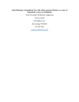
- 1. Facial Phenotypes of prepubertal boys with autism spectrum disorders as a cause of hypokalemic sensory overstimulation Yuval H Levental, BS Electrical Engineering Yuval Levental 636 Tarleton Ave. East Lansing, MI 48823 517-492-7881 yuval.levental@edu.esiee.fr
- 2. Abstract The brain develops in concert and in coordination with the developing facial tissues, with each influencing the development of the other and sharing genetic signaling pathways. Studies have shown that autistics have different facial feature lengths from typically developing children. These different lengths cause differences in muscle activity levels, with greater constant muscle activity in the center of the face for certain autistics to offset the smaller midface area. Because the greater muscle activity is in a smaller area, the activity depletes more potassium from intracellular membranes, and long-term hypokalemic sensory overstimulation occurs.
- 3. Background Autism Spectrum Disorder (ASD) comprises a group of complex neuropsychiatric disorders of childhood, diagnosed on the basis of the behavioral phenotype. The ASD phenotype is characterized by social deficits, impaired communication, and restricted and repetitive behavior patterns [1]. In 2011, a research study was done, showing that autistics have different facial features. The midface was shorter than that of a TD child, and the eye regions and mouth were longer than that of a TD child (Fig. 1). The face sizes were measured to be the same [2]. A follow-up research study in 2014 also produced the same results, with three classified subgroups of autistic facial phenotypes. One of the facial alterations include facial muscle structure [3]. Fig. 1. Results of Euclidean Distance Matrix Analysis analyses of landmark coordinate data collected from 3dMD images. White lines are statistically significantly increased in boys with autism spectrum disorder (ASD), and black lines are statistically significantly reduced in boys with ASD relative to typically developing (TD) boys. Hypothesis Facial distance lengths are indicative of underlying muscle activity. The muscles on the eye and mouth regions of the faces are longer, and the muscles in the middle of the face are shorter when
- 4. compared with a TD child [2]. Because of the shorter distance, the muscles in the middle of the face of an autistic child are more active, causing greater potassium depletion which leads to hypokalemic sensory overstimulation. Hypokalemic sensory overstimulation has been described as similar to hypokalemic periodic paralysis. The phenotype has also been described as overlapping with ADHD [4], and the phenotype of ADHD has also been described as overlapping with Autism spectrum disorders [5]. Evaluation of the Hypothesis According to the Euclidean distance matrix analysis from the Aldridge study, it is speculated that the nasalis muscle, procerus muscle, and the labii superioris muscles in the facial center are shorter than average. The orbicularis oculi muscles, Infraorbital head muscles, and Zygomatic head muscles are longer than average. Additionally, the quadratus labii inferioris and orbicularis oris muscles are horizontally longer than average [2]. The shorter central midface muscles exert more activity compared to a TD child, due to the need to offset the reduced size (Fig. 2). Fig. 2. The superficial layer of the facial muscles and the neighboring muscles of the neck seen from the side and slightly from in front. Several studies have shown that exercise results in contracting muscles, which result in a release of K+ ions from the muscles. This produces a decrease in intracellular K+ concentrations and an increase in plasma K+ concentrations. The flow reverses, and the K+ in the blood is expunged through sweat, causing a lower overall level of potassium [6]. Hypokalemic sensory overstimulation has been described as similar to hypokalemic periodic paralysis. The sensory overload has been treated with potassium gluconate, with onset of the effect in 20 minutes. The effect of potassium is reminiscent of its effect in the channelopathies underlying hypokalemic periodic paralysis. The phenotype has also been described as overlapping with ADHD [4], and the phenotype of ADHD has also been described as overlapping with Autism spectrum disorders [5].
- 5. To test this hypothesis, facial electromyography would be conducted on ASD subjects and TD subjects. The aforementioned muscles would be tested and the electrical impulses would be measured. Another possible test is a potassium blood test, where ASD subjects with the specified facial phenotypes and TD subjects would have potassium levels measured in their blood streams, while on a controlled diet. One anecdote from an autistic individual who consulted with a plastic surgeon based on this hypothesis states that the surgeon claimed the muscle above of his nose was working too hard. This anecdote was published on Cortical Chauvinism, the blog of Dr. Manuel Casanova, a University of South Carolina neurologist. The surgeon recommended botox to relieve pressure in the affected area [7]. Arguments against the hypothesis are the claim that facial muscle activity in autistic children with facial phenotypes are not powerful enough to cause measurable potassium depletion. Another argument is that the facial phenotypes may be the result of other components in the body. Consequences of the hypothesis If the hypothesis were to be confirmed, potassium supplements or a diet high in potassium and low in sodium could possibly be recommended as one treatment for a child with Autism spectrum disorder and associated facial features. Another possible treatment would be injecting botox into the midface region to reduce the activity of the muscles in this area. Clinical trials would have to be run to confirm those new hypotheses. Funding None Conflict of interest statement The autistic individual whose anecdote was featured was myself. I have frequently published autism-related opinions and anecdotes on Cortical Chauvinism.
- 6. References [1] American Psychiatric Association. Diagnostic and statistical manual of mental disorders (5th ed.). Arlington, VA: American Psychiatric Publishing. (2013). [2] Aldridge et al.: Facial phenotypes in subgroups of prepubertal boys with autism spectrum disorders are correlated with clinical phenotypes. Molecular Autism. (2011). 2:15. [3] Obafemi-Ajayi, T., Miles, J. H., Takahashi, T. N., Qi, W., Aldridge, K., Zhang, M., et al. Facial structure analysis separates autism spectrum disorders into meaningful clinical subgroups. Journal of Autism Developmental Disorders. (2014). 45(5), 1302–1317. [4] Segal, M. M., G. F. Rogers, H. L. Needleman, and C. A. Chapman. "Hypokalemic Sensory Overstimulation." Journal of Child Neurology 22.12 (2007): 1408-410. [5] Leitner, Yael. “The Co-Occurrence of Autism and Attention Deficit Hyperactivity Disorder in Children – What Do We Know?” Frontiers in Human Neuroscience 8 (2014): 268. [6] Lindinger MI, Sjøgaard G. Potassium regulation during exercise and recovery. Sports Med. (1991). 11(6):382-401. [7] Levental Y. Yuval Levental: Plastic Surgery and Autism. Cortical Chauvinism. (2016). <https://corticalchauvinism.com/2016/07/21/yuval-levental-plastic-surgery-and-autism/>.