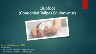
2. DEFINITION
Congenital deformity of the foot and
ankle characterized by equinus deformity
at the ankle, inversion at the subtalar,
adduction ad the midtarsal joint, cavus
and internal tibial torsion
Complex, congenital, contractural
malalignment of the bones and joints of
the foot and ankle
3. Type of Clubfoot
Idiopathic variety (most common type)
Found in otherwise normal children
Does not resolve without intensive
treatment
Postural variety
Resolves completely without
intervention, or with manipulation alone,
or with one or two casts
Neurogenic clubfoot
Myelomeningocele
Syndromic clubfoot
Children with other anomalieS
4. Clinical features
C A V E (Typical)
Cavus (plantar flexion of the forefoot on
the hindfoot)
Adductus of the forefoot on the
midfoot
Varus (or inversion) of the subtalar joint
complex
Equinus of the hindfoot
5. PATHOLOGICAL ANATOMY:
1. BONES
• TALUS
Head and neck deviated medially and downward.
Medial And Plantar Deviation Of Navicular Articulation.
Body Rotated Externally And Is In Equinus Of Neck In Ankle
Mortise.
Body Extruded Anteriorly
Smaller Than Normal
Neck- Body Angle Is 90-110* (Normal- 150*)
Dislocation Of Head Of Talus Out Of Its Socket.
6. • NAVICULAR
Medially and plantar
displacement
Close to medial malleolus
Articulates with medial surfacfe
Of dysmorphic talus
Talonavicular joint subluxation
7. • CALCANEUS
Often small in size
Medially rotated
Anterior portion lies beneath the head of talus
causing varus and equinus of heel.
Sustentaculum tali is underdeveloped.
• CUBOID
Medially
head
Subluxated Over CALCANEUS
8. 2. MUSCLES AND TENDONS
Atrophy Of Peroneal Group Of Muscles
Contracture Of Tricep Surae,tibialis
Posterior,flexor Digitorum Longus And Flexor
Hallucis Lungus.
Number Of Fibres In Muscle Is Normal But
Are Smaller In Size.
Thickening And Contracture Of Tendon
Sheaths Especially Of Tibialis Posterior And
Peroneal.
9. 3. LIGAMENTS
THICKENING AND CONTRACTURES ARESEEN
IN :
Calcaneofibular ligament
Talofibular ligament
Deltoid ligament
Long and short plantar ligament
Spring ligament
Bifurcate ligament
Interosseous talo calcaneum ligament
Master knot of henry
10. 4. JOINTS CAPSULE AND FASCIA
• CONTRACTURES ARE SEEN IN
Posterior ankle capsule
Subtalar capsule
Talonavicular joint capsule
Calcaneocuboid joint capsule
Plantar fascia contracture are seen which is responsible cavus deformity
11. Clinical features
Others finding clubfoot; atypical
Posterior ankle skin crease; single
Empty Heelpad sign
Tibial torsion
Deep transverse skin crease crosses the midfoot and
extends under the longitudinal arch
Hyperextended great toe; appear short
The calf smaller than contralateral side
Increased internal hip rotation
Looked and palpated of head talus on the
dorsolateral aspect of the midfoot/hindfoot just
anterior to the ankle joint solid fulcrum
In idiopathic clubfoot; The navicular will not fully
align with the head of the talus and displace the
examiner’s thumb
13. Pirani scoring system
Simple & Reliable to determine severity
Monitor progress of treatment
6 Signs are assesed :
3 signs in midfoot (MFS)
Assesses severity of supination, adductus, and cavus
0 (normal) to 3 (severe)
3 signs in hindfoot (HFS)
Assesses severity of equinus
0 (normal) to 3 (severe)
Total Score (TS) is the sum of HFS and MFS
It assesses severity of the clubfoot as a whole with a score range from 0
(normal) to 6 (severe)
15. Pirani scoring system
• MFS signs ; assess subtalar supination, midfoot adduction, and
cavus
16. Radiographic Features
No consensus on the role of radiographs in the diagnosis and
management
To demonstrate the relationships between bones
To confirm correction or to identify the sites of residual deformity
has been undergoing serial manipulation and casting
Surgical planning
17. Plain x-ray
Talo-calcaneal angle (Kite’s
Angle)
• AP View
• Mid-talar line (through to
medial base 1st metatarsal) &
Mid-calcaneal line (through to
base 4th metatarsal)
• 20 - 40° (Normal)
• <20° (CTEV)
• Lateral
• 35 - 50° (Normal)
• <35° (CTEV)
18. Other Imaging Studies
Arthrography, CT, MRI
May have a role in research or in
the evaluation of postsurgical
deformities
Do not have a role in the routine
assessment of the idiopathic
clubfoot
USG
Intrauterine diagnosis
Accuracy is 12 weeks of
gestational age
19. Natural history
The untreated clubfoot persists as a rigid
Develops of Callused bursa over the
dorsolateral aspect
In the most extreme cases, the toes point
backward
City-dwelling adolescents and adults
with untreated clubfoot experience pain
& disability
20. NON OPERATIVE
Treatment of Clubfoot
Goal :
Achieve a plantigrade, supple, painless foot that looks normal,
although it is not technically normal, and provides good function
Achieve good pressure distribution on the skin and no need for special
or modified shoes wear
Kite (1939)
Presented cast correction but required a lengthy period of immobilization,
often >1.5 to 2 years
Ponseti and Smoley (1940)
Developed a casting method for clubfoot that differed significantly from
Kite’s
21. PONSETI TREATMENT
Gold standard treatment
Based on pathoanatomy of deformity
The efficacy due to :
Viscoelasticity
Rate-dependent
Behavior of the collagen in the
ligaments & tendons
22. Ponseti method
Old standard : POP
Semirigid fiberglass more superior in
Durability
Convenience
Performance
Ease of removal
But more challenging in molding
Remove cast before return to clinic
Maintenance the cast until 5 -7 days
If not achieve good correction in 3
months : Operative
Full correction Cavus, adductus & varus
deformity : 90%
Equinus deformity achieved by 10° of
ankle dorsiflexion
If not achieved : Percutaneous complete
transection of the Achilles tendon
23. PONSETI CAST
CORRECTION :
CAVUS
Forefoot supination relative to the hindfoot
Pronation of the first metatarsal at the first session
the forefoot is simultaneously supinated and
abducted
The cavus is almost always corrected with the first
cast
VARUS, ADDUCTION, INVERSION
Correct simultaneously
Abduction foot in supination
EQUINUS
The last correction
Residual equinus need Tenotomy
26. Continues passive movement
Developed by Masse & Bensahel et al. In
1970 (France)
Dynamic method of management
Utilizing physiotherapist-implemented
exercises by Adhesive Taping
Begun immediately after birth
Stretch the tight plantar-medial structures
(posterior tibial tendon and plantar soft
tissues
Strengthen the peroneal muscles
Allows some functional motion that is not
permitted in rigid casts
0-2 months (daily treatment)
2-6 months (3x / weeks)
Continued : Physical therapy & night
splinting (for 2 to 3 years)
27. Percutaneus tenotomy
Integral step in Ponseti technique
HS > 1, MS < 1, Head talus is covered
PROCEDURE :
Incision 1 cm above insertion of achilles on the calcaneus
Used a small blade, small cataract knife, Needle
Insert the blade from medial side of the heel
perpendicular to the medial border of the foot
The blade parallel and directed to the Achilles tendon
Slowly moved the blade anteriorly until it slips past the
anterior border of the tendon
This technique will help ensure that the blade does
not pass near the posterior tibial neurovascular
bundle
Turned the blade to a 90° posteriorly Section the
tendon
The foot : 20° of dorsiflexion & 70° of abduction
Cast for 3 weeks
28. BOTULINUM TOXIN
INJECTION
By Alvarex et.al (2005)
Injected into triceps surae muscle complex to weaken its
function
Minimal scar & deep tissue fibrosis
Need more experiment
29. AFTER CORRECTION
EVALUATE :
In the final cast :
15 - 20° of dorsiflexion
70 - 75° of external rotation of the foot relative to the thigh
MANAGEMENT AFTER CORRECTION :
Semirigid shoes connected together by bar
(Foot Abduction Orthosis / Denis Browne bar & shoes)
70° of external foot rotation (45° for a contralateral normal foot)
5-10° of dorsoflexion
For 3 – 4 months, 23 hours per day worn at nap and nighttime
for 2 to 4 years
But, challenging to maintain nightime in 4 years children
Ankle dorsiflexion stretching exercises for at least 1 minute at
least 3x/day
30.
31.
32. Surgical management
INDICATION :
Failed with non-operative
Resistant, persistant, relapsed
Neglected case
Others secondary deformity
Early surgery not recommended ;
stimulated Myofibroblast poor
outcome
Avoid multiple operation
COMPLICATION :
Wound-healing problems
Neurovascular injury
bone/cartilage damage
avascular necrosis of the talus and
navicular
Pain
Stiffness
Weakness
residual deformity
recurrent deformity
dorsal bunion
overcorrection at the talonavicular,
talocalcaneal, and
talocalcaneonavicular joints
34. Timing of surgical procedure
After failed nonoperative management
12 months
Structure were larger
Anatomy clearly
Tendon lengthening repair more
secure
Only little advantage surgery < 12
monts
Because weight bearing &
standing position will be delayed
by the postoperative
immobilization
Soft tissue release : 1 – 4 years old
Soft tissue release + Osteotomy :
4 – 11 years old
Salvage procedure (Triple
arthrodesis,
Talectomy/astragalectomy) : > 11
years old
37. Turco approach
Popular in 1970s
Hockey-stick posteromedial
incison
Straight from base 1st
metatarsal under medial
malleolus until reached
Achilles
Crosses the skin creases on
the medial side
Identified all medial
neurovascular structures and
tendons
38. cincinnati approach
Circumferential incision
Problems with skin edges
Limited exposure of achilles
Extensile
Cosmetic
Safe (as long as, placed at least 1
cm at proximal ankle crease)
Lower placement high risk slough
of the heel pad
39. Carrol’s approach
Double incision technique (posterior &
medial)
Base of triangle :
Center of calcaneus
Front of medial malleolus
Base of 1st metatarsal
Center incision paralel of the base
triangle
Proximal part toward the center of the
heel
Distal part crosses over the dorsum of
the foot
Safe
Extensile
Less cosmetic
40. Suggested technique
Prone position
Cincinnati incision
Successful comprehensive release : involving multiple anatomic
steps, exposure
Better place : posterolateral corner of the ankle
After identified & protected the sural nerve & lesser saphenous
vein opened the peroneal sheath to allow full anterior
retraction of the two tendons
POSTERIOR RELEASE :
The calcaneofibular and lateral subtalar ligaments, avoiding
blind peroneal tendon injury
Longitudinal exposure of the Achilles tendon permits a long Z-
lengthening
The posterior talofibular ligament
MEDIAL PLANTAR RELEASE :
The posterior & medial subtalar joint capsule (leaving the
interosseus ligament intact
Talonavicular joint capsulotomy (including spring ligament &
bifurcatio Y ligament)
Medial calcaneocuboid joint capsulotomy
42. Soft tissue surgery;
Anterior Tibial Tendon
Transfer
4cm incision over of tibialis anterior extend from its
insertion to proximally
Sharply incision of tibialis anterior tendon sheath
Dissected the insertion as far distally
Avoid injury to the 1st metatarsal growth plate
Avoid bow stringing tendon
Used absorsable suture (vicryl 0) (Bunel type fashion)
4cm incision over 3rd cuneiform (proximal from 3rd
metatarsal; between EDL & peroneus tertius)
Insert small gauge (confirm with minifluoroscopy)
Make a drill hole on the 3rd cuneiform tract to tibialis
anterior tendon meet to ankle retinaculum
The foot position : maximal dorsoflexion & evertion
Suture periosteum of 3rd cuneiform with two interrupted
absorsable suture
43. Soft tissue surgery;
Transfer for Insufficient Triceps Surae
(Calcaneus Gait)
Overlengthening of the Achilles tendon or triceps insufficiency
secondary to inadequate excursion from scarring is notoriously
difficult to reconstruct and is best prevented rather than
reconstructed
The surgeon must diagnose plantar flexion weakness as early as
possible if muscle transfer is to have any chance of being effective
Muscles for transfer to reconstruct :
Peroneals
Tibialis posterior
Long toe flexors
44. Soft tissue surgery;
Transfer for Insufficient Triceps Surae (Calcaneus
Gait)
LATERALLY
The peroneus brevis can be divided distal to the fibula
and the proximal end rerouted to the calcaneus
tuberosity (Tendon-to-Bone transfer)
Drill hole on the calcaneal tuberosity – to – button on
plantar heel
The distal stump of the brevis tenodesed side to side
to the longus to maintain eversion power
MEDIALLY
The tibialis posterior or flexor hallucis longus
rerouted in a similar fashion & interwoven with the
residual Achilles tendon, if present, or anchored to
bone
Immobilized with NWB 6-8 weeks
45. Bony surgery;
Lateral Column Shortening
EVANS PROCEDURE
Standard technique for recurrent clubfoot
deformity
Calcaneocuboid fusion
4 - 8 years old (<4 ; large amount of cartilage
difficult to fusion)
Recurrent deformity : medial contracture &
excessive length of lateral collumn
Combines posteromedial release & lateral column
shortening in one stage
Concepts of midfoot (talonavicular and
calcaneocuboid) dislocation by allowing
reduction of the navicular on the talar head by
lateral column shortening avoid
recurrence/relapsed
46. Bony surgery;
Lateral Column Shortening
LICHTBLAU PROCEDURE
Concept : overgrowth lateral part of
calcaneus
Recommended for >6 years old
Calcaneocuboid arthroplasty
Resection of the anterior end of the
calcaneus
Shortening of the calcaneal neck proximal
to the calcaneocuboid joint
47. Bony surgery;
Lateral Column Shortening
GOLDNER PROCEDURE
Less commonly used
Ideal age range is unknown
Close wedge osteotomy of the
anterior calcaneus
Preserve cuboid
Goldner
48. Bony surgery;
Lateral Column Shortening
CUBOID DECANCELLATION PROCEDURE
Can be used at any age
Wedge resection
Preserve articular surface
49. Bony surgery;
Lateral Column Shortening
Fixation with small staples or a
Kirschner wire
NWB short leg cast
Cast and pin are removed after 6
weeks
50. Bony surgery;
calcaneal osteotomy
Advantage : Preserve subtalar motion (Dwyer)
Can be combined with other procedure; Triple
arthrodesis
Open or closed wedge osteotomy
Better perform at 10 years old
Open wedge;
Medial approach ; wound closure can be
compromised
Stabilized with bone graft (tricortical iliac crest
graft) increase height of heel required
more achilles
Delayed weight bearing
Closed wedge;
Less wound healing morbidity
Decreased height of heel impingement
51. Bony surgery;
Supramalleolar Osteotomy
Persistent in toeing gait
Cause by Muscle imbalance (abnormal
histopatology peroneal muslce)
Failed with 2 years observe
Supramalleolar external rotation
osteotomy
Effective
Not contribute to stiffness
Goldner;
up to 35° of external rotation
Correction at level proximal and
distal tibial physis
Using 2 pin between level of
osteotomy
52. Bony surgery;
Supramalleolar Osteotomy
For severe valgus deformity of ankle
Wiltse’s technique
Anterior approach to the distal tibial
metaphysis at the level of the metadiaphyseal
junction
A triangular piece of bone is removed from the
region of the distal tibial metadiaphyseal
junction
The apex of the cut is centered on the
longitudinal axis of the tibia
The magnitude of the angle of the lateral
portion of the triangle should be equal in size
to the magnitude of the deformity to be
corrected
Stabilized by a plate and screws or Kirschner
wires
A cast is placed as below
53. Triple arthrodesis (Astragalectomy)
Fusion : talocalcaneal, calcaneocuboid &
talonavicular joints
Indicated for salvage procedure or 'last resort’ in
severe, rigid deformities of the hindfoot that are
unresponsive / resistant to less invasive methods of
treatment
Considered : >10 years of age
Used for varus or overcorrected valgus feet
The goal of surgery
To achieve a plantigrade foot by restoring the
anatomic relationships between the affected
bones or regions of the foot
Relieve pain
• Positioning : Supine
• Approach
• Single lateral
• Ollier
• Most common
• Anterolateral
• Medial
• Useful for calcaneuovalgus foot
• Lambrinudi procedure, for severe
equinus deformity
• Combined medial & lateral
54. Lambrinudi
triple arthrodesis
Incision begin 1 cm distal to tip fibula
It curves dorsolaterally and extends to the lateral border of the
talonavicular joint
Retract Extensor tendons to medially
Mobilized and protected of peroneal tendons
Retract Extensor digitorum brevis to distally
Exposing sinus tarsi, calcaneocuboid joint & lateral aspect of
talonavicular joint
The sinus tarsi is cleared of soft tissue to expose the anterior,
middle & posterior facets of the subtalar joints
55. Lambrinudi
triple arthrodesis
Make a sequential osteotomies
1st osteotomy :
at the inferior part of the talus perpendicular to the
long axis of the tibia in both planes
2nd osteotomy :
at the superior part of the calcaneus parallel to the
sole of the foot in both the longitudinal and
transverse planes
3rd osteotomy :
at the distal end of the calcaneus at a right angle to
the long axis of the calcaneus
The final cut is made along the proximal end of the
cuboid at a right angle to the longitudinal axis of the
56. PENNY'S MODIFIED LAMBRINUDI
TRIPLE ARTHODESIS
Special attention triple arthrodesis in Neglected clubfoot in adolescent
Lengthening achilles is required in first step
The main components :
hindfoot equinus and varus, midfoot cavus, and forefoot adduction
Significant obliquity of the calcaneocuboid joint requires a specially oriented
lateral wedge excision of the calcaneocuboid joint
Typically severely plantarflexed
Need aggressive resection of the talar head to correct the midfoot cavus &
plantigrade position
57. PENNY'S MODIFIED LAMBRINUDI
TRIPLE ARTHODESIS
INCISION & DISSECTION :
1 cm distal tip of fibula
Curve dorsolaterally & extend to lateral border of talonavicular joint
Medially retracted of extensor tendon
Mobilized & protected peroneal tendon
Identified & protected sural nerve
Elevated off extensor digitorum brevis from the origin & reflected distally
Exposed the sinus tarsi, calcaneocuboid joint, & lateral aspect of talonavicular
joint
Cleared soft tissue from sinus tarsi
Identified facet of subtalar joint
58. PENNY'S MODIFIED LAMBRINUDI
TRIPLE ARTHODESIS
Removing a lateral wedge (calcaneocuboid resection)
Transverse cut perpendicular to the long axis of the lower leg
Removes the joint surface of the cuboid and should be
conservative (several millimeters)
Removing an anteriorly based wedge from the anterior process of the
calcaneus to correct equinus
Resecting a portion of the head & neck talus
The cut begins at the dorsal articular margin of the talus and
extends in a proximal and plantar direction through the posterior
subtalar joint
This cut is oriented perpendicular to the long axis of the tibia
Conservative resection of the articular surface of navicular, as well as
removal of the tuberosity of the navicular
A notch is made in the inferior articular surface of the navicular to
accept the anterior portion of the talus
With the surfaces of the talus and calcaneus apposed, the anterior end
of the talus is pushed into the notch under the navicular while
abducting the forefoot
59. TRIPLE ARTHRODESIS USING
A SINGLE MEDIAL INCISION
A 2-cm longitudinal incision is made over the peroneal
tendons 10 em above the level of the ankle joint, and
both tendons are delivered using a mosquito clamp
and divided sharply
An 8-cm medial longitudinal incision extends from the
undersurface of the posterior medial malleolus across
the talonavicular joint
The talonavicular joint is exposed, and the tibialis
posterior tendon is released from its insertion. The
talonavicular capsule is released. Flexor digitorum
longus tendon, flexor hallucis tendon, and
neurovascular bundle are protected by a retractor
The talocalcaneal interosseous ligament is divided, and
the anterior, middle, and posterior facets of the
subtalar joint are visualized
The subtalar and talonavicular joint surfaces are
denuded and prepared
The calcaneocuboid joint capsule and bifurcate
ligaments are released sharply, and a lamina spreader
is inserted to facilitate removal of the joint surfaces
60. BEAK TRIPLE ARTHRODESIS
FOR SEVERE CAWS
DEFORMITY
A lateral approach is employed, as outlined above
Wedges to be removed
The articular cartilage of the subtalar and
calcaneocuboid joints is denuded
The talar neck is osteotomized from inferior to
superior, forming a beak superiorly
The soft tissue structures on the superior aspect of the
talus anterior to the ankle are left undisturbed
The dorsal cortex of the navicular is excised
The forefoot is displaced plantarward and the
navicular is locked beneath the remaining part of the
talar head and neck
Stability can be maintained while plaster is applied by
slight upward pressure under the forefoot. a staple
may be used for fixation
61. the Ilizarov in ctev
Neglected or recurrent deformity
Combining multiple-plane corrections through the use of
hinged distraction between a tibial & foot frame
Corection slow enough to correction soft tissue
Correction of the focus of deformity
Simultaneus three-dimensional, multilevel correction
Deformity correction without shortening the foot
Ring are fixed to the tibia connected to half ring for the
calcaneus and the forefoot
Asymetric distraction corrects the various deformity
Bony deformity not severe (<8 years); unconstrained
frame
Severe deformities (>8 years); distraction osteogenesis
through osteotomies using constrained frame with hinges
Adductus & Equinus Correction