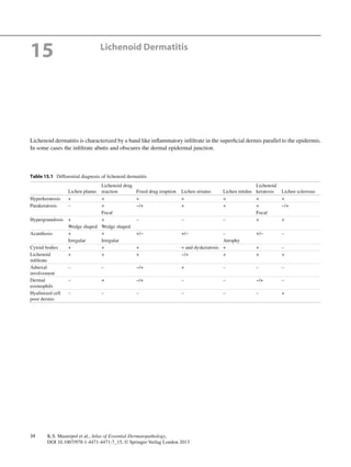
2. 3515 Lichenoid Dermatitis
Fig. 15.1 Lichenoid dermatitis. There is wedge-shaped hypergranulo-
sis with irregular epidermal hyperplasia and a band of inflammation
along the epidermal base
Fig. 15.2 Colloid bodies in lichen planus. The eosinophilic globules
are extracellular and may be present in the epidermis or the superficial
dermis, also known as Civatte bodies
Fig. 15.3 Lichen sclerosus. There is eosinophilic homogenization of
the subepidermal dermis with an underlying band of inflammation
Fig. 15.4 Fixed drug reaction. There is extensive epidermal dyskerato-
sis and interface dermatitis that may have a dense lichenoid
appearance