
Recommended
More Related Content
What's hot
What's hot (20)
Viewers also liked
Viewers also liked (12)
Similar to Radiological assessment – Part 2
Similar to Radiological assessment – Part 2 (20)
More from SpinePlus
More from SpinePlus (20)
Recently uploaded
Recently uploaded (20)
Radiological assessment – Part 2
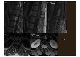
- 1. 60F T2 T1 T1FS con T1 T1FS con
- 2. 70M T2 T1 T1FS con T1 T1FS con
- 3. 35M PBA T2 T2 T1
- 5. • 72 year old male • Non mechanical back pain • Known prostate Ca: – Raised PSA (20) – Nodule on DRE – +ve on biopsy • Staging investigations
- 6. What is the most appropriate imaging modality for the spine? 1. Plain film 2. CT 3. Scintigraphy (bone scan) 4. MRI
- 10. 64F Breast Ca
- 11. T2 T1 T1FS con 76M CRC
- 12. T2 54M RCC
- 14. • 62 year old male • Severe low back pain of rapid onset • Febrile and unwell • 4 weeks ago underwent abdominal surgery for perforated diverticulitis
- 15. What is the most likely diagnosis? 1. Acute disc herniation 2. Discitis/ osteomyelitis 3. Crush fracture secondary to osteoporosis 4. Metastatic cancer
- 16. What is the most appropriate imaging modality? 1. Plain film 2. CT 3. Scintigraphy (bone scan) 4. MRI
- 17. T2 T1 T1FS con
- 18. T2 T1FS con
- 20. • 37 year old male • Low back and buttock pain, increasingover several months • Worse in morning; reduced by activity
- 21. What is the most likely diagnosis? 1. Acute disc herniation 2. Facet joint degeneration 3. Inflammatory spondyloarthropathy 4. Metastatic cancer
- 23. Seronegative spondyloarthropathies (SpA) • European Spondyloarthropathy Study Group (ESSG) Arthritis Rheum 1991;34:1218-1227 – Ankylosing spondylitis – Reactive arthritis – Arthritis spondylitis with inflammatory bowel disease – Arthritis spondylitis with psoriasis – Undifferentiated spondyloarthropathy (uSpA) • Clinical features + HLA-B27 • Rheumatoid factor –ve = seronegative
- 24. ANKYLOSING SPONDYLITIS • Chronic inflammatory disease, primarily affecting spine and sacroiliac joints • Osteitis: – Bone erosions; sclerosis; ankylosis • Peripheralarthritis: – Asymmetrical; lower limb • Enthesopathy: – Plantar fasciitis – Distal Achilles tendonosis and paratendonitis
- 25. DIAGNOSIS OF AS • Radiographic grading of sacroiliitis 0-4 Kellegren Atlas of Standard Radiographs in Arthritis, Oxford 1963 • Grade 0 = normal • Grade 1 = suspicious (mild blurring) • Grade 2 = minimal sclerosis, some erosions • Grade 3 = severe erosions, joint widening, partial ankylosis • Grade 4 = complete ankylosis
- 26. Radiographic grading of AS • Grade 0 • Grade 1 • Grade 2 • Grade 3 • Grade 4
- 27. Radiographic grading of AS • Grade 0 • Grade 1 • Grade 2 • Grade 3 • Grade 4
- 28. Radiographic grading of AS • Grade 0 • Grade 1 • Grade 2 • Grade 3 • Grade 4
- 29. Radiographic grading of AS • Grade 0 • Grade 1 • Grade 2 • Grade 3 • Grade 4
- 30. Radiographic grading of AS • Grade 0 • Grade 1 • Grade 2 • Grade 3 • Grade 4
- 31. Radiographic grading of AS • Grade 0 • Grade 1 • Grade 2 • Grade 3 • Grade 4
- 32. Dx of AS: Modified New York criteria • Arthritis Rheum 1984;27:361-368 • Clinical: 1. LBP & stiffness > 3/12 improved by exercise 2. ↓ motion lumbar spine sagittal and frontal 3. ↓ chest expansion for age & sex • Radiological: – Grade ≥ 2 bilateral – Grade 3-4 unilateral • AS = 2/3 clinical + radiological
- 33. Problems with radiographic grading • May take years for radiographic changes to develop – Early cases excluded from research and treatment • Most radiographic signs in AS reflect healing processes, not disease activity – cf erosions in RA • Most radiographic signs in AS irreversible • Radiographs do not detect inflammation
- 34. T2FS
- 35. T1 STIR STIR
- 36. Response to DMARD eg infliximab – Braun Ann Rheum Dis 2002;61:iii51-iii60
- 38. • 45 year old male • 2 weeks post discectomy L4/5 • Recurrent bilateral leg pain
- 39. What is the most appropriate imaging modality? 1. Plain film 2. CT 3. Scintigraphy (bone scan) 4. MRI
- 40. T2 T1
- 41. T2 T1FS con T2
- 42. T1FS con
- 43. • Dx: recurrent disc: – Central herniation + huge sequestration virtually filling the spinal canal • Note peripheral enhancement pattern • DD: fibrosis
- 45. • 51 year old female • Left sciatica – Intermittent pain and paraesthesia
- 46. T2 T1 T1FS con
- 47. What is the most likely diagnosis? 1. Massive disc sequestration 2. Discitis complicated by abscess 3. Synovial cyst 4. Benign peripheral nerve sheath tumour
- 48. T2 T1 T1FS con
- 49. • Dx: benign peripheral nerve sheath tumour (BPNST) of left L3 nerve root – Many clinicians use the term ‘neuroma’ • Pathologically imprecise term – Most are benign • Schwannoma or neurofibroma • Difficult (impossible) to differentiate on imaging – BPNST is probably the best terminology – Associated with NF1 and ‘NF2’ (MISME)
- 51. • 66 year old female • Severe lower back pain on and off for years • More recent (2 months) development of right sciatica
- 53. What is the most likely diagnosis? 1. Massive disc sequestration 2. Discitis complicated by abscess 3. Synovial cyst 4. Benign peripheral nerve sheath tumour
- 54. L4/5
- 55. • Severe OA of facet (zygoapophyseal) joints • Round heterogeneouslesion projecting into right spinal canal • Note: close relationship to facet joint • Dx: synovial cyst
- 56. Synovial cyst lumbar facet joint • Fairly common • Key is relationship to degenerate facet joint • Density may vary from pure cyst to varying levels of calcification and heterogeneity • Usually present clinically with intractable sciatica • May respond to aspiration and steroid injection, but usually treated surgically
- 57. T2 T1
- 58. T2 T1
- 59. Image interpretation: spine • Anatomy • Cross sectional techniques: – CT – MRI • Nomenclature of disc herniationsand spinal stenosis • A few cases