

Recommended
More Related Content
What's hot
What's hot (20)
Similar to Burns dressings.pptx
Similar to Burns dressings.pptx (20)
More from Shubhanshu Gaurav
More from Shubhanshu Gaurav (11)
Recently uploaded
Recently uploaded (20)
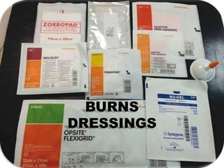
Burns dressings.pptx
- 2. Burns Devastating incident with prolonged outcome with respect to • Wound healing. • Repeated infections. • Prolonged hospitalization. • Morbidity /mortality. • Physical handicapped. • Mental/psychological disturbances . • Additional morbidity to patients (donor areas ) • Surgeon challenges with respect to closure of wounds /donor areas • Once the initial resuscitation of burns patient done , focus shifts onto wound management.
- 3. Classification of burn wounds
- 5. Local appearance of burn wounds • Extensive burnt raw areas/blisters. • Exudates containing serum/tissue fluids • Dry Eschar wounds.(3rd degree burns) • Pus /slough (infected wounds) . • Surrounding skin erythema. • Thrombosis of superficial veins. • Singeing of hairs. • Extremely Painful
- 6. Aim of burns dressings • Proper covering of the burn wound. • Prevent dryness of the wound by providing moisture • Reduce the pain • Reduce contamination of wound/bacterial invasion • Reduce evaporative losses • Joint should be immobilized to prevent contractures..
- 7. Principles of burns dressings 4 basic principles- • Desiccated wound needs to be kept moist. • Excess exudate need to be absorbed • Slough/pus/necrotic tissue to be removed by autolytic/chemical/surgical removal. • Infected wound needs to be tackled- topical or systemic antibiotics.
- 8. What makes the dressing material IDEAL • Provide moist environment. • Effective barrier for microbes/foreign bodies. • Prevent body tissue fluid loss • Allows gaseous exchange • Protect against shearing forces(adhesive) • Non traumatic / non irritant • Eliminate dead space. • Helps in early epithelization. • Easy for application • Cost effective
- 9. Initial care of wound • Immediate after burns wound is sterile. • Hydrotherapy- wound washed with running water / normal saline. • Encircling objects/ornaments should be removed. • Blisters- ruptured/deroofed/aspirated. • managed later with either exposure mtd(open technique)/closed mtd
- 10. Open method of dressings • Introduced by Wallace 1948. • Face/neck/perineum wounds treated by this mtd. • Used in >90% non salvageable pts . • Wound left open->exudates dries up-> forms a coating over wounds->prevents bacterial colonization underneath • Paraffin wax impregnated gauzes can be placed on the wound
- 11. Advantages • Less man power/less materials/easy wound examination-re-assessment/early physiotherapy Disadvantages • Wound kept dry delayed epithelization. • Over the joint repeated break in epithelium/scab causes bleeding. • Eschar takes long time to separate. • Unsighty and uncomfortable for patient and attenders. • Evaporative loss increases->shock • Hypothermia .
- 12. Bed side conventional dressings • It can be dry or wet , used in full thickness wounds • Should contain first layer of antimicrobials • Second layer of mesh/non adherent gauze • Third layer of absorbent ->gamzee/pads. • In dry technique- Simple gauze can be used which is easily available/inexpensive/highly absorbent.
- 13. Bed side conventional dressings • In wet technique- saline with gauze or paraffin wax/jellonet/tulli-grass/bacti-grass can be used – Keeps the wound moist . – Easy to remove doesn’t adhere to tissues. – Does not damage underlying new epithelium • Disadvantage – porus/permiable to exogenous bacteria and high infection rates – Repeated changing in heavy exudative wounds
- 15. Topical antimicrobials used in burn wounds . • History- variety of local medications used in past. Tincture(600 B.C) vinegar soaked dressing (430 BC) greasy dressing(1596) tannic acid by Edward Davidson (1904) Gentian violet by Aldridge in 1933 Petroleum gauze -Harvey Allen in 1942. Carbolic acid, mercurial compounds and aniline dyes were used as inner layer of burn dressing( WW-2 )
- 16. Ideal topical antimicrobial agent • Should have broad spectrum antimicrobial property especially effective against psuedomonas/staphaylococcus/klebshiella/enterococci. • Penetrate eschar • Less systemic absorption. • Non allergic/irritant. • Effective –minimal emergence of resistance. • Cost effective. • Minimal side effects. • Easy application.
- 17. Silver sulphadiazene • Synthesized by Fox-1968 • White crystalline, insoluble,water based 1% cream. • Most commonly used topical agent. • MOA-silver ion and sulfadiazene moeity released at the wound site.silver ions destroy bacterial cell wall ,also damages DNA directly • Sulfadiazene-PABA inhibitor,interupts bacterial replication. • Primary bacteriostatic,delayed colonization by 2 weeks • Effective against Gm positive/negative/anerobes.
- 18. • Advantages – Easy application – Reduces pain, intermidiate eschar penetration – Minimal systemic absorption – Less incidence of resistance • Disadvantages – Transient leucopenia. – Metheamoglobinemia(sulpha moiety) – Neoepithelial damage – Hypersensitivity reactions
- 19. Silver nitrate • Used during WW-2 (10% solution)->toxicity. • Recent years - 0.5% solution used • MOA- similar to SSD, action is by Silver Ion. • Bacteriostatic ,Effective against –Gm neg. • 6-8 layer dressing done after wash and Dressing to be soaked once in 2-3 hrs with silver nitrate.
- 20. • Advantages – Less evaporative loss/ wound kept moist. – Early granulation because of early separation of eschar/necrotic tissue. – Painless – Less resistance • Disadvantages – Inability to penetrate. – Precipitation into silver chloride/sulpide ->staining – Distilled water use as vehicle for application- >dyselectrolytemia. – Enterobacter species ->metabloise nitrate to nitrite and cause metheamoglobinemia
- 22. Mefenide (11.1%) • Methylated sulfonamide introduced in 1964 • Effective against psuedomonas,clostridium and gm neg organisms and minimal antifungal activity. • Good penetration in eschars. Applied twice daily. • Occlusive dressings not advised. • Significant carbonic anhydrase inhibition property • Banned in many countries due to its toxicity
- 23. • Advantage – Effective in established wound infection – Eschar penetration • Disadvantage – Significant pain. – Metabolic acidosis and renal bicarbonate excretion – Hyperventilation->resp distress – Damage neo-epithelium – Hypersensitivity raections – Hemolytic anemia(rare)
- 24. Other local application agents • Gentamycin-aminoglycoside(0.1%),effective against pseudomonas /gm neg organisms./active eschar penetration. S/E -Oto/nephrotoxicity. • Bacitracin • Chlorhexidine • Povidone iodine-effective against staphylococcus,fungi. • Polymixin-B • Framycetin • Soframycin • Nanocrystalline silver based ointments
- 25. Temporary skin substitutes • Biological • synthetic
- 26. Permanent skin substitutes • Epidermal • Dermal • composite
- 27. Biological dressings and skin substitutes • The ultimate aim in the treatment of burns is to achieve wound closure. • Wound closure in partial thickness burns occurs by epithelialization. • In extensive burns (30-40%) the wounds need to be covered temporarily with some biological dressing material or skin substitute till autografts are available. • Once the epithelialization is complete, the biological dressings and skin substitutes either come out of their own or are peeled off
- 28. Temporary skin substitute • Biological – Amniotic membrane – Collagen – Allografts – Xeno-grafts • Synthetic – Polyurethane Film – Hydrogel – Hydro fiber – Hydrocolloid – Alginates – Foams(silver impregnated )
- 29. Amniotic membrane • Placenta is procured from normal or cesarean section deliveries. • placental membrane dissected from the blood clots with a sterile gauze swab. • washed with normal saline /antibiotic sprays and used to cover burn wounds.. • Both human and bovine amnion has been used • Acts as excellent temporary cover for a few days • Used in clean second degree burns or donor areas of split skin grafts. • Amnion can be stored for 2-3 days in sterile bottles containing 0.25% sodium hypochlorite solution
- 30. • Treated with 0.25 percent sodium hypochlorite and 200,000 units of penicillin, sterilized, dried and stored at room temperature up to nine months • Advantages – It relieves pain – Avoids discomfort during dressing change, – reduces oozing – protects underlying regenerating epithelium. – No vascularization ->no rejection/reaction • Disadvantages – carries the risk of transmitting diseases like HIV/CMV
- 31. Amnion
- 32. Collagen • Abundant protein/ 25-30% of body protein constitutes of collagen .(mainly type -1) • Fibrous protein of vertebrates and forms main constituent of connective tissue. • It can be isolated from tissues ,purified and preserved wet and dried sheets , powdered form. • Preservative used iso-prophyl alcohol.(wash with saline or distilled water prior to use) • It is hydrophilic and adheres firmly to raw wounds after getting dried • It has very low antigenicity.
- 33. • Sterilized by GAMMA radiation. • Collagen powder and granules are available. • Precaution to take while applying sheet – To wash alcohol /rinse – Contains porus sheet with thin nylon woven fibril.
- 34. • When healing is complete, the dried collagen automatically falls off. • provides good biological temporary cover in superficial and deep partial thickness burns, SSG donor sites • Reduces pain and evaporative losses • Forms a barrier for microorganisms and thus reduces chances of wound infection. • No vascularization -> no rejection/reaction
- 35. Allograft/homograft • In 1881 Girdner treated burns with skin harvested from suicide victim. • Availability of skin banks makes it possible to get the donor preserved skin harvested from cadaver or live donors. • Freeze dried and treated with glycerol and stored for several years in acellular form • Can be used as temporary cover in partial thickness burns preventing evaporative loss of proteins and electrolytes • Reduces pain
- 36. • HLA match being done in western countries • Patient after allograft started on steroids and immunosuppresents to avoid early rejection • Rejection starts between 3-10 weeks by immune system . • Allografts vascularise and thus immune/inflammatory repose seen and bacterial load reduces. • Mean time granulation tissue develops from beneath and wound will be ready for auto-grafting. • Disadvantage- – immuno-supression can increase infection. – Transmission of HIV/CMV/fungal infection – Rejection (HLA II )
- 37. Indications- • Extensive wound when local skin not available. • Covering wide meshed autografts • 2nd degree deep and 3rd degree burns • Steven johnsons/TEN • Waiting period for granulation to grow • template for delayed application of keratinocytes
- 38. Xenografts • Bromerg and song popularized porcine skin graft in 1965. • Procured from pork/canine/bovine skin. • Acts in similar manner to allografts. • Does not get vascularized->no rejection.
- 39. Synthetic temporary skin substitutes – Polyurethane Film – Hydrogel – Hydro fiber – Hydrocolloid – Alginates – Foams(silver impregnated )
- 40. Polyurethane Film • Semi permeable /transperent dressing materials. • Made up of polyurethane/polyethelene • Adhesive coating on one side • Allows water vapour/gases,impermeable to water /bacteria • Combined with antibiotic local applicants or gauze • Applied to 2nd degree sup and deep burns/ssg donor areas
- 41. • Advantage – Easy applicable – Inspect wounds – Maintain moist environment – Reduces pain – Adheres to surface ,elastic • Disadvantage – Non absorbant – Cant use for infected wounds – Strips off neo-epithelium
- 43. hydrogels • Transparent polyethylene dressings • Available as sheets and fillers /composition 90% water • Non adhesive, needs covering over this layer • Water released from gel helps in softening the necrotic tissue and helps in auto debridement and de-sloughing. • Disadvantage- wound maceration
- 44. • Used in dry, necrotic, 3rd degree burns wounds • Pressure sores • Donor sites of ssg • Radiation injuries
- 46. Hydro-colloid • Contains adhesive, hydrophlic,gel forming particles-gelatin covered with outer layer of foam. • Absorbant,impermeable to bacteria,water proof,painless. • Helps in autolytic debridement
- 47. • Encourages faster healing and re-epithelization • Used in 2nd and 3rd degree burns wounds with exudate and slough. • Can be left in place for 5-7 days if minimal to moderate soakage is present.
- 49. Alginates • Derived from Calcium salts of alginates (sea weeds) • Sodium and calcium ions react with exudate and form gel • Provide moist environment available as pastes,sheets ,powders • Highly absorptive • Used in moderate to high exudate wounds/necrotic wounds with slough
- 50. Alginates
- 51. Hydrofibres • Made up of sodium carboxy-methyl cellulose with calcium ions • Similar to alginates and similar properties. • Excellent Ability to absorb the exudates. • Can be left for 4-7 days • Used in 2nd degree burns • Ex-AQUACEL
- 53. Foams • Made of polyurethene polymers • Thick and thin /adhesive and non adhesive foams with excellent absorptive capacity • Helps in autolytic debridement • Can be combined with antimicrobials/silver impregnated. • Used in moderate to high exudate wounds/necrotic wounds with slough • Ex- biotin AG/mepilex AG
- 54. Foams
- 55. Biobrane • It is a two layered temporary, synthetic skin substitute. • outer layer -ultrathin layer of silicon rubber(semi-permeable and permits the exit of water vapors but prevents entry of bacteria from the exterior). • The inner layer -tightly woven nylon fabric. • Inner layer has porcine collagen peptides covalently bonded. • Silicone sheet is removed and later grafted.
- 56. Biobrane
- 57. • Opsite-made up of modified PVC • Trancyte-bilayered – dermal:collagen synthesised from human neonatal fibroblast+ nylon mesh – Epidermal:silicon sheet
- 58. Temporary skin substitute • Biological – Amniotic membrane – Collagen – Allografts – Xeno-grafts • Synthetic – Polyurethane Film – Hydrogel – Hydro fiber – Hydrocolloid – Alginates – Foams(silver impregnated )
- 59. Permanent skin substitute • Dermal replacement – Synthetic: integra / derma graft – Biological: alloderm • Epidermal replacement – Cultured epidermal allograft(CEA) – Cultured keratinocytes • Composite replacement • Dermal + epidermal cultured grafts.
- 60. Integra • Synthetic bilayered dermal substitute. • Outer- silicone layer • Inner- bovine tendon collagen arranged in porous matrix+cross linked shark cartilage gylcosaminoglycans • Chondroitin maintains structural integrity and maintains porus matrix for ingrowth of cellular structures • Wound debridement done and integra applied. • Dermal layer infiltrated with fibroblast and collagen tissue + capillary invasion
- 61. • Silicone layer separates out in 2-4 weeks • Outer layer grafted later. • Less hypertrophic scarring /contractures • Disadvantage – expensive/2 staged procedure.
- 62. Dermagraft • Contains bio absorbable polyglactin mesh with allogenic neonatal fibroblasts • Applied after tangential excision. • More resistant to contamination • After 1 month mesh gets absorbed and ssg can be done on the dermal matrix
- 63. Alloderm • Acellular dermal matrix • Biological dermal substitute. • Derived from cadaveric graft. • Used in deep 2nd degree or 3rd degree wounds • Harvested graft processed , de-epithelised , freeze dried after screening for HIV/CMV/HBsAg. • Applied on raw wound and grafted in single sitting.
- 64. Cultured keratinocytes • Cultured epidermal keratinocytes(CEA) • Rheinwald and Green in 1975 -irradiated mice fibroblasts can support the growth of human fibroblasts. • technique -culture medium of fetal calf serum is used to grow keratinocytes into sheets of epidermis from two to eight layers thick. • sub-culturing, a three square centimeter sheet can be expanded five thousand times to cover enough epithelium to cover the whole body • Disadv-time consuming /high cost /sheering /lack of adherence.(weak adhesive fibrils and enzymatic separation )
- 66. Composite graft • Epidermal + dermal components (APLIGRAF) • Apligraf- – Dermal:neonatal fibroblast +bovine type 1collagen lattice – Epidermal:human foreskin derived neonatal keratinocytes (CEA) • Used in 2nd degree deep/3rd degree burns • In trials /not available for clinical use
- 69. • Applied after thorough debridement • Used in bedsore/2nd and 3rd degree burns/chronic wounds/electric burns/fasciotomy wounds • Exposed vessels and vital structures to be avoided with VAC application.
- 70. Recent advances • Micro-grafting technique- grafts of size 0.8X 0.8 mm harvested under LA,OPD basis .using xpansion micrografting tecnique • Regenerative capacity of 1:100 • Technically demanding /expensive • Fractional skin grafting- large no. of skin full thickness microscopic skin harvested (700 mic) using hypodermic syringe and under microscope/micro-sheets placed on raw areas randomly. • no donor site morbidity/faster healing/technically demanding • Autologous non cultured cell therapy (ReCell) – thin ssg harvested /incubated , after mechanical agitation keratinocytes separated ,suspended in lactate solution , treated with antibiotics and sprayed over wound. • Good expansion 1:80/expensive/mechanical damage to cells
- 71. Conclusion • 1st degree-silver sulphadiazine/bacitracin/mupirocin LA • 2nd degree superficial- paraffin gauze/silver nitrate/Collagen/amnion/biofilms/hydro- fibres/biobrane/trancyte/CEA • 2nd degree deep hydrocolloid/foam/alginates/autograft/xenograft/biobrane/integra dermagraft. • 3rd degree mefenide/alginate/foams/alloderm/composite dermal+ epidermal substitutes.
- 72. References • Total burn care, David.Herdon ed.5 • Principles and practice of burns care –sujtha sarabai ed.1 • Grabb and Smith’s plastic surgery ed.8 • Handbook of burns,vol-2 P.kamolz,M.G.Jeschke ,ed.2
- 73. • Thank you…