1. Gilpin 1
Shelby Gilpin
ANS 124; Dr. Hovey
Effects of Tamoxifen, Estrogen, Domperidone, and Progesterone on Male and
Female Mammary Gland Development
Introduction
Mammary gland morphology is essentially determined by the stroma and the
parenchyma. Interplay between the two compartments heavily influences the mammary
gland’s development throughout life. Much of this interplay occurs via hormones. Even
during embryogenesis the hormonal impact of testosterone controls the extent of the
mammary gland development as well as its future functionality. Androgen receptors in
the mesenchyme around the neck of the mammary sprout respond by closing the sprout
when exposed to androgens. During puberty in a female, growth hormone and estrogen
work together to stimulate further ductal growth into the fat pad, local production of IGF-
1, and act to increase stromal estrogen receptors to promote the hormonal response. At
sexual maturity estrogen and progesterone work together to initiate tertiary branching –
making ducts receptive to adding alveoli in the event that pregnancy occurs. During
pregnancy, prolactin, growth hormone, insulin, glucocorticoids and placental lactogen
work to prepare the mammary gland for lactaion. Prolactin stimulates the formation of
budding structures and induces casein transcription, as well as induce golgi swelling.
Blocking prolactin or the glucocorticoids leads to an unsuccessful lactation after birth.
Finally, lactation requires prolactin for milk synthesis, oxytocin for milk letdown, as well
as growth hormone, thyroid hormone, corticosteroids, and insulin for metabolic effects.
Tamoxifen (TAM) is a selective estrogen receptor modulator (SERM) that is
often used in treatment and prevention of breast cancer. Studies are still being carried out
to comprehend the full effect of TAM, however it is understood that it decreases
mammary epithelial cell proliferation, has little effect on mammary blood flow (Zoma Et.
al. 2002), and at full dose causes increased levels of FSH and its cascading consequences
(Bernades Et. al. 1999 )
Understanding the importance of hormones and how they affect development is
critically important throughout all aspects of biology. More specifically, understanding
mammary gland development and the hormones involved is crucial in human and
veterinary medicine. SERMs can be used in a variety of patients such as those at risk for
breast cancer. Tamoxifen can also be used to treat post-menopausal patients at risk of
bone degradation, when estrogen concentrations are too low to induce the bone-sparing
effect, without increasing their risk for breast cancer. Also, tamoxifen may have some
conservation applications in that it shows potential in the induction of ovulation when
used in concert with a gonadotropin primer in salmon and possibly other fish (Donaldson
Et. al. 1981)
In this experiment, observed the effects of TAM on adult male mouse mammary
development. We did this by maintaining a control group, which remained unaffected by
outside hormones in order to monitor what would occur under normal circumstances. In a
male mouse of the same strain we inserted a hormonal pellet and allowed the mouse to
develop over the course of two weeks. At the end of this time, a necropsy was performed
on the experimental and control mouse. Whole mounts of the two mice were made and
analyzed to compare, contrast, and record findings. I suspected that tamoxifen would
have little to no effect on the male mammary gland due to its mammary estrogen
2. Gilpin 2
inhibiting trait combined with the usual lack of activity in the mammary tissue, but other
tissues such as bone would develop as usual.
Methods and Materials
We performed this experiment with several different groups of mice. In total the
study included 6 different treatments, each performed on males and females. Our
specific mouse was a male treated with tamoxifen. All pellets were made of
cholesterol (source: Sigma Aldrich) and were about 10 mg mass each, and were gas
sterilized before use. The control pellet consisted of cholesterol and nothing else. The
tamoxifen pellets consisted of 1mg of tamoxifen each. The estrogen pellets contained 1µg
of 17-beta estradiol each. The estrogen+tamoxifen pellets contained 1mg of tamoxifen
and 1µg of estradiol each. The estrogen+domperidone pellets contained 1µg of estradiol
and 1mg of domperidone each. The estrogen+progesterone+domperidone pellets were
composed of 1µg of estradiol, 1mg of progesterone, and 1mg of domperidone. The
variation in female group size was the result of surgical complications.
Treatment Number of
Males
Number of
Females
Control 5 4
Tam 6 11
Estrogen 5 10
E+Tam 5 10
E+D 5 12
E + P + D 5 13
Figure 1: Overall study design showing number of mice (males and
females) used for each Treatment type
Each mouse was treated for two weeks before being euthanized and
examined. The mice were housed with ad libitum access to standard laboratory
rodent chow and water, and were monitored daily. The light cycle consisted of 12
hours of light and 12 hours of dark. Wound clips from the pellet surgery were
removed seven days following the surgery. All procedures of the experiment were in
accordance with Institutional Animal Care and Use Committee (IACUC) regulations
and approval.
We began by anesthetizing a male FVB strain mouse with a ‘cocktail’ of mg of
ketamine and mg of xylazine (60/10 mg/kg respectively). Our mouse was given
108.12 µl of the cocktail via an intraperitoneal injection. Once he was sedated, we
began surgery preparation by shaving a quarter sized area at the nape of the neck
and sanitizing this area with alternating swipes of betadine and ethanol for a total of
6 swipes (3 iodine and 3 alcohol). Ophthalmic lubricant was also placed in the
mouse’s eyes to maintain hydration throughout the time it was under anesthesia.
Periodic toe-pinch tests were performed to monitor the level of anesthesia. The
surgeon prepped by thoroughly washing arms and hands and donning hair net, face
mask, sterile gown, and sterile gloves. The mouse was placed in the sterile field
3. Gilpin 3
under a drape where the surgeon used forceps and surgical scissors to make a small
incision on the nape of the neck and insert a pellet of tamoxifen into the mouse’s
neck. Once the pellet was inserted, forceps were used to pinch the incision site and
surgical clamps were used to close the wound. A single dose of 0.05 mg/kg of
buprenorphine was administered subcutaneously immediately post-surgery. The
mouse was then monitored until he made a safe recovery from the anesthetic.
The fourth mammary glands were dissected and prepared as whole mounts.
The mouse was weighed prior to dissection. For the dissection, the ventral region of
the mouse was opened in an inverted-Y fashion so that the incision extended
between the fourth and fifth nipple and down the legs. Blunt dissection was used to
separate the skin from the abdominal tissues allowing the whole mammary gland
beginning at the far end of the mammary fat pad to be carefully removed.
The glands were then mounted onto two separate microscope slides and
allowed to dry for five minutes at room temperature before being immersed in 50ml
of Carnoy’s fixative for 60 minutes at room temperature. Then the glands were
rinsed in tap-distilled water for five minutes. The glands were then stained in
Carmine alum overnight at room temperature and then washed with water for
fifteen minutes. Finally they were washed in 70%, then 80%, then 95%, and finally
100% ethanol for fifteen minutes each, to dehydrate the glands. Once all this had
been completed the mounts were transferred to citrisolve for at least 3 hours, after
which a cover slip was applied to the mount with cytoseal mounting media.
The whole mount was then analyzed under a microscope where a
photograph of the ductal system in the gland was taken and printed. The number of
branch structures and end buds were counted and recorded. End buds were defined
as a bud that was twice the width of the duct from which it stemmed. The length of
the mammary gland elongation was also measured from the teat structure to the
furthest end bud. This whole mount was then compared to the control’s mammary
gland whole mount.
Results
On average the overall weight of males was more than that of the females
with relatively small deviation, while the reproductive organs of both genders
weighed about the same. Mammary gland elongation, number of end buds, and
number of branch points vary greatly between the sexes. They are significantly
smaller in males than they are in females. An approximate ratio for males to females
for elongation, number of end buds, and number of branch points are 10:1, 70:1, and
23:1 respectively. The same is true of all the factors for the males and females
treated with tamoxifen with approximate rations being 3:1, 7:1, and 4:1. Estrogen
treatments resulted with slightly smaller differences between the sexes with ratios
of 2:1, 5:1, and 2:1. E+Tam treatments resulted similarly to the tamoxifen
treatments in both males and females (~4:1, 8:1, and 5:1). E+D+P average ratios
were ~3:1, 6:1, and 3:1. Finally, E+P treatment average ratios were approximately
2:1, 5:1, and 4:1. Overall females had higher numbers in all treatments for
elongation, number of end buds, and number of branch points, while male numbers
did increase with treatment but never reached the magnitude of females. Average
reproductive organ weight remains about equal between the sexes until the E+P+D
4. Gilpin 4
treatment where females’ organs weight exceeds the average weight of the males’
organs. This difference is also true for females and males treated with P+E. Figures
2-6 show the measured variables and standard deviations of each variable.
Discussion
Tamoxifen affected male mouse mammary gland development as expected.
The mammary glands of the control male and that of the TAM treated mouse have
relatively small differences in all of the measured variables. In figure 2 the
bodywieght of the treated mouse is slightly smaller than that of the control. This
could be due to the bone-sparing effect of TAM as an increased in bone deposition
would lead to an increased metabolic demand of the body. Seeing as the weight
changes are fairly minimal it could be that these mice were, on average, smaller.
However, looking at other treatments, this seems unlikely because the pattern is
also seen for E and E+TAM treated mice and the mice strain and age were all
relatively the same. Average mammary gland elongation did slightly increase in
TAM-treated males, however when compared to the elongation of the males treated
with estrogen there is a stark difference thus demonstrating the suppressive effects
of TAM (especially when compared to female mammary development when treated
with E and then with TAM). The slight increase could possibly be due to increased
testosterone, caused by the effects TAM has on FSH, and a subsequent increase in
aromatase activity leading to a small increase in estrogen concentrations. The
number of branch points and end buds present in untreated males were relatively
the same in those of TAM treated males which is an expected effect of a SERM.
Finally, testes weight decreases with TAM treatment. Overall, I’m uncertain of the
cause of this effect as TAM has an FSH increasing effect, which would lead to an
increase in testosterone, and testosterone is usually linked with increased testes
weight.
These results of TAM treated males both reflect its estrogen-inhibiting effects
(as seen in E+TAM treated males compared to just E treated males) but also raise a
few questions as to some effects on male testes and body weight as well as
mammary gland elongation. Further research is necessary to understand the full
effects of TAM on male physiology and development, however our results support
the use of TAM in patients with breast cancer as it does have an overall decreasing
effect on mammary gland growth.
5. Gilpin 5
References:
Bernardes, J.R.M, Jr., S. Nonogaki, M.T Seixas, G. Rodrigues De Lima, E.R Baracat, and
L.H Gebrim. "Effect of a Half Dose of Tamoxifen on Proliferative Activity in
Normal Breast Tissue." International Journal of Gynecology & Obstetrics 67.1
(1999): 33-38. Print.
Brisken, C., and B. O'malley. "Hormone Action in the Mammary Gland." Cold Spring
Harbor Perspectives in Biology 2.12 (2010): A003178. Print.
Donaldson, Edward M., George A. Hunter, and Helen M. Dye. "Induced Ovulation in
Coho Salmon (Oncorhynchus Kisutch). III. Preliminary Study on the Use of the
Antiestrogen Tamoxifen." Aquaculture 26.1-2 (1981): 143-54. Print.
Hovey, Russell. Lectures and Lecture Slides of ANS 124. (2014). *I’d like to reference
Lectures but not sure how*
Speroni, Lucia, Gregory S. Whitt, Joanna Xylas, Kyle P. Quinn, Adeline Jondeau-
Cabaton, Clifford Barnes, Irene Georgakoudi, Carlos Sonnenschein, and Ana M.
Soto. "Hormonal Regulation of Epithelial Organization in a Three-Dimensional
Breast Tissue Culture Model." Tissue Engineering Part C: Methods 20.1 (2014):
42-51. Print.
Woditschka, S., J. D. Haag, R. Sullivan, and M. N. Gould. "A Short-term Rat Mammary
Carcinogenesis Model for the Prevention of Hormonally Responsive and
Nonresponsive In Situ Carcinomas." Cancer Prevention Research 2.2 (2009):
153-60. Print.
Zoma, Willie D., Scott R. Baker, Gideon Kopernik, John L. Mershon, and Kenneth E.
Clark. "Differential Effects of Selective Estrogen Receptor Modulators and
Estrogens on Mammary Blood Flow in the Ovine." American Journal of
Obstetrics and Gynecology 187.6 (2002): 1555-560. Print.
6. Gilpin 6
0
5
10
15
20
25
30
Male Female
Bodyweight
Weight(g)
Figure 2: Bodywieght of Males and
Females
Control
Tam
E
E + Tam
E + P + D
E + P
0
5
10
15
20
25
30
Male Female
Elongation
Length
Figure 3: Mammary Gland Elongation of
Males and Females
Control
Tam
E
E + Tam
E + P + D
E + P
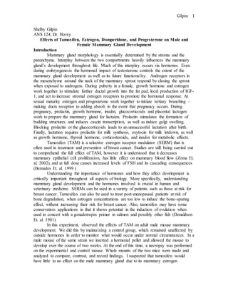
7. Gilpin 7
0
100
200
300
400
500
600
700
Male Female
# Branch Points
NumberofBranchPoints
Figure 4: Number of Mammary Gland
Branch Points in Males and Females
Control
Tam
E
E + Tam
E + D + P
E + P
0
50
100
150
200
250
Male Female
# End Buds
NumberofEndBuds
Figure 5: Number of End Buds in Males
and Females
Control
Tam
E
E + Tam
E + D + P
E + P